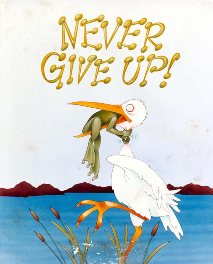
Pic of the day: Dopamine from a non-dopamine place

147 lc da ppvn - Copy

Reference: Beas BS, Wright BJ, Skirzewski M, Leng Y, Hyun JH, Koita O, Ringelberg N, Kwon HB, Buonanno A, & Penzo MA (Jul 2018, Epub 18 Jun 2018). The locus coeruleus drives disinhibition in the midline thalamus via a dopaminergic mechanism. Nature Neuroscience,21(7):963-973. PMID: 29915192, PMCID: PMC6035776 [Available on 2018-12-18], DOI:10.1038/s41593-018-0167-4. ARTICLE

Locus Coeruleus in mania

From all the mental disorders, bipolar disorder, a.k.a. manic-depressive disorder, has the highest risk for suicide attempt and completion. If the thought of suicide crosses your mind, stop reading this, it’s not that important; what’s important is for you to call the toll-free National Suicide Prevention Lifeline at 1-800-273-TALK (8255).

The bipolar disorder is defined by alternating manic episodes of elevated mood, activity, excitation, and energy with episodes of depression characterized by feelings of deep sadness, hopelessness, worthlessness, low energy, and decreased activity. It is also a more common disease than people usually expect, affecting about 1% or more of the world population. That means almost 80 million people! Therefore, it’s imperative to find out what’s causing it so we can treat it.

Unfortunately, the disease is very complex, with many brain parts, brain chemicals, and genes involved in its pathology. We don’t even fully comprehend how the best medication we have to lower the risk of suicide, lithium, works. The good news is the neuroscientists haven’t given up, they are grinding at it, and with every study we get closer to subduing this monster.

One such study freshly published last month, Cao et al. (2018), looked at a semi-obscure membrane protein, ErbB4. The protein is a tyrosine kinase receptor, which is a bit unfortunate because this means is involved in ubiquitous cellular signaling, making it harder to find its exact role in a specific disorder. Indeed, ErbB4 has been found to play a role in neural development, schizophrenia, epilepsy, even ALS (Lou Gehrig’s disease).

Given that ErbB4 is found in some neurons that are involved in bipolar and mutations in its gene are also found in some people with bipolar, Cao et al. (2018) sought to find out more about it.

First, they produced mice that lacked the gene coding for ErbB4 in neurons from locus coeruleus, the part of the brain that produces norepinephrine out of dopamine, better known for the European audience as nor-adrenaline. The mutant mice had a lot more norepinephrine and dopamine in their brains, which correlated with mania-like behaviors. You might have noticed that the term used was ‘manic-like’ and not ‘manic’ because we don’t know for sure how the mice feel; instead, we can see how they behave and from that infer how they feel. So the researchers put the mice thorough a battery of behavioral tests and observed that the mutant mice were hyperactive, showed less anxious and depressed behaviors, and they liked their sugary drink more than their normal counterparts, which, taken together, are indices of mania.

Next, through a series of electrophysiological experiments, the scientists found that the mechanism through which the absence of ErbB4 leads to mania is making another receptor, called NMDA, in that brain region more active. When this receptor is hyperactive, it causes neurons to fire, releasing their norepinephrine. But if given lithium, the mutant mice behaved like normal mice. Correspondingly, they also had a normal-behaving NMDA receptor, which led to normal firing of the noradrenergic neurons.

So the mechanism looks like this (Jargon alert!):

No ErbB4 –> ↑ NR2B NMDAR subunit –> hyperactive NMDAR –> ↑ neuron firing –> ↑ catecholamines –> mania.

In conclusion, another piece of the bipolar puzzle has been uncovered. The next obvious step will be for the researchers to figure out a medicine that targets ErbB4 and see if it could treat bipolar disorder. Good paper!

142 erbb4 - Copy

P.S. If you’re not familiar with the journal eLife, go and check it out. The journal offers for every study a half-page summary of the findings destined for the lay audience, called eLife digest. I’ve seen this practice in other journals, but this one is generally very well written and truly for the lay audience and the non-specialist. Something of what I try to do here, minus the personal remarks and in parenthesis metacognitions that you’ll find in most of my posts. In short, the eLife digest is masterly done. As my continuous struggles on this blog show, it is tremendously difficult for a scientist to write concisely, precisely, and jargonless at the same time. But eLife is doing it. Check it out. Plus, if you care to take a look on how science is done and published, eLife publishes all the editor’s rejection notes, all the reviewers’ comments, and all the author responses for a particular paper. Reading those is truly a teaching moment.

REFERENCE: Cao SX, Zhang Y, Hu XY, Hong B, Sun P, He HY, Geng HY, Bao AM, Duan SM, Yang JM, Gao TM, Lian H, Li XM (4 Sept 2018). ErbB4 deletion in noradrenergic neurons in the locus coeruleus induces mania-like behavior via elevated catecholamines. Elife, 7. pii: e39907. doi: 10.7554/eLife.39907. PMID: 30179154 ARTICLE | FREE FULLTEXT PDF

By Neuronicus, 14 October 2018

Treatment for lupus

Science has trends, like everything else. Some are longer or shorter lived, depending on how many astonishing discoveries are linked to that given subject. The 2000’s were unquestionably the years of the DNA. Many a grant have been written (and granted) for whole-genome surveys of this and that. Alternative splicing followed. The ’10s saw the rise of various -omics: transcriptomics, metabolomics, proteomics etc. Then everybody and his mamma got on the cart of epigenetics. With a side of immune stuff. Now, move aside epigenetics, here comes the microbiome. And CRISPR.

That is not to say that the not so hip subjects of the bygone years are thoroughly squeezed of knowledge and we throw them aside like some dry dead end and never touch them again. Not at all, not a bit. The trends only mark the momentary believes of the purse holders about which direction the next panaceum universalis will jump from.

Here comes a groundbreaking paper on the gut microbiome. It’s groundbreaking because it comes with a cure for systemic lupus erythematosus (SLE). Possibly autoimmune hepatitis and others autoimmune diseases as well.

An autoimmune disease is a terrible malady that is often incurable and sometimes deadly. It happens when the immune system starts attacking the body. One hypothesis as to why that happens posits that after a particular infection, maybe a particularly nasty one, the immune system doesn’t stop attacking, but now in the absence of an enemy it turns on its own body in genetically susceptible individuals.

Vieira et al. (2018) worked with genetically susceptible mice. And the bombshell comes right there in the first page: after treatment with an oral antibiotic (vancomycin or ampicillin, but not neomycin), mice genetically designed to develop lupus had lower “mortality, lupus-related autoantibodies, and autoimmune manifestations” (p. 1156). Then the researchers took a closer look at the bodies of these mice and observed that 82% of the mice had spleens and livers infected with Enterococcus gallinarum, a gut bacterium that should stay in the gut. But this bacterium is capable of weakening the gut barriers by loosening the tightness of the junctions between gut cells and then migrate to liver, spleen, and lymph nodes. Its high abundance in these places triggers a systemic immune response. Then the authors force-fed some germ-free mice with E. galinarum and saw that the mice developed systemic autoimmune pathology.

As if that’s not enough of a news story, the researchers developed a vaccine against this bacterium. The vaccine is very specific (being made of heat-killed E. gallinarum) and results in reduced levels of serum autoantibodies and prolonged survival rate in the lupus-prone mice.

So people don’t quibble, and rightly so, that those are rodents and humans are not (well, most of them, anyway), the authors looked at the liver biopsies of three humans with SLE and five with autoimmune hepatitis (AIH). They were positive for E. gallinarum, but the controls, i. e. healthy humans, were not. Also, when healthy human liver cells were stimulated with E. gallinarum they displayed autoimmune responses, just like in the murine cells. Finally, you don’t have to undergo a liver biopsy to see if you’re infected with E. gallinarum, just a specific blood test to see if you have increased antibody titers against this bug (or its RNA) as most SLE and AIH patients did.

Needless to say, I am extremely happy with this paper. Who wouldn’t be?! It’s a cure paper! I know, I know, they don’t say that, but what does this sound to you?:

“Administration of oral vancomycin or an intramuscular vaccine against E. gallinarum prevent translocation, Th17/Tfh cell induction, autoantibody production and autoimmune-related mortality (Supplemental, p. 62).”

Call it a very promising cure or a highly effective treatment if you like, but it stares you in the face for what it is as it did the researchers who already patented their stuff and are currently conducting clinical trials.

Most of the paper is in the Supplemental material, not in the 4 pages and a bit in Science. So even if the paper is under the paywall, the Supplementals are not. Be ready for a 71 page worth of 167 MB of data though.

134 lupus treat.jpg

REFERENCE: Manfredo Vieira S, Hiltensperger M, Kumar V, Zegarra-Ruiz D, Dehner C, Khan N, Costa FRC, Tiniakou E, Greiling T, Ruff W, Barbieri A, Kriegel C, Mehta SS, Knight JR, Jain D, Goodman AL, Kriegel MA (9 Mar 2018). Translocation of a gut pathobiont drives autoimmunity in mice and humans.  Science, 359(6380):1156-1161. doi: 10.1126/science.aar7201. PMID: 29590047, DOI: 10.1126/science.aar7201. ARTICLE |  Supplemental Material | Yale press release

By Neuronicus, 8 April 2018

Aging and its 11 hippocampal genes

Aging is being quite extensively studied these days and here is another advance in the field. Pardo et al. (2017) looked at what happens in the hippocampus of 2-months old (young) and 28-months old (old) female rats. Hippocampus is a seahorse shaped structure no more than 7 cm in length and 4 g in weight situated at the level of your temples, deep in the brain, and absolutely necessary for memory.

First the researchers tested the rats in a classical maze test (Barnes maze) designed to assess their spatial memory performance. Not surprisingly, the old performed worse than the young.

Then, they dissected the hippocampi and looked at neurogenesis and they saw that the young rats had more newborn neurons than the old. Also, the old rats had more reactive microglia, a sign of inflammation. Microglia are small cells in the brain that are not neurons but serve very important functions.

After that, the researchers looked at the hippocampal transcriptome, meaning they looked at what proteins are being expressed there (I know, transcription is not translation, but the general assumption of transcriptome studies is that the amount of protein X corresponds to the amount of the RNA X). They found 210 genes that were differentially expressed in the old, 81 were upregulated and 129 were downregulated. Most of these genes are to be found in human too, 170 to be exact.

But after looking at male versus female data, at human and mouse aging data, the authors came up with 11 genes that are de-regulated (7 up- and 4 down-) in the aging hippocampus, regardless of species or gender. These genes are involved in the immune response to inflammation. More detailed, immune system activates microglia, which stays activated and this “prolonged microglial activation leads to the release of pro-inflammatory cytokines that exacerbate neuroinflammation, contributing to neuronal loss and impairment of cognitive function” (p. 17). Moreover, these 11 genes have been associated with neurodegenerative diseases and brain cancers.

112hc-copy

These are the 11 genes: C3 (up), Cd74  (up), Cd4 (up), Gpr183 (up), Clec7a (up), Gpr34 (down), Gapt (down), Itgam (down), Itgb2 (up), Tyrobp (up), Pld4 (down).”Up” and “down” indicate the direction of deregulation: upregulation or downregulation.

I wish the above sentence was as explicitly stated in the paper as I wrote it so I don’t have to comb through their supplemental Excel files to figure it out. Other than that, good paper, good work. Gets us closer to unraveling and maybe undoing some of the burdens of aging, because, as the actress Bette Davis said, “growing old isn’t for the sissies”.

Reference: Pardo J, Abba MC, Lacunza E, Francelle L, Morel GR, Outeiro TF, Goya RG. (13 Jan 2017, Epub ahead of print). Identification of a conserved gene signature associated with an exacerbated inflammatory environment in the hippocampus of aging rats. Hippocampus, doi: 10.1002/hipo.22703. ARTICLE

By Neuronicus, 25 January 2017

Save

Save

Drink before sleep

Among the many humorous sayings, puns, and jokes that one inevitably encounters on any social medium account, one that was popular this year was about the similarity between putting a 2 year old to bed and putting your drunk friend to bed, which went like this: they both sing to themselves, request water, mumble and blabber incoherently, do some weird yoga posses, cry, hiccup, and then they pass out. The joke manages to steal a smile only if someone has been through both situations, otherwise it looses its appeal.

Being exposed to both situations, I thought that while the water request from the drunk friend is a response to the dehydrating effects of alcohol, the water request from the toddler is probably nothing more than a delaying tactic to postpone bedtime. Whether there may or may not be some truth to my assumption in the case of the toddler, here is a paper to show that there is definitely more to the water request than meets the eye.

Generally, thirst is generated by the hypothalamus when its neurons and neurons from organum vasculosum lamina terminalis (OVLT) in the brainstem sense that the blood is either too viscous (hypovolaemia) or too salty (hyperosmolality), both phenomena indicating a need for water. Ingesting water would bring these indices to homeostatic values.

More than a decade ago, researchers observed that rodents get a good gulp of water just before going to sleep. This surge was not motivated by thirst because the mice were not feverish, were not hungry and they did not have a too viscous or a too salty blood. So why do it then? If the rodents are restricted from drinking the water they get dehydrated, so obviously the behavior has function. But is not motivated by thirst, at least not the way we know it. Huh… The authors call this “anticipatory thirst”, because it keeps the animal from becoming dehydrated later on.

Since the behavior occurs with regularity, maybe the neurons that control circadian rhythms have something to do with it. So Gizowski et al. (2016) took a closer look at  the activity of clock neurons from the suprachiasmatic nucleus (SCN), a well known hypothalamic nucleus heavily involved in circadian rhythms. The authors did a lot of work on SCN and OVLT neurons: fluorescent labeling, c-fos expression, anatomical tracing, optogenetics, genetic knockouts, pharmacological manipulations, electrophysiological recordings, and behavioral experiments. All these to come to this conclusion:

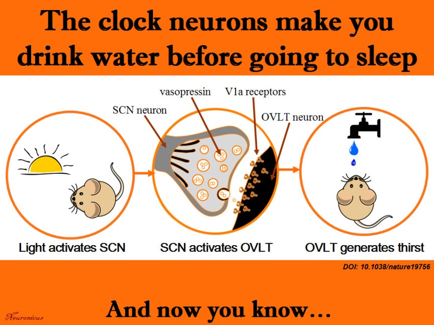
SCN neurons release vasopressin and that excites the OVLT neurons via V1a receptors. This is necessary and sufficient to make the animal drink the water, even if it’s not thirsty.

That’s a lot of techniques used in a lot of experiments for only three authors. Ten years ago, you needed only one, maybe two techniques to prove the same point. Either there have been a lot of students and technicians who did not get credit (there isn’t even an Acknowledgements section. EDIT: yes, there is, see the comments below or, if they’re missing, the P.S.) or these three authors are experts in all these techniques. In this day and age, I wouldn’t be surprised by either option. No wonder small universities have difficulty publishing in Big Name journals; they don’t have the resources to compete. And without publishing, no tenure… And without tenure, less research… And thus shall the gap widen.

Musings about workload aside, this is a great paper, shedding light on yet another mysterious behavior and elucidating the mechanism behind it. There’s still work to be done though, like answering how accurate is the SCN in predicting bedtime to activate the drinking behavior. Does it take its cues from light only? Does ambient temperature play a role and so on. This line of work can help people that work in shifts to prevent certain health problems. Their SCN is out of rhythm and that can influence deleteriously the activity of a whole slew of organs.

scn-h2o-copy
Summary of the doi: 10.1038/nature19756 findings. 1) The light is a cue for suprachiasmatic nulceus (SCN) that bedtime is near. 2) The SCN vasopressin neurons that project to organum vasculosum lamina terminalis (OVLT) are activated. 3) The OVLT generates the anticipatory thirst. 4) The animal drinks fluids.

Reference: Gizowski C, Zaelzer C, & Bourque CW (28 Sep 2016). Clock-driven vasopressin neurotransmission mediates anticipatory thirst prior to sleep. Nature, 537(7622): 685-688. PMID: 27680940. DOI: 10.1038/nature19756. ARTICLE

By Neuronicus, 5 October 2016

EDIT (12 Oct 2016): P.S. The blog comments are automatically deleted after a period of time. In case of this post that would be a pity because I have been fortunate to receive comments from at least one of the authors of the paper, the PI, Dr. Charles Bourque and, presumably under pseudonym, but I don’t know that for sure, also the first author, Claire Gizowski. So I will include here, in a post scriptum, the main idea of their comments. Here is an excerpt from Dr. Bourque’s comment:

“Let me state for the record that Claire accomplished pretty much ALL of the work in this paper (there is a description of who did what at the end of the paper). More importantly, there were no “unthanked” undergraduates, volunteers or other parties that contributed to this work.”

My hat, Ms. Gizowski. It is tipped. To you. Congratulations! With such an impressive work I am sure I will hear about you again and that pretty soon I will blog about Dr. Gizowski.

Who invented optogenetics?

Wayne State University. Ever heard of it? Probably not. How about Zhuo-Hua Pan? No? No bell ringing? Let’s try a different approach: ever heard of Stanford University? Why, yes, it’s one of the most prestigious and famous universities in the world. And now the last question: do you know who Karl Deisseroth is? If you’re not a neuroscientist, probably not. But if you are, then you would know him as the father of optogenetics.

Optogenetics is the newest tool in the biology kit that allows you to control the way a cell behaves by shining a light on it (that’s the opto part). Prior to that, the cell in question must be made to express a protein that is sensitive to light (i.e. rhodopsin) either by injecting a virus or breeding genetically modified animals that express that protein (that’s the genetics part).

If you’re watching the Nobel Prizes for Medicine, then you would also be familiar with Deisseroth’s name as he may be awarded the Nobel soon for inventing optogenetics. Only that, strictly speaking, he did not. Or, to be fair and precise at the same time, he did, but he was not the first one. Dr. Pan from Wayne State University was. And he got scooped.98.png

The story is at length imparted to us by Anna Vlasits in STAT and republished in Scientific American. In short, Dr. Pan, an obscure name in an obscure university from an ill-famed city (Detroit), does research for years in an unglamorous field of retina and blindness. He figured, quite reasonably, that restoring the proteins which sense light in the human eye (i.e. photoreceptor proteins) could restore vision in the congenitally blind. The problem is that human photoreceptor proteins are very complicated and efforts to introduce them into retinas of blind people have proven unsuccessful. But, in 2003, a paper was published showing how an algae protein that senses light, called channelrhodopsin (ChR), can be expressed into mammalian cells without loss of function.

So, in 2004, Pan got a colleague from Salus University (if Wayne State University is a medium-sized research university, then Salus is a really tiny, tiny little place) to engineer a ChR into a virus which Pan then injected in rodent retinal neurons, in vivo. After 3-4 weeks he obtained the expression of the protein and the expression was stable for at least 1 year, showing that the virus works nicely. Then his group did a bunch of electrophysiological recordings (whole cell patch-clamp and voltage clamp) to see if shining light on those neurons makes them fire. It did. Then, they wanted to see if ChR is for sure responsible for this firing and not some other proteins so they increased the intensity of the blue light that their ChR is known to sense and observed that the cell responded with increased firing. Now that they saw the ChR works in normal rodents, next they expressed the ChR by virally infecting mice who were congenitally blind and repeated their experiments. The electrophysiological experiments showed that it worked. But you see with your brain, not with your retina, so the researchers looked to see if these cells that express ChR project from the retina to the brain and they found their axons in lateral geniculate and superior colliculus, two major brain areas important for vision. Then, they recorded from these areas and the brain responded when blue light, but not yellow or other colors, was shone on the retina. The brain of congenitally blind mice without ChR does not respond regardless of the type of light shone on their retinas. But does that mean the mouse was able to see? That remains to be seen (har har) in future experiments. But the Pan group did demonstrate – without question or doubt – that they can control neurons by light.

All in all, a groundbreaking paper. So the Pan group was not off the mark when they submitted it to Nature on November 25, 2004. As Anna Vlasits reports in the Exclusive, Nature told Pan to submit to a more specialized journal, like Nature Neuroscience, which then rejected it. Pan submitted then to the Journal of Neuroscience, which also rejected it. He submitted it then to Neuron on November 29, 2005, which finally accepted it on February 23, 2006. Got published on April 6, 2006. Deisseroth’s paper was submitted to Nature Neuroscience on May 12, 2005, accepted on July, and published on August 14, 2005… His group infected rat hippocampal neurons cultured in a Petri dish with a virus carrying the ChR and then they did some electrophysiological recordings on those neurons while shining lights of different wavelengths on them, showing that these cells can be controlled by light.

There’s more on the saga with patent filings and a conference where Pan showed the ChR data in May 2005 and so on, you can read all about it in Scientific American. The magazine is just hinting to what I will say outright, loud and clear: Pan didn’t get published because of his and his institution’s lack of fame. Deisseroth did because of the opposite. That’s all. This is not about squabbles about whose work is more elegant, who presented his work as a scientific discovery or a technical report or whose title is more catchy, whose language is more boisterous or native English-speaker or luck or anything like that. It is about bias and, why not?, let’s call a spade a spade, discrimination. Nature and Journal of Neuroscience are not caught doing this for the first time. Not by a long shot. The problem is that they are still doing it, that is: discriminating against scientific work presented to them based on the name of the authors and their institutions.

Personally, so I don’t get comments along the lines of the fox and the grapes, I have worked at both high profile and low profile institutions. And I have seen the difference not in the work, but in the reception.

That’s my piece for today.

Source:  STAT, Scientific American.

References:

1) Bi A, Cui J, Ma YP, Olshevskaya E, Pu M, Dizhoor AM, & Pan ZH (6 April 2006). Ectopic expression of a microbial-type rhodopsin restores visual responses in mice with photoreceptor degeneration. Neuron, 50(1): 23-33. PMID: 16600853. PMCID: PMC1459045. DOI: 10.1016/j.neuron.2006.02.026. ARTICLE | FREE FULLTEXT PDF

2) Boyden ES, Zhang F, Bamberg E, Nagel G, & Deisseroth K. (Sep 2005, Epub 2005 Aug 14). Millisecond-timescale, genetically targeted optical control of neural activity. Nature Neuroscience, 8(9):1263-1268. PMID: 16116447. DOI: 10.1038/nn1525. doi:10.1038/nn1525. ARTICLE 

By Neuronicus, 11 September 2016

Save

Save

Save

Save

Painful Pain Paper

There has been much hype over the new paper published in the latest Nature issue which claims to have discovered an opioid analgesic that doesn’t have most of the side effects of morphine. If the claim holds, the authors may have found the Holy Grail of pain research chased by too many for too long (besides being worth billions of dollars to its discoverers).

The drug, called PZM21, was discovered using structure-based drug design. This means that instead of taking a drug that works, say morphine, and then tweaking its molecular structure in various ways and see if the resultant drugs work, you take the target of the drug, say mu-opioid receptors, and design a drug that fits in that slot. The search and design are done initially with sophisticated software and there are many millions of virtual candidates. So it takes a lot of work and ingenuity to select but a few drugs that will be synthesized and tested in live animals.

Manglik et al. (2016) did just that and they came up with PZM21 which, compared to morphine, is:

1) selective for the mu-opioid receptors (i.e. it doesn’t bind to anything else)
2) produces no respiratory depression (maybe a touch on the opposite side)
3) doesn’t affect locomotion
4) produces less constipation
5) produces long-lasting affective analgesia
6) and has less addictive liability

The Holy Grail, right? Weeell, I have some serious issues with number 5 and, to some extent, number 6 on this list.

Normally, I wouldn’t dissect a paper so thoroughly because, if there is one thing I learned by the end of GradSchool and PostDoc, is that there is no perfect paper out there. Consequently, anyone with scientific training can find issues with absolutely anything published. I once challenged someone to bring me any loved and cherished paper and I would tear it apart; it’s much easier to criticize than to come up with solutions. Probably that’s why everybody hates Reviewer No. 2…

But, for extraordinary claims, you need extraordinary evidence. And the evidence simply does not support the 5 and maybe 6 above.

Let’s start with pain. The authors used 3 tests: hotplate (drop a mouse on a hot plate for 10 sec and see what it does), tail-flick (give an electric shock to the tail and see how fast the mouse flicks its tail) and formalin (inject an inflammatory painful substance in the mouse paw and see what the animal does). They used 3 doses of PZM21 in the hotplate test (10, 20, and 40 mg/Kg), 2 doses in the tail-flick test (10 and 20), and 1 dose in the formalin test (20). Why? If you start with a dose-response in a test and want to convince me it works in the other tests, then do a dose-response for those too, so I have something to compare. These tests have been extensively used in pain research and the standard drug used is morphine. Therefore, the literature is clear on how different doses of morphine work in these tests. I need your dose-responses for your new drug to be able to see how it measures up, since you claim it is “more efficacious than morphine”. If you don’t want to convince me there is a dose-response effect, that’s fine too, I’ll frown a little, but it’s your choice. However, then choose a dose and stick with it! Otherwise I cannot compare the behaviors across tests, rendering one or the other test meaningless. If you’re wondering, they used only one dose of morphine in all the tests, except the hotplate, where they used two.

Another thing also related to doses. The authors found something really odd: PZM21 works (meaning produces analgesia) in the hotplate, but not the tail-flick tests. This is truly amazing because no opiate I know of can make such a clear-cut distinction between those two tests. Buuuuut, and here is a big ‘BUT” they did not test their highest dose (40mg/kg) in the tail-flick test! Why? I’ll tell you how, because I am oh sooo familiar with this argument. It goes like this:

Reviewer: Why didn’t you use the same doses in all your 3 pain tests?

Author: The middle and highest doses have similar effects in the hotplate test, ok? So it doesn’t matter which one of these doses I’ll use in the tail-flick test.

Reviewer: Yeah, right, but, you have no proof that the effects of the two doses are indistinguishable because you don’t report any stats on them! Besides, even so, that argument applies only when a) you have ceiling effects (not the case here, your morphine hit it, at any rate) and b) the drug has the expected effects on both tests and thus you have some logical rationale behind it. Which is not the case here, again: your point is that the drug DOESN’T produce analgesia in the tail-flick test and yet you don’t wanna try its HIGHEST dose… REJECT AND RESUBMIT! Awesome drug discovery, by the way!

So how come the paper passed the reviewers?! Perhaps the fact that two of the reviewers are long term publishing co-authors from the same University had something to do with it, you know, same views predisposes them to the same biases and so on… But can you do that? I mean, have reviewers for Nature from the same department for the same paper?

Alrighty then… let’s move on to the stats. Or rather not. Because there aren’t any for the hotplate or tail-flick! Now, I know all about the “freedom from the tyranny of p” movement (that is: report only the means, standard errors of mean, and confidence intervals and let the reader judge the data) and about the fact that the average scientist today needs to know 100-fold more stats that his predecessors 20 years ago (although some biologists and chemists seem to be excused from this, things either turn color or not, either are there or not etc.) or about the fact that you cannot get away with only one experiment published these days, but you need a lot of them so you have to do a lot of corrections to your stats so you don’t fall into the Type 1 error. I know all about that, but just like the case with the doses, choose one way or another and stick to it. Because there are ANOVAs ran for the formalin test, the respiration, constipation, locomotion, and conditioned place preference tests, but none for the hotplate or tailflick! I am also aware that to be published in Science or Nature you have to strip your work and wordings to the bare minimum because the insane wordcount limits, but you have free rein in the Supplementals. And I combed through those and there are no stats there either. Nor are there any power analyses… So, what’s going on here? Remember, the authors didn’t test the highest dose on the tail-flick test because – presumably – the highest and intermediary doses have indistinguishable effects, but where is the stats to prove it?

And now the thing that really really bothered me: the claim that PZM21 takes away the affective dimension of pain but not the sensory. Pain is a complex experience that, depending on your favourite pain researcher, has at least 2 dimensions: the sensory (also called ‘reflexive’ because it is the immediate response to the noxious stimulation that makes you retract by reflex the limb from whatever produces the tissue damage) and the affective (also called ‘motivational’ because it makes the pain unpleasant and motivates you to get away from whatever caused it and seek alleviation and recovery). The first aspect of pain, the sensory, is relatively easy to measure, since you look at the limb withdrawal (or tail, in the case of animals with prolonged spinal column). By contrast, the affective aspect is very hard to measure. In humans, you can ask them how unpleasant it is (and even those reports are unreliable), but how do you do it with animals? Well, you go back to humans and see what they do. Humans scream “Ouch!” or swear when they get hurt (so you can measure vocalizations in animals) or humans avoid places in which they got hurt because they remember the unpleasant pain (so you do a test called Conditioned Place Avoidance for animals, although if you got a drug that shows positive results in this test, like morphine, you don’t know if you blocked the memory of unpleasantness or the feeling of unpleasantness itself, but that’s a different can of worms). The authors did not use any of these tests, yet they claim that PZM21 takes away the unpleasantness of pain, i.e. is an affective analgesic!

What they did was this: they looked at the behaviors the animal did on the hotplate and divided them in two categories: reflexive (the lifting of the paw) and affective (the licking of the paw and the jumping). Now, there are several issues with this dichotomy, I’m not even going to go there; I’ll just say that there are prominent pain researchers that will scream from the top of their lungs that the so-called affective behaviors from the hotplate test cannot be indexes of pain affect, because the pain affect requires forebrain structures and yet these behaviors persist in the decerebrated rodent, including the jumping. Anyway, leaving the theoretical debate about what those behaviors they measured really mean aside, there still is the problem of the jumpers: namely, the authors excluded from the analysis the mice who tried to jump out of the hotplate test in the evaluation of the potency of PZM21, but then they left them in when comparing the two types of analgesia because it’s a sign of escaping, an emotionally-valenced behavior! Isn’t this the same test?! Seriously? Why are you using two different groups of mice and leaving the impression that is only one? And oh, yeah, they used only the middle dose for the affective evaluation, when they used all three doses for potency…. And I’m not even gonna ask why they used the highest dose in the formalin test… but only for the normal mice, the knockouts in the same test got the middle dose! So we’re back comparing pears with apples again!

Next (and last, I promise, this rant is way too long already), the non-addictive claim. The authors used the Conditioned Place Paradigm, an old and reliable method to test drug likeability. The idea is that you have a box with 2 chambers, X and Y. Give the animal saline in chamber X and let it stay there for some time. Next day, you give the animal the drug and confine it in chamber Y. Do this a few times and on the test day you let the animal explore both chambers. If it stays more in chamber Y then it liked the drug, much like humans behave by seeking a place in which they felt good and avoiding places in which they felt bad. All well and good, only that is standard practice in this test to counter-balance the days and the chambers! I don’t know about the chambers, because they don’t say, but the days were not counterbalanced. I know, it’s a petty little thing for me to bring that up, but remember the saying about extraordinary claims… so I expect flawless methods. I would have also liked to see a way more convincing test for addictive liability like self-administration, but that will be done later, if the drug holds, I hope. Thankfully, unlike the affective analgesia claims, the authors have been more restrained in their verbiage about addiction, much to their credit (and I have a nasty suspicion as to why).

I do sincerely think the drug shows decent promise as a painkiller. Kudos for discovering it! But, seriously, fellows, the behavioral portion of the paper could use some improvements.

Ok, rant over.

EDIT (Aug 25, 2016): I forgot to mention something, and that is the competing financial interests declared for this paper: some of its authors already filed a provisional patent for PZM21 or are already founders or consultants for Epiodyne (a company that that wants to develop novel analgesics). Normally, that wouldn’t worry me unduly, people are allowed to make a buck from their discoveries (although is billions in this case and we can get into that capitalism-old debate whether is moral to make billions on the suffering of other people, but that’s a different story). Anyway, combine the financial interests with the poor behavioral tests and you get a very shoddy thing indeed.

Reference: Manglik A, Lin H, Aryal DK, McCorvy JD, Dengler D, Corder G, Levit A, Kling RC, Bernat V, Hübner H, Huang XP, Sassano MF, Giguère PM, Löber S, Da Duan, Scherrer G, Kobilka BK, Gmeiner P, Roth BL, & Shoichet BK (Epub 17 Aug 2016). Structure-based discovery of opioid analgesics with reduced side effects. Nature, 1-6. PMID: 27533032, DOI: 10.1038/nature19112. ARTICLE 

By Neuronicus, 21 August 2016
Fructose bad effects reversed by DHA, an omega-3 fatty acid

Despite alarm signals raised by various groups and organizations regarding the dangers of the presence of sugars – particularly fructose derived from corn syrup – in almost every food in the markets, only in the past decade there has been some serious evidence against high consumption of fructose.

A bitter-sweet (sic!) paper comes from Meng et al. (2016) who, in addition to showing some bad things that fructose does to brain and body, it also shows some rescue from its deleterious effects by DHA (docosahexaenoic acid), an omega-3 fatty acid.

The authors had 3 groups of rodents: one group got fructose in their water for 6 weeks, another group got fructose and DHA, and another group got their normal chow. The amount of fructose was calculated to be ecologically valid, meaning that they fed the animals the equivalent of 1 litre soda bottle per day (130 g of sugar for a 60 Kg human).

The rats that got fructose had worse learning and memory performance at a maze test compared to the other two groups.

The rats that got fructose had altered gene expression in two brain areas: hypothalamus (involved in metabolism) and hippocampus (involved in learning and memory) compared to the other two groups.

The rats that got fructose had bad metabolic changes that are precursors for Type 2 diabetes, obesity and other metabolic disorders (high blood glucose, triglycerides, insulin, and insulin resistance index) compared to the other two groups.

86 - Copy.jpg

The genetic analyses that the researchers did (sequencing the RNA and analyzing the DNA methylation) revealed a whole slew of the genes that had been affected by the fructose treatment. So, they did some computer work that involved Bayesian modeling  and gene library searching and they selected two genes (Bgn and Fmod) out of almost a thousand possible candidates who seemed to be the drivers of these changes. Then, they engineered mice that lacked these genes. The resultant mice had the same metabolic changes as the rats that got fructose, but… their learning and memory was even better than that of the normals? I must have missed something here. EDIT: Well… yes and no. Please read the comment below from the Principal Investigator of the study.

It is an ok paper, done by the collaboration of 7 laboratories from 3 countries. But there are a few things that bother me, as a neuroscientist, about it. First is the behavior of the genetic knock-outs. Do they really learn faster? The behavioral results are not addressed in the discussion. Granted, a genetic knockout deletes that gene everywhere in the brain and in the body, whereas the genetic alterations induced by fructose are almost certainly location-specific.

Which brings me to the second bother: nowhere in the paper (including the supplemental materials, yeas, I went through those) are any brain pictures or diagrams or anything that can tell us which nuclei of the hypothalamus the samples came from. Hypothalamus is a relatively small structure with drastically different functional nuclei very close to one another. For example, the medial preoptic nucleus that deals with sexual hormones is just above the suprachiasmatic nucleus that deals with circadian rhythms and near the preoptic is the anterior nucleus that deals mainly with thermoregulation. The nuclei that deal with hunger and satiety (the lateral and the ventromedial nucleus, respectively) are located in different parts of the hypothalamus. In short, it would matter very much where they got their samples from because the transcriptome and methylome would differ substantially from nucleus to nucleus. Hippocampus is not so complicated as that, but it also has areas with specialized functions. So maybe they messed up the identification of the two genes Bgn and Fmod as drivers of the changes; after all, they found almost 1 000 genes altered by fructose. And that mess-up might have been derived by their blind hypothalamic and hippocampal sampling. EDIT: They didn’t mess up,  per se. Turns out there were technical difficulties of extracting enough nucleic acids from specific parts of hypothalamus for analyses. I told you them nuclei are small…

Anyway, the good news comes from the first experiment, where DHA reverses the bad effects of fructose. Yeay! As a side note, the fructose from corn syrup is metabolized differently than the fructose from fruits. So you are far better off consuming the equivalent amount on fructose from a litre of soda in fruits. And DHA comes either manufactured from algae or extracted from cold-water oceanic fish oils (but not farmed fish, apparently).

If anybody that read the paper has some info that can help clarify my “bothers”, please do so in the Comment section below. The other media outlets covering this paper do not mention anything about the knockouts. Thanks! EDIT: The last author of the paper, Dr. Yang, was very kind and clarified a bit of my “bothers” in the Comments section. Thanks again!

Reference: Meng Q, Ying Z, Noble E, Zhao Y, Agrawal R, Mikhail A, Zhuang Y, Tyagi E, Zhang Q, Lee J-H, Morselli M, Orozco L, Guo W, Kilts TM, Zhu J, Zhang B, Pellegrini M, Xiao X, Young MF, Gomez-Pinilla F, Yang X (2016). Systems Nutrigenomics Reveals Brain Gene Networks Linking Metabolic and Brain Disorders. EBioMedicine, doi: 10.1016/j.ebiom.2016.04.008. Article | FREE fulltext PDF | Supplementals | Science Daily cover | NeuroscienceNews cover

By Neuronicus, 24 April 2016

Autism cure by gene therapy

shank3 - Copy

Nothing short of an autism cure is promised by this hot new research paper.

Among many thousands of proteins that a neuron needs to make in order to function properly there is one called SHANK3 made from the gene shank3. (Note the customary writing: by consensus, a gene’s name is written using small caps and italicized, whereas the protein’s name that results from that gene expression is written with caps).

This protein is important for the correct assembly of synapses and previous work has shown that if you delete its gene in mice they show autistic-like behavior. Similarly, some people with autism, but by far not all, have a deletion on Chromosome 22, where the protein’s gene is located.

The straightforward approach would be to restore the protein production into the adult autistic mouse and see what happens. Well, one problem with that is keeping the concentration of the protein at the optimum level, because if the mouse makes too much of it, then the mouse develops ADHD and bipolar.

So the researchers developed a really neat genetic model in which they managed to turn on and off the shank3 gene at will by giving the mouse a drug called tamoxifen (don’t take this drug for autism! Beside the fact that is not going to work because you’re not a genetically engineered mouse with a Cre-dependent genetic switch on your shank3, it is also very toxic and used only in some form of cancers when is believed that the benefits outweigh the horrible side effects).

In young adult mice, the turning on of the gene resulted in normalization of synapses in the striatum, a brain region heavily involved in autistic behaviors. The synapses were comparable to normal synapses in some aspects (from the looks, i.e. postsynaptic density scaffolding, to the works, i.e. electrophysiological properties) and even more so in others (more dendritic spines than normal, meaning more synapses, presumably). This molecular repair has been mirrored by some behavioral rescue: although these mice still had more anxiety and more coordination problems than the control mice, their social aversion and repetitive behaviors disappeared. And the really really cool part of all this is that this reversal of autistic behaviors was done in ADULT mice.

Now, when the researchers turned the gene on in 20 days old mice (which is, roughly, the equivalent of the entering the toddling stage in humans), all four behaviors were rescued: social aversion, repetitive, coordination, and anxiety. Which tells us two things: first, the younger you intervene, the more improvements you get and, second and equally important, in adult, while some circuits seem to be irreversibly developed in a certain way, some other neural pathways are still plastic enough as to be amenable to change.

Awesome, awesome, awesome. Even if only a very small portion of people with autism have this genetic problem (about 1%), even if autism spectrum disorders encompass such a variety of behavioral abnormalities, this research may spark hope for a whole range of targeted gene therapies.

Reference: Mei Y, Monteiro P, Zhou Y, Kim JA, Gao X, Fu Z, Feng G. (Epub 17 Feb 2016). Adult restoration of Shank3 expression rescues selective autistic-like phenotypes. Nature. doi: 10.1038/nature16971. Article | MIT press release

By Neuronicus, 19 February 2016

Save

A third way of neuronal communication

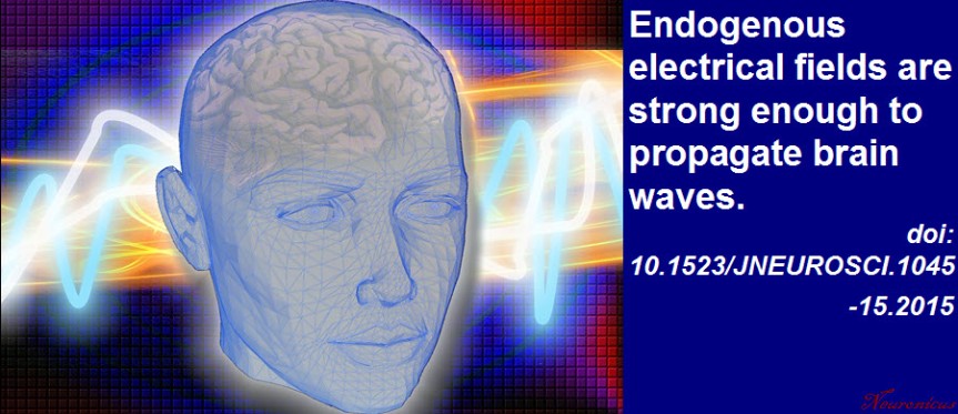
electrical fields - CopyEvery neuroscience or biology textbook will tell you that neurons communicate with one another in two ways: via chemical synapses (one neuron releases some substances that change the membrane potential of the receiving neuron) or via electrical synapses (neurons physically share some membrane proteins that allows the electrical impulse to go from one neuron to another). (Nota bene: for the taxonomic nitpickers, I decided that the other non-conventional forms of communication, like diffusion, fall in one or the other of the two categories above).
Now Qiu et al. (2015) have some evidence that there may be another way of neuronal chatting. Long ago (in 1924), Hans Berger observed that neurons have rhythmic and spontaneous electrical activity. The rhythmic activity of an entire population of neurons is called a wave and can be detected with rudimentary tools such as EEG. Qiu et al. reasoned that the speed of such a traveling wave is too slow to be explained by conventional neuronal communication (of which we know quite a lot).

So the researchers ran some computer simulations to test if diffuse electrical fields can explain the speed of a wave, instead of the conventional fast communications. In other words, can local, weak, slow endogenous fields sweep the brain by recruiting nearby neurons? The computer simulations said it is possible, with a very slow speed of 10 cm/s.

Then they recorded the electrical activity of mouse hippocampus isolated in a Petri dish. When the researchers blocked the electrical field there was a decrease in the speed of the wave, meaning the field is required for the wave speed observed.

To be fair, this is not a new idea; even in the early eighties these fields were suggested, because the wave persisted even when the other two ways of communicating have been blocked. And in the early noughts it was shown that a neural network is much more sensitive to electrical field manipulation than individual neurons. What makes this paper interesting is that the authors show that the endogenous electrical fields are not too weak to underlie the wave, as previously thought. The work has implications for the study of epilepsy, sleep, and memory.

Reference: Qiu C, Shivacharan RS, Zhang M, & Durand DM (2 December 2015). Can Neural Activity Propagate by Endogenous Electrical Field?, Journal of Neuroscience, 35(48): 15800-15811; doi: 10.1523/JNEUROSCI.1045-15.2015 Article | Full Text via Research Gate | ScienceAlert cover

By Neuronicus, 11 February 2016

CCL11 found in aged but not young blood inhibits adult neurogenesis

vil - Copy
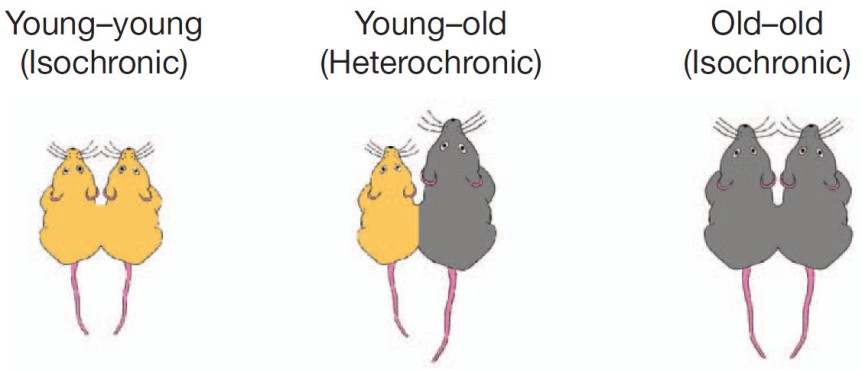
Portion of Fig. 1 from Villeda et al. (2011, doi: 10.1038/nature10357) describing the parabiosis procedure. Basically, under complete anesthesia, the peritoneal membranes and the skins of the two mice were sutured together. The young mice were 3–4 months (yellow) and old mice were 18–20 months old (grey).

My last post was about parabiosis and its sparse revival as a technique in physiology experiments. Parabiosis is the surgical procedure that joins two living animals allowing them to share their circulatory systems. Here is an interesting paper that used the method to tackle blood’s contribution to neurogenesis.

Adult neurogenesis, that is the birth of new neurons in the adult brain, declines with age. This neurogenesis has been observed in some, but not all brain regions, called neurogenic niches.

Because these niches occur in blood-rich areas of the brain, Villeda et al. (2011) wondered if, in addition with the traditional factors required for neurogenesis like enrichment or running, blood factors may also have something to do with neurogenesis. The authors made a young and an old mouse to share their blood via parabiosis (see pic.).

Five weeks after the parabiosis procedure, the young mouse had decreased neurogenesis and the old mouse had increased neurogenesis compared to age-matched controls. To make sure their results are due to something in the blood, they injected plasma from an old mouse into a young mouse and that also resulted in reduced neurogenesis. Moreover, the reduced neurogenesis was correlated with impaired learning as shown by electrophysiological recordings from the hippocampus and from behavioral fear conditioning.

So what in the blood does it? The authors looked at 66 proteins found in the blood (I don’t know the blood make-up, so I can’t tell if 66 is a lot or not ) and noticed that 6 of these had increased levels in the blood of ageing mice whether linked by parabiosis or not. Out of these six, the authors focus on CCL11 (unclear to me why that one, my bet is that they tried the others too but didn’t have enough data). CCLL11 is a small signaling protein involved in allergies. So the authors injected it into young mice and Lo and Behold! there was decreased neurogenesis in their hippocampus. Maybe the vampires were onto something, whadda ya know? Just kidding… don’t go around sucking young people’s blood!

This paper covers a lot of work and, correspondingly, has no less than 23 authors and almost 20 Mb of supplemental documents! The story it tells is very interesting and as complete as it gets, covering many aspects of the problems investigated and many techniques to address those problems. Good read.

Reference: Villeda SA, Luo J, Mosher KI, Zou B, Britschgi M, Bieri G, Stan TM, Fainberg N, Ding Z, Eggel A, Lucin KM, Czirr E, Park JS, Couillard-Després S, Aigner L, Li G, Peskind ER, Kaye JA, Quinn JF, Galasko DR, Xie XS, Rando TA, Wyss-Coray T. (31 Aug 2011). The ageing systemic milieu negatively regulates neurogenesis and cognitive function. Nature. 477(7362):90-94. doi: 10.1038/nature10357. Article | FREE Fulltext PDF

By Neuronicus, 6 January 2016

Your blood is better than my blood

pictures-004
Siamese tomatoes. Taken from here.

Parabiosis is a surgical procedure that lets two animals to share the same blood; it’s a case of reverse conjoined twins restricted to the circulatory system.

The procedure is over 150 years old and is a useful technique in physiology, though rarely used, probably due to the perceived cruelty towards the animals, although today is performed under anesthesia and aseptic condition. It delivered good data; for example, it was a parabiosis experiment with rodents that showed is not the sugar in the blood that causes cavities but the sugar in the mouth. Similarly, parabiosis has been proven useful in cancer, diabetes, and ageing research.

Scudellari (2015) wrote a News piece for Nature describing some advancements in the ageing field using the parabiosis technique. Namely, by joining the circulatory systems of a young and an old mouse, researchers have observed that the old mouse is faster, smarter, with rejuvenated muscles and glossier fur. Now the race is to find out what in the blood does it. Researchers caution that the young blood is not effectively reversing ageing, but may have factors circulating in it that promote tissue repair. Already a muscle-rejuvenating protein has been identified.

I am not going through the original papers themselves as I usually do (they are provided as links in the Reference paper). Instead, I am featuring the news piece by Scudellari because in addition of looking at parabiosis and ageing result, it also provides a nice historical account of the use of parabiosis. Enjoy!

Reference: Scudellari, M. (22 Jan 2015). Ageing research: Blood to blood. Nature, 517: 426-429. Article | FREE Fulltext PDF

By Neuronicus, 4 January 2015

Prostaglandins in the sickness syndrome

63woman-698962_960_720When you’re sick you also feel awful: no appetite, weak, sleepy, feverish, achy, and so on. This is called, appropriately so, the sickness syndrome.
Saper, Romanovsky & Scammell (2012) wrote a beautiful review of the neural circuits underlying this collection of symptoms. In a nutshell, the immune system releases cytokines to fight the inflammation, which in turn stimulate the release of prostaglandins. Prostaglandins bind to various areas in the brain to produce the sickness syndrome symptoms. Below are outlined four simplified brain circuits which the non-specialists can skip entirely.

  1. Prostaglandins in the median preoptic nucleus lead to a cascade involving dorsomedial hypothalamus, rostral medullary raphe and finally the spinal cord to produce fever by activating the brown adipose tissue.
  2. Prostaglandins in the preoptic area lead to the inhibition of the brain’s analgesic system involving the descending projections of the periaqueductal grey to spinal cord, thus promoting achiness.
  3. Prostaglandins in the meninges result in adenosine release in nucleus accumbens and ventrolateral preoptic nucleus which, downstream, end in inhibiting the arousal system to produce sleepiness.
  4. Prostaglandins in the arcuate nucleus lead to inhibition of several hypothalamic nuclei involved in promoting feeding, thereby producing anorexia.

The sickness syndrome and the role prostaglandins play in it has tremendous adaptive role, as it promotes rest and recuperation. So don’t blame them too much. And if you’re really done feeling sick, take some non-steroid anti-inflammatory drugs, like aspirin, which inhibit the prostaglandins’ synthesis very effectively. That’s how and why NSAIDs work.

Reference: Saper CB, Romanovsky AA & Scammell TE (26 Jul 2012). Neural Circuitry Engaged by Prostaglandins during the Sickness Syndrome. Nature Neuroscience, 15(8):1088-95. doi: 10.1038/nn.3159. Article | FREE Fulltext PDF

By Neuronicus, 21 December 2015

The culprit in methamphetamine-induced psychosis is very likely BDNF

Psychoses. Credit: NIH (Publication Number 15-4209) & Neuronicus.
Psychoses. Credit: NIH (Publication Number 15-4209) & Neuronicus. License: PD.

Methamphetamine prolonged use may lead to psychotic episodes in the absence of the drug. These episodes are persistent and closely resemble schizophrenia. One of the (many) molecules involved in both schizophrenia and meth abuse is BDNF (brain derived neurotrophic factor), a protein mainly known for its role in neurogenesis and long-term memory.

Lower BDNF levels have been observed in schizophrenia, therefore Manning et al. (2015) wondered if it’s also involved in meth-induced psychosis. So they got normal mice and mice that were genetically engineered to express lower levels of BDNF. They gave them meth for 3 weeks, with escalating doses form one week to the next. Interestingly, no meth on weekends, which made me rapidly scroll to the beginning of the paper and confirm my suspicion that the experiments were not done in USA; if they were, the grad students would not have had the weekends off and mice would have received meth every day, including weekends. Look how social customs can influence research! Anyway, social commentary aside, after the meth injections, the researchers let the mice untroubled for 2 more weeks. And then they tested them on a psychosis test.

How do you measure psychosis in rodents? By inference, since the mouse will not grab your coat and tell you about the newly appeared hypnotizing wall pattern and the like. Basically, it was observed that psychotic people have a tendency to walk in a disorganized manner when given the opportunity to explore, a behavior that was also observed in rodents on amphetamines. This disorganized walk can be quantifies into an entropic index, which is thought to reflect occurrence of psychosis (I know, a lot of inferring. But you come up with a better model of psychosis in rodent!).

Manning et al. (2015) gave their mice amphetamine to mimic psychosis and then observed their behavior. And the results were that the genetically engineered mice to express less BDNF showed reduced psychosis (i.e. had a lower entropic index). In conclusion, the alteration of the BDNF pathway may be responsible for the development of psychosis in methamphetamine users.

Reference: Manning EE, Halberstadt AL, & van den Buuse M. (Epub 9 Oct 2015). BDNF-Deficient Mice Show Reduced Psychosis-Related Behaviors Following Chronic Methamphetamine. International Journal of Neuropsychopharmacology, 1–5. doi: 10.1093/ijnp/pyv116. Article | FREE FULLTEXT PDF

By Neuronicus, 9 November 2015

Hope for a new migraine medication

Headache clipart. Courtesy of ClipArtHut.
Headache. Courtesy of ClipArtHut.

The best current anti-migraine medication are triptans (5-HT1B/1D receptor agonists). Because these medications are contra-indicated in patients with a variety of other diseases (cardiovascular, renal, hepatic, etc.), the search for alternative drugs continues.

The heat- and pain-sensitive TRPV1 receptors (Transient Receptor Potential Vanilloid 1) localized on the trigeminal terminals (the fifth cranial nerve) have been implicated in the production of headaches. That is, if you activate them by, say, capsaicin, the same substance that gives the chili peppers their hotness, you get headaches (you’d have to eat an awful lot of peppers to get the migraine, though). On the other hand, if you block these receptors by triptans, you alleviate the migraines. All good and well, so let’s hunt for some TRPV1 antagonists, i.e. blockers. But, as theory often doesn’t meet practice, the first two antagonists that were tried were dropped in the clinical trials for lack of efficiency.

Meents et al. (2015) are giving another try to two different TRPV1 antagonists, by their fetching names of JNJ-38893777 and JNJ-17203212, respectively. Because you cannot ask a rat if it has a headache, it is very difficult to have a rodent model for migraine. Instead, researchers focused on giving rats some inflammatory soup directly into the subarachnoid space or capsaicin directly into the carotid artery, actions which they have reasons to believe produce severe headaches and some biological changes, like increase in a certain gene expression (c-fos, if you must know) in the trigeminal brain stem complex and release of the neurotransmitter calcitonin gene-related peptide (CGRP).

JNJ-17203212 got rid of all those physiological changes in a dose-dependent manner, presumably of the migraine, too. The other drug, JNJ-38893777, was effective only on the highest dose. Give these drugs a few more tests to pass, and off to the clinical trials with them. I’m joking, it takes a lot more research than just a paper between discovery and human drug trials.

Reference: Meents JE, Hoffmann J, Chaplan SR, Neeb L, Schuh-Hofer S, Wickenden A, & Reuter U (December 2015, Epub 24 June 2015). Two TRPV1 receptor antagonists are effective in two different experimental models of migraine. The Journal of Headache and Pain. 16:57. doi: 10.1186/s10194-015-0539-z. Article | FREE FULLTEXT PDF

By Neuronicus, 8 November 2015

How long does it take for environmental enrichment to show effects?

From funnyvet.
From funnyvet.

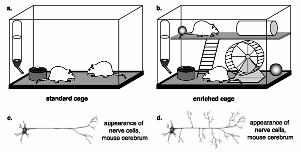
Environmental enrichment is a powerful way to give a boost to neurogenesis and alleviate some anxiety and depression symptoms. For the laboratory rodents, who spend their lives in cages with water and food access, environmental enrichment can refer to as little as a toy or two or as much as large room colonies with different size tubes, different levels to explore, nesting materials, plenty of toys with various shapes, textures, and colors, exercise wheels, and even the occasional fruit or peanut butter snack. But for how long does a mouse need to be exposed to enrichment to show cognitive and emotional improvement?

Leger et al. (2015) ran several anxiety, depression, and long-term memory tests in mice who have been exposed to environmental enrichment for 24 h, 1, 3, or 5 weeks. Although 24 h exposure was enough to improve memory, only after 3-week exposure some anxiety behaviors were attenuated. No effect on depressive behaviors or coticosterone levels, which may be due to that particular strain of mouse (several other studies found that environmental enrichment ameliorates depressive symptoms in other mice strains and rats). The 3-week exposure also increased the levels of serotonin in the frontal cortex. Only after 5-eweek exposure there was a significant survival rate of the hippocampal new cells. Of note, these were normal mice, i.e. they were not suffering from any disorder prior to exposure.

Mice raised in an impoverished environment (a) show less dendrite growth (c) than do mice raised in an enriched environment (b, d). Copyright: BSCS.

The findings give us a nice timeline for environmental enrichment to show its desired effects. But… if there are differences in the timeline and effects of environmental enrichment exposure from mouse strain to mouse strain, then what can we say for humans? Probably not much, unfortunately. As the ad nauseam overused phrase goes at the end of so many papers, ‘more research is needed to elucidate this problem’.

Reference: Leger M, Paizanis E, Dzahini K, Quiedeville A, Bouet V, Cassel JC, Freret T, Schumann-Bard P, & Boulouard M. (Nov 2015, Epub 5 Jun 2014). Environmental Enrichment Duration Differentially Affects Behavior and Neuroplasticity in Adult Mice. Cerebral Cortex, 25(11):4048-61. doi: 10.1093/cercor/bhu119. Article | FREE PDF

By Neuronicus, 1 November 2015

Grooming only half side of the body

Grooming only half side of the body. Credit: http://www.bajiroo.com/2013/04/23-guys-with-half-shaved-beard/

Contrary to popular belief, rats and mice are very fastidious animals; they keep themselves scrupulously clean by engaging in a very meticulous routine of self-grooming. The routine is so rigorous that allows the researchers to divide the grooming sequence into four different phases, starting with the nose and whiskers and ending with the genitalia and tail. It is also a symmetrical behavior (no whisker left ungroomed, no paw unlicked).

Grooming is sensitive to dopaminergic manipulations, so Pelosi, Girault, & Hervé (2015) sought to see what happens if they destroy the dopamine fibers in the mouse brain. So they lesioned the medial forebrain bundle, which is a bunch axon fibers that contains over 80% of the midbrain dopaminergic axons. But they were tricky, they lesioned only one side.

And the results were that the lesioned mice not only exhibited less self-grooming on the opposite side to the lesion, but the behavior was rescued by L-DOPA, which is medication for Parkinson’s. That is, they gave the mice some L-DOPA and they began to merrily self-groom again on both sides of the body. The authors discuss in depths other findings, like the changes (or absence thereof) in grooming bouts, grooming time, grooming bouts, completeness of grooming etc.

The findings have significance in the Parkinson’s research, where the mild to moderate phases of the disease often present with asymmetrical motor behavior.

Reference: Pelosi A, Girault J-A, & Hervé D (23 Sept 2015). Unilateral Lesion of Dopamine Neurons Induces Grooming Asymmetry in the Mouse. PLoS One. 2015; 10(9): e0137185. doi: 10.1371/journal.pone.0137185. PMCID: PMC4580614. Article | FREE FULLTEXT PDF

By Neuronicus, 29 October 2015

Fat & afraid or slim & brave (Leptin and anxiety in ventral tegmental area)

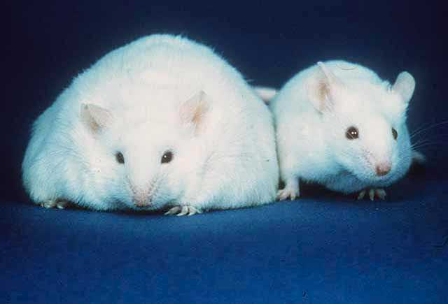
A comparison of a mouse unable to produce leptin thus resulting in obesity (left) and a normal mouse (right). Courtesy of Wikipedia. License: PD

Leptin is a small molecule produced mostly by the adipose tissue, whose absence is the cause of morbid obesity in the genetically engineered ob/ob mice. Here is a paper that gives us another reason to love this hormone.

Liu, Guo, & Lu (2015) build upon their previous work of investigating the leptin action(s) in the ventral tegmental area of the brain (VTA), a region that houses dopamine neurons and widely implicated in pleasure and drug addiction (among other things). They did a series of very straightforward experiments in which the either infused leptin directly into the mouse VTA or deleted the leptin receptors in this region (by using a virus in genetically engineered mice). Then they tested the mice on three different anxiety tests.

The results: leptin decreases anxiety; absence of leptin receptors increases anxiety. Simple and to the point. And also makes sense, given that leptin receptors are mostly located on the VTA neurons that project to the central amygdala, a region involved in fear and anxiety (curiously, the authors cite the amygdala papers, but do not comment on the leptin-VTA-dopamine-amygdala connection). For the specialists, I would say that they are a little liberal with their VTA hit assessment (they are mostly targeting the posterior VTA) and their GFP (green fluorescent protein) is sparsely expressed.

Reference: Liu J, Guo M, & Lu XY (Epub ahead of print 5 Oct 2015). Leptin/LepRb in the Ventral Tegmental Area Mediates Anxiety-Related Behaviors. International Journal of Neuropsychopharmacology, 1–11. doi:10.1093/ijnp/pyv115. Article | FREE PDF

By Neuronicus, 28 October 2015

You were not my first choice either!

lego
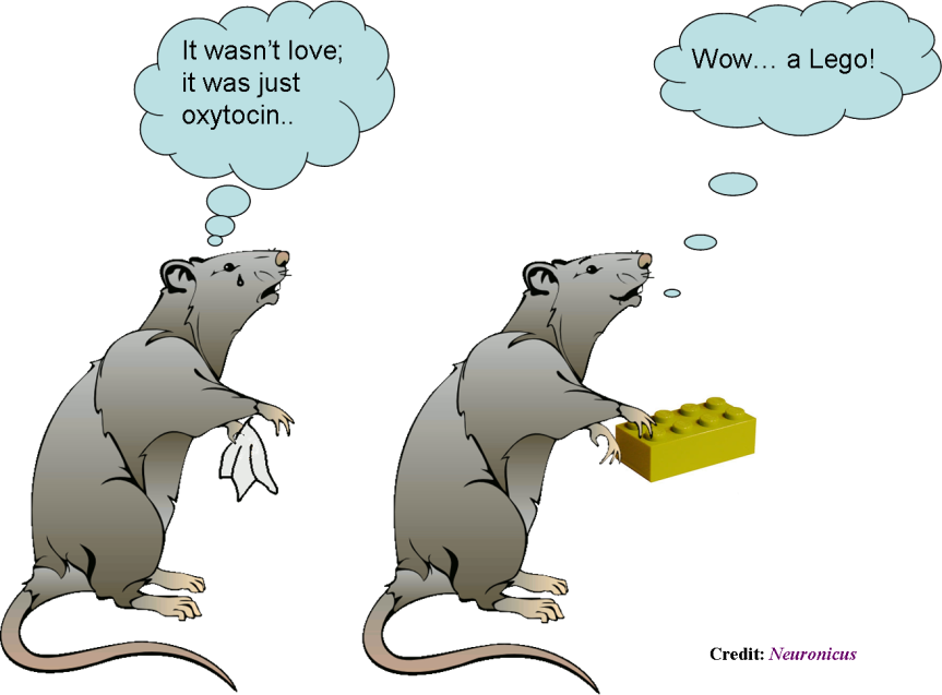
Sexually receptive female mice prefer a Lego brick over a male if their prefrontal oxytocin neurons are silenced.

Over the past five years or so, dopamine stepped down from the role of the “love molecule” in favor of oxytocin, a hormone previously known mostly for its crucial role in pregnancy, labor, delivery, lactation, and breastfeeding. Since some interesting discoveries in monogamous vs. polygamous voles (a type of rodent) pointing to oxytocin as essential for bonding, many studies implicated the chemical in all sorts of behaviors, from autistic to trusting, from generosity to wound healing.

Nakajima, Görlich, & Heintz (2015) add to that body of knowledge by finding that only a small group of cells in the medial prefrontal cortex express oxytocin receptors: a subpopulation of somatostatin cortical interneurons. Moreover, these neurons are gender dimorphic, meaning they differ from male to female: the female ones have twice as many action potentials upon application of oxytocin as compared to male’s.

And here is the more interesting part:
– Females in the sexually receptive phase of their estrus whose oxytocin neurons were silenced preferred to interact with a Lego brick over a male mouse (which, as you might have guessed, in not what they typically choose).
– Females that were not in their sexually receptive phase when their oxytocin neurons were silenced still preferred to interact with a mouse (male or female) over the Lego brick.
– Silencing of other neurons had no effect on their choice.
– Silencing had no effect on the males.

Hm… there are such things out there as oxytocin inter-nasal sprays… How soon do you think until the homeopaths, naturopaths, and other charlatans market oxytocin as a potent aphrodisiac? And it will take some deaths until the slow machine of beaurocracy turns its wheels and tightens the regulations on the accessibility to the hormone. Until then… as the cartoons say, don’t try this at home! Go buy some flowers or something for your intended one… it would work better, trust me on this.

Reference: Nakajima M, Görlich A, & Heintz N (9 October 2014). Oxytocin modulates female sociosexual behavior through a specific class of prefrontal cortical interneurons. Cell. 159(2): 295–305. doi:10.1016/j.cell.2014.09.020. Article | FREE FULLTEXT PDF

By Neuronicus, 23 October 2015

Giving up? Your parvalbumin neurons may have something to do with it

Cartoon from http://i393.photobucket.com/albums/pp20/saisi24/dontgivedup.jpg, licensing unknown
Cartoon from Photobucket, licensing unknown.

One of the most ecologically-valid rodent models of depression is the learned helplessness paradigm. You get a rat or a mouse and you confine it in a cage with an electrified grid. Then you apply mild foot shocks at random intervals and of random duration for an hour (which is one session). The mouse initially tries to escape, but there is no escape; the whole floor is electrified. After a couple of sessions, the mouse doesn’t try to escape anymore; it gives up. Even when you put the mouse in a cage with an open door, so it can flee to no-pain freedom, it doesn’t attempt to do so. The interpretation is that the mouse has learned that it cannot control the environment, no matter what he does, he’s helpless, so why bother? Hence the name of the behavioral paradigm: learned helplessness.

All antidepressants on the market have been tested at one point or another against this paradigm; if the drug got the mouse to try to escape more, then the drug passed the test.

Just like in the higher vertebrate realm, there are a few animals who keep trying to escape longer than the others, before they too finally give up; we call these resilient.

Perova, Delevich, & Li (2015) looked at a type of neuron that may have something to do with the capacity of some of the mice to be resilient; the parvalbumin interneurons (PAI) from the medial prefrontal cortex (mPFC). These neurons produce GABA, the major inhibitory neurotransmitter in the brain, and modulates the activity of the nearby neurons. Thanks to the ability to genetically engineer mice to have a certain kind of cell fluoresce, the researchers were able to identify and subsequently record from and manipulate the function of the PAIs. These PAIs’ response to stimulation was weaker in helpless animals compared to resilient or controls. Also, inactivation of the PAI via a designer virus promotes helplessness.

Reference: Perova Z, Delevich K, & Li B (18 Feb 2015). Depression of Excitatory Synapses onto Parvalbumin Interneurons in the Medial Prefrontal Cortex in Susceptibility to Stress. The Journal of Neuroscience, 35(7):3201–3206. doi: 10.1523/JNEUROSCI.2670-14.2015. Article | FREE FULLTEXT PDF

By Neuronicus, 21 October 2015