Stress can kill you and that’s no metaphor

The term ‘heartbreak’ is used as a metaphor to describe the intense feeling of loss, sometimes also called emotional pain. But what if the metaphor has roots into something more tangible than a feeling, that of the actual muscular organ giving signs of failure?

Although there have been previous reports that found stress causes cardiovascular problems, including myocardial infarction, Graff et al. (2016) conducted the largest study to date that investigated this link: they had almost 1 million subjects. That’s right, 1 million people (well, actually 974 732). Out of these, almost 20% of them had a partner who died between 1995 and 2014. The chosen stressor was the loss of a loved one because “the loss of a partner is considered one of the most severely stressful life events and is likely to affect most people, independently of coping mechanisms” (p. 1-2). The authors looked at Danish hospital records for people who were diagnosed with atrial fibrillation (AF) for the first time and correlated that data with bereavement information. AF increases the risk of death due to stroke or heart failure.

The people who underwent loss had an increased risk to develop AF for 1 year after the loss. The risk was more pronounced in the first 8-14 days after the loss, the bereaved people having a 90% higher risk of developing AF than non-bereaved people. By the end of the first month the risk had declined, but still was a whooping 41% higher than the average. Only 1 year after the loss the risk of developing AF was similar to that of non-bereaved people.

The risk was even higher in young people or if the death of the partner was unexpected. The authors also looked to see if other variables play a role in the risk, like gender, civil status, education, diabetes, or cardiovascular medication and none influenced the results.

I suspect the number of people that have heart problems after major stress is actually a lot higher because of the under-reporting bias. In other words, not everybody who feels their heart aching would go to the hospital, particularly in the first couple of weeks after losing a loved one.

As for the mechanism, there is some data pointing to some stress hormones (like adrenaline or cortisol) which can damage the heart. Other substances released in abundance during stress and likely to act in concert with the stress hormones are proinflamatory cytokines which also can lead to arrhythmias.

85heart - Copy

Reference: Graff S, Fenger-Grøn M, Christensen B, Søndergaard Pedersen H, Christensen J, Li J, & Vestergaard M (2016). Long-term risk of atrial fibrillation after the death of a partner. Open Heart, 3: e000367. doi:10.1136/openhrt-2015-000367. Article  | FREE FULTEXT PDF

By Neuronicus, 16 April 2016

The Firsts: Anandamide (1992)

seedling cannabis-1062908_1920
Cannabis, the plant whose psychoactive tetrahydrocannabinol (THC) binds to the same receptors in the brain as anandamide.

A rare tragedy took place in France a few days ago when a Phase I clinical trial for a new drug destined to improve mood and alleviate pain has resulted in one person dead and five other hospitalized. Phase I means that the drug successfully passed all animal tests and was being tried for the first time in humans to test its safety (efficacy and potency are tested in phase II and III, respectively).

The trial has been suspended and an investigation is on the way. So far, it appears that both the manufacturer (Bial) and the testing company (Biotrial) have followed all the guidelines and regulations. The running hypothesis is that the drug (BIA 10-2474) is acting on an unexpected target. What does that mean?

BIA 10-2474 is a FAAH inhibitor (fatty acid amide hydrolase). This enzyme breaks down anandamide, which is an endocannabinoid. In other words, is a neurotransmitter in the brain that binds to the same receptors as THC, the main active component of marijuana. So, if you give someone BIA 10-2474, the result would be an increase in the availability of anandamide, presumably with anxiolytic and analgesic effects (yes, similar to smoking weed).

There are other FAAH inhibitors out there that had been previously tried in humans and they were never marketed not because they were unsafe, but because they were ineffective in producing the desired results, i.e. less pain and/or anxiety.

So we don’t know yet why BIA 10-2474 killed people, but the bet is that in addition to FAAH, it also binds to some other protein. Why they didn’t discover this in animal trials, is a mystery; perhaps the unknown protein is unique to humans? By the looks of the drug’s structure, I think is computer generated, meaning is composed of a bunch of functional groups that someone put together in the hopes that it would fit neatly on the target binding site; but so many functional groups thrown in together might bind unexpectedly to other places than the intended. More on the story in Nature.

Anyway, that was the very long intro to today’s featured paper: the discovery of anandamide. Which happened very recently, in 1992, by the Mechoulam group at the Hebrew University of Jerusalem, Israel. Anandamide is the first endocannabinoid to be isolated. Mechoulam’s postodcs, William Devane and Lumir Hanus, used mass spectroscopy and NMR (nuclear magnetic resonance, MRI is an application of the same principles) to identify and isolate the molecule in a pig brain. And then they named it, fittingly, the “amide of bliss”…

Of note, members of the same Mechoulam group identified two more of the six known endocannabinoids. The three pages paper is highly technical, but I am assured (by a chemist) that is an easy-peasy read for any organic chemist.

Reference: Devane WA, Hanus L, Breuer A, Pertwee RG, Stevenson LA, Griffin G, Gibson D, Mandelbaum A, Etinger A, & Mechoulam R (18 Dec 1992). Isolation and structure of a brain constituent that binds to the cannabinoid receptor. Science, 258(5090):1946-9. PMID: 1470919, DOI: 10.1126/science.1470919.  Article | Research Gate Full Text

By Neuronicus, 18 January 2016

Fat & afraid or slim & brave (Leptin and anxiety in ventral tegmental area)

A comparison of a mouse unable to produce leptin thus resulting in obesity (left) and a normal mouse (right). Courtesy of Wikipedia. License: PD

Leptin is a small molecule produced mostly by the adipose tissue, whose absence is the cause of morbid obesity in the genetically engineered ob/ob mice. Here is a paper that gives us another reason to love this hormone.

Liu, Guo, & Lu (2015) build upon their previous work of investigating the leptin action(s) in the ventral tegmental area of the brain (VTA), a region that houses dopamine neurons and widely implicated in pleasure and drug addiction (among other things). They did a series of very straightforward experiments in which the either infused leptin directly into the mouse VTA or deleted the leptin receptors in this region (by using a virus in genetically engineered mice). Then they tested the mice on three different anxiety tests.

The results: leptin decreases anxiety; absence of leptin receptors increases anxiety. Simple and to the point. And also makes sense, given that leptin receptors are mostly located on the VTA neurons that project to the central amygdala, a region involved in fear and anxiety (curiously, the authors cite the amygdala papers, but do not comment on the leptin-VTA-dopamine-amygdala connection). For the specialists, I would say that they are a little liberal with their VTA hit assessment (they are mostly targeting the posterior VTA) and their GFP (green fluorescent protein) is sparsely expressed.

Reference: Liu J, Guo M, & Lu XY (Epub ahead of print 5 Oct 2015). Leptin/LepRb in the Ventral Tegmental Area Mediates Anxiety-Related Behaviors. International Journal of Neuropsychopharmacology, 1–11. doi:10.1093/ijnp/pyv115. Article | FREE PDF

By Neuronicus, 28 October 2015

The F in memory

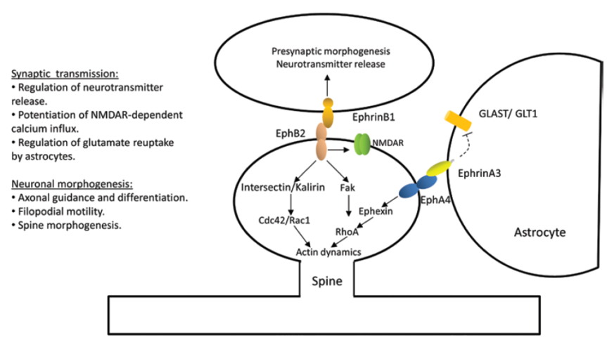
"Figure 2. Ephs and ephrins mediate molecular events that may be involved in memory formation. Evidence shows that memory formation involves alterations of presynaptic neurotransmitter release, activation of glutamate receptors, and neuronal morphogenesis. Eph receptors regulate synaptic transmission by regulating synaptic release, glutamate reuptake from the synapse (via astrocytes), and glutamate receptor conductance and trafficking. Ephs and ephrins also regulate neuronal morphogenesis of axons and dendritic spines through controlling the actin cytoskeleton structure and dynamics" (Dines & Lamprecht, 2015, p. 3).
“Figure 2. Ephs and ephrins mediate molecular events that may be involved in memory formation. Evidence shows that memory formation involves alterations of presynaptic neurotransmitter release, activation of glutamate receptors, and neuronal morphogenesis. Eph receptors regulate synaptic transmission by regulating synaptic release, glutamate reuptake from the synapse (via astrocytes), and glutamate receptor conductance and trafficking. Ephs and ephrins also regulate neuronal morphogenesis of axons and dendritic spines through controlling the actin cytoskeleton structure and dynamics” (Dines & Lamprecht, 2015, p. 3).

When thinking about long-term memory formation, most people immediately picture glutamate synapses. Dines & Lamprecht (2015) review the role of a family of little known players, but with big roles in learning and long-term memory consolidation: the ephs and the ephrines.

Ephs (the name comes from erythropoietin-producing human hepatocellular, the cancer line from which the first member was isolated) are transmembranal tyrosine kinase receptors. Ephrines (Eph receptor interacting protein) bind to them. Ephrines are also membrane-bound proteins, which means that in order for the aforementioned binding to happen, cells must touch each other, or at least be in a very very cozy vicinity. They are expressed in many regions of the brain like hippocampus, amygdala, or cortex.

The authors show that “interruption of Ephs/ephrins mediated functions is sufficient for disruption of memory formation” (p. 7) by reviewing a great deal of genetic, pharmacologic, and electrophysiological studies employing a variety of behavioral tasks, from spatial memory to fear conditioning. The final sections of the review focus on the involvement of ephs/ephrins in Alzheimer’s and anxiety disorders, suggesting that drugs that reverse the impairment on eph/ephrin signaling in these brain diseases may lead to an eventual cure.

Reference: Dines M & Lamprecht R (8 Oct 2015, Epub 13 Sept 2015). The Role of Ephs and Ephrins in Memory Formation. International Journal of Neuropsychopharmacology, 1-14. doi:10.1093/ijnp/pyv106. Article | FREE FULLTEXT PDF

By Neuronicus, 26 October 2015

Nettles are good for you in more ways than one

Urtica Urens (the small nettle). Photo by H. Zell, released under CC BY-SA 3.0. Courtesy of Wikipedia.

Many cultures, specially East-European and North-African, use nettles in their cuisine, as soups, creams, or teas. Nettles’ taste resemble spinach. Now Doukkali et al. (2015) discovered a new use for the plant.

The authors harvested Urtica urens from north Morocco, dried the plants, and prepared a methanolic extract (see p. 2 for the procedure. Don’t drink methanol!). Then, they gave the extract in 3 different doses to mice and assessed its effects in two anxiety and one locomotor test against saline (the control) and diazepam (Valium), a powerful anxiolytic from the benzodiazepine class. Like diazepam, the plant extract had anxiolytic properties, but unlike diazepam, it did not induce any locomotor effects. And this is where the big thing is: ALL benzos on the market have significant side effects in the form of drowsiness, impaired coordination, sedation and so on. Having an anxiolytic without motor impairment would be wonderful.

This is a short, simple to read paper, clearly written, and covers some classic aspects of new drug discovery (like dose-response and lethal dose assessment). The reasons why I think it did not make it to one of the big journals is the small sample size, the relatively moderate effect, and the lack of identifying the active compound (there are virtually no straight-forward behavioral studies published in Nature or Science any more; you’ve got to have the molecules, or proteins, or cells, or what-have-yous as proof that you mean hard-science business).

Or, the fact that it does not have any graphs, all data is presented in tables, which I personally enjoy, as it is oh so easy to manipulate with a graph; and the fancier looking the image, the better chances few people get it anyway. Give me tables with standard deviation any day, as I suspect is the position of the authors of the paper too. But, for the visually inclined, I made a Fig. with some of their data, took only 15 minutes in Excel.

Fig. 1. The effect of saline (S), diazepam (D), and nettle extract (N) on the light-dark test (left) and hole board test (right). Data from Doukkali et al. (2015) , graph by Neuronicus.

Or, not to put a too fine point to it, the authors are from Morocco, so they don’t come shrouded in the A-list universities glamour. In any case, the next obvious step is to isolate the active compound and replicate its anxiolytic effects on other tests and other species.

Reference: Doukkali Z, Taghzouti K, Bouidida EL, Nadjmouddine M, Cherrah Y, & Alaoui K. (24 April 2015). Evaluation of anxiolytic activity of methanolic extract of Urtica urens in a mice model. Behavioral and Brain Functions, 11(19): 1-5. doi: 10.1186/s12993-015-0063-y. Article | FREE PDF

By Neuronicus, 16 October 2015

Cell phones give you hallucinations

A young businessman in a suit screaming at a cell phone. By: Benjamin Miller. License FSP Standard FreeStockPhotos.biz
Photo by Benjamin Miller. License: FSP Standard FreeStockPhotos.biz

Medical doctors (MD) are overworked, particularly when they are hatchlings (i.e. Medical School students) and fledglings (interns and residents). So overworked, that in many countries is routine to have 80-hour weeks and 30-hour shifts as residents and interns. This is a concern as it has been shown that sleep deprivation impairs learning (which is the whole point of residency) and increases the number of medical mistakes (the lack of which is the whole point of their profession).

Lin et al. (2013) show that it can do more than that. Couple internship and cell phones and you get… hallucinations. That’s right. The authors asked 73 medical interns to complete some tests before their internship, then every third, sixth, and twelfth months of their internship, and after the internship. The questionnaires were on anxiety, depression, personality, and cell phone habits and hallucinations. That is: the sensation that your cell phone is vibrating or ringing when, in fact, it is not (which fully corresponds to the definition of hallucination). And here is what they found:

 Before internship, 78% of MDs experienced phantom vibration and 27% experienced phantom ringing.
 During their 1-year internship, about 85 to 95% of MDs experienced phantom vibration and phantom ringing.
 After the internship when the MDs did no work for two weeks, 50% still had these hallucinations.

Composite figure from Lin et al. (2015) showing the interns' depression (above) and anxiety (below) scores before, during, and after internship. The differences are statistically significant.
Fig. 1. Composite figure from Lin et al. (2015) showing the interns’ depression (above) and anxiety (below) scores before, during, and after internship. The differences are statistically significant.

The MDs’ depression and anxiety were also elevated more during the internship than before or after (see Fig. 1), but there was no correlation between the hallucinations and the depression and anxiety scores.

These findings are disturbing on so many levels… Should we be worried that prolonged exposure to cell phones can produce hallucinations? Or that o good portion of the MDs have hallucinations before going to internship? Or that 90% the people in charge with your life or your child’s life are so overworked that are hallucinating on a regular basis? Fine, fine, believing that your phone is ringing or vibrating may not be such a big deal of a hallucination, compared with, let’s say, “the voices told me to give you a lethal dose of morphine”, but as a neuroscientist I beg the question: is there a common mechanism between these two types of hallucinations and, if so, what ELSE is the MD hallucinating about while reassuring you that your CAT scan is normal? Or, forget about the hallucinations, should we worry that your MD is probably more depressed and anxious than you? Or, the “good” news, that the medical interns provide “a model of stress-induced psychotic symptoms” better that previous models, as the authors put it (p. 5)? I really wish there was more research on positive things (… that was a pun; hallucinations are a positive schizophrenic symptom, look it up 🙂 ).

Reference: Lin YH, Lin SH, Li P, Huang WL, & Chen CY. (10 June 2013). Prevalent hallucinations during medical internships: phantom vibration and ringing syndromes. PLoS One, 8(6): e65152. doi: 10.1371/journal.pone.0065152. Article | FREE PDF | First time the phenomenon was documented in press

By Neuronicus, 14 October 2015

Stressed out? Blame your amygdalae

amygdala
Clipart: Royalty free from http://www.cliparthut.com. Text: Neuronicus.

Sooner or later, everyone is exposed to high amounts of stress, whether it is in the form losing someone dear, financial insecurity, or health problems and so on. Most of us manage to bounce right up and continue with our lives, but there is a considerable segment of the population who do not and develop all sorts of problems, from autoimmune disorders to severe depression and anxiety. What makes those people more susceptible to stress? And, more importantly, can we do something about it (yeah, besides making the world a less stressful place)?

Swartz et al. (2015) scanned the brain of 753 healthy young adults (18-22 yrs) while performing a widely used paradigm that elicits amygdalar activation (brain structure, see pic): the subjects had to match a face appearing in the upper part of the screen with one of the faces in the lower part of the screen. The faces looked fearful, angry, surprised, or neutral and amygdalae are robustly activated when matching the fearful face. Then the authors had the participants fill out questionnaires regarding their life events and perceived stress level every 3 months over a period of 2 years (they say 4 years everywhere else in the paper minus Methods & Results, which are the sections that count if one wants to replicate; maybe this is only half of the study and they intend to follow-up to 4 years?).

The higher your baseline amygdalar activation, the higher the risk to develop anxiety disorders later on if expossed to life stressors. Yellow = amygdala. Photo credit: https://www.youtube.com/watch?v=JD44PbAOTy8, presumably copyrighted to Duke University.

The finding of the study is this: baseline amygdalar activation can predict who will develop anxiety later on. In other words, if your natural, healthy, non-stressed self has a an overactive amygdala, you will develop some anxiety disorder later on if exposed to stressors (and who isn’t?). The good news is that knowing this, the owner of the super-sensitive amygdalae, even if s/he may not be able to protect her/himself from stressors, at least can engage in some preventative therapy or counseling to be better equipped with adaptive coping mechanisms when the bad things come. Probably we could all benefit from being “better equipped with adaptive coping mechanisms”, feisty amygdalae or not. Oh, well…

Reference: Swartz, J.R., Knodt, A.R., Radtke, S.R., & Hariri, A.R. (2015). A neural biomarker of psychological vulnerability to future life stress. Neuron, 85, 505-511. doi: 10.1016/j.neuron.2014.12.055. Article | PDF | Video

By Neuronicus, 12 October 2015

Stress can get you fat. And then kill you.

stress meSome people lose weight under stressful conditions and some gain weight. How does that play into the risk for the cardiovascular disease and subsequent mortality? Medical doctors keep warning us that fat people are at risk for diabetes and heart disease. Turns out that being a little on the heavy side might actually not be that bad. It all depends on what kind of fat and where it is.

The paper featured today reviews a series of interesting articles with surprising results. Peters & McEwen (2015) identify three distinct phenotypes:

1) The good stress leads to well-proportionate body shape. People who live in safe environments, they do well socioeconomically, they have good self-esteem, and they have a fulfilling social and family life. They experience low levels of stress, they are well proportionate, and have a low mortality rate due to cardiovascular disease. Might as well call these ones the lucky ones.

2) The tolerable stress leads to corpulent-but-narrow-waisted body shape. People who experience stress but in order to cope with it they supply the brain with more energy by eating more. So they become more corpulent, gaining subcutaneous fat, but their cardiovascular mortality risk remains low.

3) The toxic stress leads to lean-but-wide-waisted body shape. People who experience prolonged stress exposure to uncertain socioeconomic conditions, poor work, or family life. They have low self-esteem, often associated with depressive periods. They are or become lean, but they accumulate large visceral fat deposits (as opposed to subcutaneous), and their cardiovascular mortality risk is the highest. They also are at risk for other physical and mental disorders. The phenotype 3 people have a wider waist relative to their body mass index and height.

Source: Peters & McEwen (2015, p.144)

Thus, the authors propose that instead or along with the body mass index, another metric should be used to identify the ones in dire need of help: the body shape index. Also, the review outlines the mechanisms responsible for these findings.

So next time you see a not so well-proportionate person, smile. Maybe even offer to help or chat; you don’t know what they’re going through.

Reference: Peters, A. & McEwen, B. S. (September 2015, Epub 3 July 2015). Stress habituation, body shape and cardiovascular mortality. Neuroscience Biobehavioral Reviews, 56:139-50. doi: 10.1016/j.neubiorev.2015.07.001. Article | FREE PDF

By Neuronicus, 5 October 2015