Plants emit sounds in the ultrasonic range when stressed

REFERENCE:

Itzhak KhaitOhad Lewin-EpsteinRaz SharonKfir SabanRevital GoldsteinYehuda AniksterYarden ZeronChen AgassyShaked NizanGayl SharabiRan PerelmanArjan BoonmanNir SadeYossi YovelLilach Hadany (2023 Mar 30). Sounds emitted by plants under stress are airborne and informative. Cell, 186(7):1328-1336.e10. doi: 10.1016/j.cell.2023.03.009, PMID: 37001499. ARTICLE | FREE FULLTEXT PDF

By Neuronicus, 5 December 2023

The FIRSTS: Lack of happy events in depression (2003)

My last post focused on depression and it reminded me of something that I keep telling my students and they all react with disbelief. Well, I tell them a lot of things to which they react with disbelief, to be sure, but this one I keep thinking it should not generate such incredulity. The thing is: depressed people perceive the same amount of negative events happening to them as healthy people, but far fewer positive ones. This seems to be counter-intuitive to non-professionals, who believe depressed people are just generally sadder than average and that’s why they see the half-empty side of the glass of life.

So I dug out the original paper who found this… finding. It’s not as old as you might think. Peeters et al. (2003) paid $30/capita to 86 people, 46 of which were diagnosed with Major Depressive Disorder and seeking treatment in a community mental health center or outpatient clinic (this is in Netherlands). None were taking antidepressants or any other drugs, except low-level anxiolytics. Each participant was given a wristwatch that beeped 10 times a day at semi-random intervals of approximately 90 min. When the watch beeped, the subjects had to complete a form within maximum 25 min answering questions about their mood, currents events, and their appraisal of those events. The experiment took 6 days, including weekend.

The results? Contrary to popular belief, people with depression “did not report more frequent negative events, although they did report fewer positive events and appraised both types of events as more stressful” (p. 208). In other words, depressed people are not seeing half-empty glasses all the time; instead, they don’t see the half-full glasses. Note that they regarded both negative and positive events as stressful. We circle back to the ‘stress is the root of all evil‘ thing.

I would have liked to see if the decrease in positive affect and perceived happy events correlates with increased sadness. The authors say that “negative events were appraised as more unpleasant, more important, and more stressful by the depressed than by the healthy participants ” (p. 206), but, curiously, the  mood was assessed with ratings on the feeling anxious, irritated, restless, tense, guilty, irritable, easily distracted, and agitate, and not a single item on depression-iconic feelings: sad, empty, hopeless, worthless.

Nevertheless, it’s a good psychological study with in depth statistical analyses. I also found thought-provoking this paragraph: “The literature on mood changes in daily life is dominated by studies of daily hassles. The current results indicate that daily uplifts are also important determinants of mood, in both depressed and healthy people” (p. 209).

152 depression and lack of happy events - Copy

REFERENCE: Peeters F, Nicolson NA, Berkhof J, Delespaul P, & deVries M. (May 2003). Effects of daily events on mood states in major depressive disorder. Journal of Abnormal Psychology, 112(2):203-11. PMID: 12784829, DOI: 10.1037/0021-843X.112.2.203. ARTICLE

By Neuronicus, 4 May 2019

Epigenetics of BDNF in depression

Depression is the leading cause of disability worldwide, says the World Health Organization. The. The. I knew it was bad, but… ‘the’? More than 300 million people suffer from it worldwide and in many places fewer than 10% of these receive treatment. Lack of treatment is due to many things, from lack of access to healthcare to lack of proper diagnosis; and not in the least due to social stigma.

To complicate matters, the etiology of depression is still not fully elucidated, despite hundreds of thousand of experimental articles published out-there. Perhaps millions. But, because hundreds of thousands of experimental articles perhaps millions have been published, we know a helluva a lot about it than, say, 50 years ago. The enormous puzzle is being painstakingly assembled as we speak by scientists all over the world. I daresay we have a lot of pieces already, if not all at least 3 out of 4 corners, so we managed to build a not so foggy view of the general picture on the box lid. Here is one of the hottest pieces of the puzzle, one of those central pieces that bring the rabbit into focus.

Before I get to the rabbit, let me tell you about the corners. In the fifties people thought that depression is due to having too little neurotransmitters from the monoamine class in the brain. This thought did not arise willy-nilly, but from the observation that drugs that increase monoamine levels in the brain alleviate depression symptoms, and, correspondingly, drugs which deplete monoamines induce depression symptoms. A bit later on, the monoamine most culpable was found to be serotonin. All well and good, plenty of evidence, observational, correlational, causational, and mechanistic supporting the monoamine hypothesis of depression. But two more pieces of evidence kept nagging the researchers. The first one was that the monoamine enhancing drugs take days to weeks to start working. So, if low on serotonin is the case, then a selective serotonin reuptake inhibitor (SSRI) should elevate serotonin levels within maximum an hour of ingestion and lower symptom severity, so how come it takes weeks? The second was even more eyebrow raising: these monoamine-enhancing drugs work in about 50 % of the cases. Why not all? Or, more pragmatically put, why not most of all if the underlying cause is the same?

It took decades to address these problems. The problem of having to wait weeks until some beneficial effects of antidepressants show up has been explained away, at least partly, by issues in the serotonin regulation in the brain (e.g. autoreceptors senzitization, serotonin transporter abnormalities). As for the second problem, the most parsimonious answer is that that archeological site called DSM (Diagnostic and Statistical Manual of Mental Disorders), which psychologists, psychiatrists, and scientists all over the world have to use to make a diagnosis is nothing but a garbage bag of last century relics with little to no resemblance of this century’s understanding of the brain and its disorders. In other words, what DSM calls major depressive disorder (MDD) may as well be more than one disorder and then no wonder the antidepressants work only in half of the people diagnosed with it. As Goldberg put it in 2011, “the DSM diagnosis of major depression is made when a patient has any 5 out of 9 symptoms, several of which are opposites [emphasis added]”! He was referring to DSM-4, not that the 5 is much different. I mean, paraphrasing Goldberg, you really don’t need much of a degree other than some basic intro class in the physiology of whatever, anything really, to suspect that someone who’s sleeping a lot, gains weight, has increased appetite, appears tired or slow to others, and feels worthless might have a different cause for these symptoms than someone who has daily insomnias, lost weight recently, has decreased appetite, is hyperagitated, irritable, and feels excessive guilt. Imagine how much more understanding we would have about depression if scientists didn’t use the DSM for research. No wonder that there’s a lot of head scratching when your hypothesis, which is logically correct, paradigmatically coherent, internally consistent, flawlessly tested, turns out to be true only sometimes because your ‘depressed’ subjects are as a homogeneous group as a pack of Trail Mix.

I got sidetracked again. This time ranting against DSM. No matter, I’m back on track. So. The good thing about the work done trying to figure out how antidepressants work and psychiatrists’ minds work (DSM is written overwhelmingly by psychiatrists), scientists uncovered other things about depression. Some of the findings became clumped under the name ‘the neurotrophic hypothesis of depression’ in the early naughts. It stems from the finding that some chemicals needed by neurons for their cellular happiness are in low amount in depression. Almost two decades later, the hypothesis became mainstream theory as it explains away some other findings in depression, and is not incompatible with the monoamines’ behavior. Another piece of the puzzle found.

One of these neurotrophins is called brain-derived neurotrophic factor (BDNF), which promotes cell survival and growth. Crucially, it also regulates synaptic plasticity, without which there would be no learning and no memory. The idea is that exposure to adverse events generates stress. Stress is differently managed by different people, largely due to genetic factors. In those not so lucky at the genetic lottery (how hard they take a stressor, how they deal with it), and in those lucky enough at genetics but not so lucky in life (intense and/or many stressors hit the organism hard regardless how well you take it or how good you are at it), stress kills a lot of neurons, literally, prevents new ones from being born, and prevents the remaining ones from learning well. Including learning on how to deal with the stressors, present and future, so the next time an adverse event happens, even if it is a minor stressor, the person is way more drastically affected. in other words, stress makes you more vulnerable to stressors. One of the ways stress is doing all these is by suppressing BDNF synthesis. Without BDNF, the individual exposed to stress that is exacerbated either by genes or environment ends up unable to self-regulate mood successfully. The more that mood is not regulated, the worse the brain becomes at self-regulating because the elements required for self-regulation, which include learning from experience, are busted. And so the vicious circle continues.

Maintaining this vicious circle is the ability of stressors to change the patterns of DNA expression and, not surprisingly, one of the most common findings is that the BDNF gene is hypermethylated in depression. Hypermethylation is an epigenetic change (a change around the DNA, not in the DNA itself), meaning that the gene in question is less expressed. This means lower amounts of BDNF are produced in depression.

After this long introduction, the today’s paper is a systematic review of one of epigenetic changes in depression: methylation. The 67 articles that investigated the role of methylation in depression were too heterogeneous to make a meta-analysis out of them, so Li et al. (2019) made a systematic review.

The main finding was that, overall, depression is associated with DNA methylation modifications. Two genes stood out as being hypermethylated: our friend BDNF and SLC6A4, a gene involved in the serotonin cycle. Now the question is who causes who: is stress methylating your DNA or does your methylated DNA make you more vulnerable to stress? There’s evidence both ways. Vicious circle, as I said. I doubt that for the sufferer it matters who started it first, but for the researchers it does.

151 bdnf 5htt people - Copy

A little disclaimer: the picture I painted above offers a non-exclusive view on the causes of depression(s). There’s more. There’s always more. Gut microbes are in the picture too. And circulatory problems. And more. But the picture is more than half done, I daresay. Continuing my puzzle metaphor, we got the rabbit by the ears. Now what to do with it…

Well, one thing we can do with it, even with only half-rabbit done, is shout loud and clear that depression is a physical disease. And those who claim it can be cured by a positive attitude and blame the sufferers for not ‘trying hard enough’ or not ‘smiling more’ or not ‘being more positive’ can bloody well shut up and crawl back in the medieval cave they came from.

REFERENCES:

1. Li M, D’Arcy C, Li X, Zhang T, Joober R, & Meng X (4 Feb 2019). What do DNA methylation studies tell us about depression? A systematic review. Translational Psychiatry, 9(1):68. PMID: 30718449, PMCID: PMC6362194, DOI: 10.1038/s41398-019-0412-y. ARTICLE | FREE FULLTEXT PDF

2. Goldberg D (Oct 2011). The heterogeneity of “major depression”. World Psychiatry, 10(3):226-8. PMID: 21991283, PMCID: PMC3188778. ARTICLE | FREE FULLTEXT PDF

3. World Health Organization Depression Fact Sheet

By Neuronicus, 23 April 2019

Pic of the day: Dopamine from a non-dopamine place

147 lc da ppvn - Copy

Reference: Beas BS, Wright BJ, Skirzewski M, Leng Y, Hyun JH, Koita O, Ringelberg N, Kwon HB, Buonanno A, & Penzo MA (Jul 2018, Epub 18 Jun 2018). The locus coeruleus drives disinhibition in the midline thalamus via a dopaminergic mechanism. Nature Neuroscience,21(7):963-973. PMID: 29915192, PMCID: PMC6035776 [Available on 2018-12-18], DOI:10.1038/s41593-018-0167-4. ARTICLE

The Mom Brain

Recently, I read an opinion titled When I Became A Mother, Feminism Let Me Down. The gist of it was that some (not all!) feminists, while empowering women and girls to be anything they want to be and to do anything a man or a boy does, they fail in uplifting the motherhood aspect of a woman’s life, should she choose to become a mother. In other words, even (or especially, in some cases) feminists look down on the women who chose to switch from a paid job and professional career to an unpaid stay-at-home mom career, as if being a mother is somehow beneath what a woman can be and can achieve. As if raising the next generation of humans to be rational, informed, well-behaved social actors instead of ignorant brutal egomaniacs is a trifling matter, not to be compared with the responsibilities and struggles of a CEO position.

Patriarchy notwithstanding, a woman can do anything a man can. And more. The ‘more’ refers to, naturally, motherhood. Evidently, fatherhood is also a thing. But the changes that happen in a mother’s brain and body during pregnancy, breastfeeding, and postpartum periods are significantly more profound than whatever happens to the most loving and caring and involved father.

Kim (2016) bundled some of these changes in a nice review, showing how these drastic and dramatic alterations actually have an adaptive function, preparing the mother for parenting. Equally important, some of the brain plasticity is permanent. The body might spring back into shape if the mother is young or puts into it a devilishly large amount of effort, but some brain changes are there to stay. Not all, though.

One of the most pervasive findings in motherhood studies is that hormones whose production is increased during pregnancy and postpartum, like oxytocin and dopamine, sensitize the fear circuit in the brain. During the second trimester of pregnancy and particularly during the third, expectant mothers start to be hypervigilent and hypersensitive to threats and to angry faces. A higher anxiety state is characterized, among other things, by preferentially scanning for threats and other bad stuff. Threats mean anything from the improbable tiger to the 1 in a million chance for the baby to be dropped by grandma to the slightly warmer forehead or the weirdly colored poopy diaper. The sensitization of the fear circuit, out of which the amygdala is an essential part, is adaptive because it makes the mother more likely to not miss or ignore her baby’s cry, thus attending to his or her needs. Also, attention to potential threats is conducive to a better protection of the helpless infant from real dangers. This hypersensitivity usually lasts 6 to 12 months after childbirth, but it can last lifetime in females already predisposed to anxiety or exposed to more stressful events than average.

Many new mothers worry if they will be able to love their child as they don’t feel this all-consuming love other women rave about pre- or during pregnancy. Rest assured ladies, nature has your back. And your baby’s. Because as soon as you give birth, dopamine and oxytocin flood the body and the brain and in so doing they modify the reward motivational circuit, making new mothers literally obsessed with their newborn. The method of giving birth is inconsequential, as no differences in attachment have been noted (this is from a different study). Do not mess with mother’s love! It’s hardwired.

Another change happens to the brain structures underlying social information processing, like the insula or fusiform gyrus, making mothers more adept at self-motoring, reflection, and empathy. Which is a rapid transformation, without which a mother may be less accurate in understanding the needs, mental state, and social cues of the very undeveloped ball of snot and barf that is the human infant (I said that affectionately, I promise).

In order to deal with all these internal changes and the external pressures of being a new mom the brain has to put up some coping mechanisms. (Did you know, non-parents, that for the first months of their newborn lives, the mothers who breastfeed must do so at least every 4 hours? Can you imagine how berserk with sleep deprivation you would be after 4 months without a single night of full sleep but only catnaps?). Some would be surprised to find out – not mothers, though, I’m sure – that “new mothers exhibit enhanced neural activation in the emotion regulation circuit including the anterior cingulate cortex, and the medial and lateral prefrontal cortex” (p. 50). Which means that new moms are actually better at controlling their emotions, particularly at regulating negative emotional reactions. Shocking, eh?

140 mom brain1 - Copy

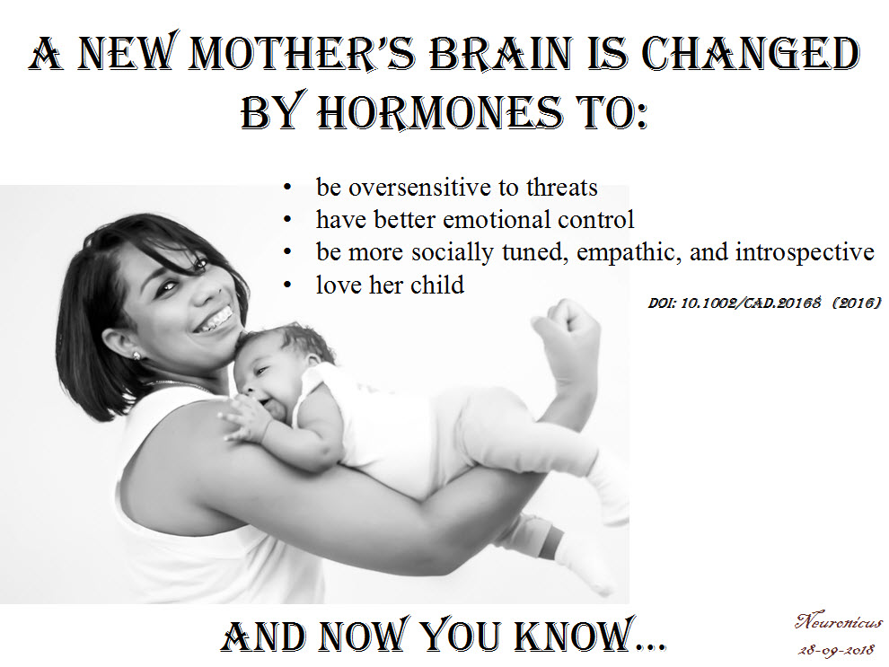
Finally, it appears that very few parts of the brain are spared from this overhaul as the entire brain of the mother is first reduced in size and then it grows back, reorganized. Yeah, isn’t that weird? During pregnancy the brain shrinks, being at its lowest during childbirth and then starts to grow again, reaching its pre-pregnancy size 6 months after childbirth! And when it’s back, it’s different. The brain parts heavily involved in parenting, like the amygdala involved in the anxiety, the insula and superior temporal gyrus involved in social information processing and the anterior cingulate gyrus involved in emotional regulation, all these show increased gray matter volume. And many other brain structures that I didn’t list. One brain structure is rarely involved only in one thing so the question is (well, one of them) what else is changed about the mothers, in addition to their increased ability to parent?

I need to add a note here: the changes that Kim (2016) talks about are averaged. That means some women get changed more, some less. There is variability in plasticity, which should be a pleonasm. There is also variability in the human population, as any mother attending a school parents’ night-out can attest. Some mothers are paranoid with fear and overprotective, others are more laissez faire when it comes to eating from the floor.

But SOME changes do occur in all mothers’ brains and bodies. For example, all new mothers exhibit a heightened attention to threats and subsequent raised levels of anxiety. But when does heightened attention to threats become debilitating anxiety? Thanks to more understanding and tolerance about these changes, more and more women feel more comfortable reporting negative feelings after childbirth so that now we know that postpartum depression, which happens to 60 – 80% of mothers, is a serious matter. A serious matter that needs serious attention from both professionals and the immediate social circle of the mother, both for her sake as well as her infant’s. Don’t get me wrong, we – both males and females – still have a long way ahead of us to scientifically understand and to socially accept the mother brain, but these studies are a great start. They acknowledge what all mothers know: that they are different after childbirth than the way they were before. Now we have to figure out how are they different and what can we do to make everyone’s lives better.

Kim (2016) is an OK review, a real easy read, I recommend it to the non-specialists wholeheartedly; you just have to skip the name of the brain parts and the rest is pretty clear. It is also a very short review, which will help with reader fatigue. The caveat of that is that it doesn’t include a whole lotta studies, nor does it go in detail on the implications of what the handful cited have found, but you’ll get the gist of it. There is a vastly more thorough literature if one would include animal studies that the author, curiously, did not include. I know that a mouse is not a chimp is not a human, but all three of us are mammals, and social mammals at that. Surely, there is enough biological overlap so extrapolations are warranted, even if partially. Nevertheless, it’s a good start for those who want to know a bit about the changes motherhood does to the brain, behavior, thoughts, and feelings.

Corroborated with what I already know about the neuroscience of maternity, my favourite takeaway is this: new moms are not crazy. They can’t help most of these changes. It’s biology, you see. So go easy on new moms. Moms, also go easy on yourselves and know that, whether they want to share or not, the other moms probably go through the same stuff. The other moms are doing better than you are either. You’re not alone. And if that overactive threat circuit gives you problems, i.e. you feel overwhelmed, it’s OK to ask for help. And if you don’t get it, ask for it again and again until you do. That takes courage, that’s empowerment.

P. S. The paper doesn’t look like it’s peer-reviewed. Yes, I know the peer-reviewing publication system is flawed, I’ve been on the receiving end of it myself, but it’s been drilled into my skull that it’s important, flawed as it is, so I thought to mention it.

REFERENCE: Kim, P. (Sept. 2016). Human Maternal Brain Plasticity: Adaptation to Parenting, New Directions for Child and Adolescent Development, (153): 47–58. PMCID: PMC5667351, doi: 10.1002/cad.20168. ARTICLE | FREE FULLTEXT PDF

By Neuronicus, 28 September 2018

The Benefits of Vacation

My prolonged Internet absence from the last month or so was due to a prolonged vacation. In Europe. Which I loved. Both the vacation and the Europe. Y’all, people, young and old, listen to me: do not neglect vacations for they strengthen the body, nourish the soul, and embolden the spirit.

More pragmatically, vacations lower the stress level. Yes, even the stressful vacations lower the stress level, because the acute stress effects of “My room is not ready yet” / “Jimmy puked in the car” / “Airline lost my luggage” are temporary and physiologically different from the chronic stress effects of “I’ll lose my job if I don’t meet these deadlines” / “I hate my job but I can’t quit because I need health insurance” / “I’m worried for my child’s safety” / “My kids will suffer if I get a divorce” / “I can’t make the rent this month”.

Chronic stress results in a whole slew of real nasties, like cognitive, learning, and memory impairments, behavioral changes, issues with impulse control, immune system problems, weight gain, cardiovascular disease and so on and so on and so on. Even death. As I told my students countless of times, chronic stress to the body is as real and physical as a punch in the stomach but far more dangerous. So take a vacation as often as you can. Even a few days of total disconnect help tremendously.

There are literally thousands of peer-reviewed papers out there that describe the ways in which stress produces all those bad things, but not so many papers about the effects of vacations. I suspect this is due to the inherent difficulty in accounting for the countless environmental variables that can influence one’s vacation and its outcomes, whereas identifying and characterizing stressors is much easier. In other words, lack of experimental control leads to paucity of good data. Nevertheless, from this paucity, Chen & Petrick (2013) carefully selected 98 papers from both academic and nonacademic publications about the benefits of travel vacations.

These are my take-home bullet-points:

  • vacation effects last no more than a month
  • vacations reduce both the subjective perception of stress and the objective measurement of it (salivary cortisol)
  • people feel happier after taking a vacation
  • there are some people who do not relax in a vacation, presumably because they cannot ‘detach’ themselves from the stressors in their everyday life (long story here why some people can’t let go of problems)
  • vacations lower the occurrence of cardiovascular disease
  • vacations decrease work-related stress, work absenteeism, & work burnout
  • vacations increase job performance
  • the more you do on a vacation the better you feel, particularly if you’re older
  • you benefit more if you do new things or go to new places instead of just staying home
  • vacations increase overall life satisfaction

Happy vacationing!

137 - Copy

REFERENCE: Chen, C-C & Petrick, JF (Nov. 2013, Epub 17 Jul. 2013). Health and Wellness Benefits of Travel Experiences: A Literature Review, Journal of Travel Research, 52(6):709-719. doi: 10.1177/0047287513496477. ARTICLE | FULLTEXT PDF via ResearchGate.

By Neuronicus, 20 July 2018

Stress can kill you and that’s no metaphor

The term ‘heartbreak’ is used as a metaphor to describe the intense feeling of loss, sometimes also called emotional pain. But what if the metaphor has roots into something more tangible than a feeling, that of the actual muscular organ giving signs of failure?

Although there have been previous reports that found stress causes cardiovascular problems, including myocardial infarction, Graff et al. (2016) conducted the largest study to date that investigated this link: they had almost 1 million subjects. That’s right, 1 million people (well, actually 974 732). Out of these, almost 20% of them had a partner who died between 1995 and 2014. The chosen stressor was the loss of a loved one because “the loss of a partner is considered one of the most severely stressful life events and is likely to affect most people, independently of coping mechanisms” (p. 1-2). The authors looked at Danish hospital records for people who were diagnosed with atrial fibrillation (AF) for the first time and correlated that data with bereavement information. AF increases the risk of death due to stroke or heart failure.

The people who underwent loss had an increased risk to develop AF for 1 year after the loss. The risk was more pronounced in the first 8-14 days after the loss, the bereaved people having a 90% higher risk of developing AF than non-bereaved people. By the end of the first month the risk had declined, but still was a whooping 41% higher than the average. Only 1 year after the loss the risk of developing AF was similar to that of non-bereaved people.

The risk was even higher in young people or if the death of the partner was unexpected. The authors also looked to see if other variables play a role in the risk, like gender, civil status, education, diabetes, or cardiovascular medication and none influenced the results.

I suspect the number of people that have heart problems after major stress is actually a lot higher because of the under-reporting bias. In other words, not everybody who feels their heart aching would go to the hospital, particularly in the first couple of weeks after losing a loved one.

As for the mechanism, there is some data pointing to some stress hormones (like adrenaline or cortisol) which can damage the heart. Other substances released in abundance during stress and likely to act in concert with the stress hormones are proinflamatory cytokines which also can lead to arrhythmias.

85heart - Copy

Reference: Graff S, Fenger-Grøn M, Christensen B, Søndergaard Pedersen H, Christensen J, Li J, & Vestergaard M (2016). Long-term risk of atrial fibrillation after the death of a partner. Open Heart, 3: e000367. doi:10.1136/openhrt-2015-000367. Article  | FREE FULTEXT PDF

By Neuronicus, 16 April 2016

Mechanisms of stress resilience

71 stress - CopyLast year a new peer-reviewed journal called Neurobiology of Stress made its debut. The journal is published by Elsevier, who, in an uncharacteristic move, has provided Open Access for its first three issues. So hurry up and download the papers.

The very first issue is centered around the idea of resilience. That is, exposed to the same stressors, some people are more likely to develop stress-induced diseases, whereas others seem to be immune to the serious effects of stress.

Much research has been carried out to uncover the effects of chronic stress or of an exposure to a single severe stressor, which vary from cardiovascular disorders, obesity, irritable bowel syndrome, immune system dysfunctions to posttraumatic stress disorder, generalized anxiety, specific phobias, or depression. By comparison, there is little, but significant data on resilience: the ability to NOT develop those nasty stress-induced disorders. Without doubt, one reason for this scarcity is the difficulty in finding such rare subjects in our extremely stressful society. Therefore most of the papers in this issue focus on animal models.

Nevertheless, there is enough data on resilience to lead to no less that twenty reviews on the subject. It was difficult to choose one as most are very interesting, tackling various aspects of resilience, from sex differences to prenatal exposure to stress, from epigenetic to neurochemical modifications, from social inequalities to neurogenesis and so on.

So I chose for today a more general review of Pfau & Russo (2015), entitled “Peripheral and central mechanisms of stress resilience”. After it introduces the reader to four animal models of resilience, the paper looks at the neruoendocrine responses to stress and identifies some possible chemical mediators of resilience (like certain hormones), then at the immune responses to stress (bad, bad cytokines), and finally at the brain responses to stress (surprisingly, not focusing on amygdala, hypothalamus or hippocampus, but on the dopamine system originating from ventral tegmental area).

I catalogue the review as a medium difficulty read because it requires a certain amount of knowledge of the stress field beforehand. But do check out the other ones in the issue, too!

Reference: Pfau ML & Russo SJ (1 Jan 2015). Peripheral and central mechanisms of stress resilience. Neurobiology of Stress, 1:66-79. PMID: 25506605, PMCID: PMC4260357, DOI: 10.1016/j.ynstr.2014.09.004. Article | FREE FULLTEXT PDF

By Neuronicus, 24 January 2016

Terrorist attacks increase the male fetal loss

effelThe odds of having a baby boy decreases after terrorist attacks, natural or man-made disasters, or economical depression. There are several studies worldwide that support this finding. This is somewhat counter-intuitive, because there are anecdotal accounts that report an increase in male births after a war, presumably to make up for the lost men.

Bruckner et al. (2010) wanted to see if this decrease in the odds of a male births, also called the secondary sex ratio, is due to a failure to conceive male babies or the male fetuses die in the womb before birth. They looked at the public databases from 1996-2002 fetal deaths and births from the U.S. National Center for Health Statistics.

The results showed that in the months following the September 11, 2001 terrorist attacks the deaths of male fetuses older that 20 weeks increased significantly. The authors make reference to the communal bereavement hypothesis, which stipulates that stress increases in persons not directly affected by a tragedy. Although the effects of stress on pregnant females is well documented, why the male fetuses seem to be more susceptible to mother’s stress is unknown.

I chose to feature this paper because of the recent Paris atrocities.

Reference: Bruckner TA, Catalano R, & Ahern J. (25 May 2010). Male fetal loss in the U.S. following the terrorist attacks of September 11, 2001. BMC Public Health.;10:273. doi: 10.1186/1471-2458-10-273. Article | FREE FULLTEXT PDF

By Neuronicus, 15 November 2015

The F in memory

"Figure 2. Ephs and ephrins mediate molecular events that may be involved in memory formation. Evidence shows that memory formation involves alterations of presynaptic neurotransmitter release, activation of glutamate receptors, and neuronal morphogenesis. Eph receptors regulate synaptic transmission by regulating synaptic release, glutamate reuptake from the synapse (via astrocytes), and glutamate receptor conductance and trafficking. Ephs and ephrins also regulate neuronal morphogenesis of axons and dendritic spines through controlling the actin cytoskeleton structure and dynamics" (Dines & Lamprecht, 2015, p. 3).
“Figure 2. Ephs and ephrins mediate molecular events that may be involved in memory formation. Evidence shows that memory formation involves alterations of presynaptic neurotransmitter release, activation of glutamate receptors, and neuronal morphogenesis. Eph receptors regulate synaptic transmission by regulating synaptic release, glutamate reuptake from the synapse (via astrocytes), and glutamate receptor conductance and trafficking. Ephs and ephrins also regulate neuronal morphogenesis of axons and dendritic spines through controlling the actin cytoskeleton structure and dynamics” (Dines & Lamprecht, 2015, p. 3).

When thinking about long-term memory formation, most people immediately picture glutamate synapses. Dines & Lamprecht (2015) review the role of a family of little known players, but with big roles in learning and long-term memory consolidation: the ephs and the ephrines.

Ephs (the name comes from erythropoietin-producing human hepatocellular, the cancer line from which the first member was isolated) are transmembranal tyrosine kinase receptors. Ephrines (Eph receptor interacting protein) bind to them. Ephrines are also membrane-bound proteins, which means that in order for the aforementioned binding to happen, cells must touch each other, or at least be in a very very cozy vicinity. They are expressed in many regions of the brain like hippocampus, amygdala, or cortex.

The authors show that “interruption of Ephs/ephrins mediated functions is sufficient for disruption of memory formation” (p. 7) by reviewing a great deal of genetic, pharmacologic, and electrophysiological studies employing a variety of behavioral tasks, from spatial memory to fear conditioning. The final sections of the review focus on the involvement of ephs/ephrins in Alzheimer’s and anxiety disorders, suggesting that drugs that reverse the impairment on eph/ephrin signaling in these brain diseases may lead to an eventual cure.

Reference: Dines M & Lamprecht R (8 Oct 2015, Epub 13 Sept 2015). The Role of Ephs and Ephrins in Memory Formation. International Journal of Neuropsychopharmacology, 1-14. doi:10.1093/ijnp/pyv106. Article | FREE FULLTEXT PDF

By Neuronicus, 26 October 2015

Cell phones give you hallucinations

A young businessman in a suit screaming at a cell phone. By: Benjamin Miller. License FSP Standard FreeStockPhotos.biz
Photo by Benjamin Miller. License: FSP Standard FreeStockPhotos.biz

Medical doctors (MD) are overworked, particularly when they are hatchlings (i.e. Medical School students) and fledglings (interns and residents). So overworked, that in many countries is routine to have 80-hour weeks and 30-hour shifts as residents and interns. This is a concern as it has been shown that sleep deprivation impairs learning (which is the whole point of residency) and increases the number of medical mistakes (the lack of which is the whole point of their profession).

Lin et al. (2013) show that it can do more than that. Couple internship and cell phones and you get… hallucinations. That’s right. The authors asked 73 medical interns to complete some tests before their internship, then every third, sixth, and twelfth months of their internship, and after the internship. The questionnaires were on anxiety, depression, personality, and cell phone habits and hallucinations. That is: the sensation that your cell phone is vibrating or ringing when, in fact, it is not (which fully corresponds to the definition of hallucination). And here is what they found:

 Before internship, 78% of MDs experienced phantom vibration and 27% experienced phantom ringing.
 During their 1-year internship, about 85 to 95% of MDs experienced phantom vibration and phantom ringing.
 After the internship when the MDs did no work for two weeks, 50% still had these hallucinations.

Composite figure from Lin et al. (2015) showing the interns' depression (above) and anxiety (below) scores before, during, and after internship. The differences are statistically significant.
Fig. 1. Composite figure from Lin et al. (2015) showing the interns’ depression (above) and anxiety (below) scores before, during, and after internship. The differences are statistically significant.

The MDs’ depression and anxiety were also elevated more during the internship than before or after (see Fig. 1), but there was no correlation between the hallucinations and the depression and anxiety scores.

These findings are disturbing on so many levels… Should we be worried that prolonged exposure to cell phones can produce hallucinations? Or that o good portion of the MDs have hallucinations before going to internship? Or that 90% the people in charge with your life or your child’s life are so overworked that are hallucinating on a regular basis? Fine, fine, believing that your phone is ringing or vibrating may not be such a big deal of a hallucination, compared with, let’s say, “the voices told me to give you a lethal dose of morphine”, but as a neuroscientist I beg the question: is there a common mechanism between these two types of hallucinations and, if so, what ELSE is the MD hallucinating about while reassuring you that your CAT scan is normal? Or, forget about the hallucinations, should we worry that your MD is probably more depressed and anxious than you? Or, the “good” news, that the medical interns provide “a model of stress-induced psychotic symptoms” better that previous models, as the authors put it (p. 5)? I really wish there was more research on positive things (… that was a pun; hallucinations are a positive schizophrenic symptom, look it up 🙂 ).

Reference: Lin YH, Lin SH, Li P, Huang WL, & Chen CY. (10 June 2013). Prevalent hallucinations during medical internships: phantom vibration and ringing syndromes. PLoS One, 8(6): e65152. doi: 10.1371/journal.pone.0065152. Article | FREE PDF | First time the phenomenon was documented in press

By Neuronicus, 14 October 2015

Stressed out? Blame your amygdalae

amygdala
Clipart: Royalty free from http://www.cliparthut.com. Text: Neuronicus.

Sooner or later, everyone is exposed to high amounts of stress, whether it is in the form losing someone dear, financial insecurity, or health problems and so on. Most of us manage to bounce right up and continue with our lives, but there is a considerable segment of the population who do not and develop all sorts of problems, from autoimmune disorders to severe depression and anxiety. What makes those people more susceptible to stress? And, more importantly, can we do something about it (yeah, besides making the world a less stressful place)?

Swartz et al. (2015) scanned the brain of 753 healthy young adults (18-22 yrs) while performing a widely used paradigm that elicits amygdalar activation (brain structure, see pic): the subjects had to match a face appearing in the upper part of the screen with one of the faces in the lower part of the screen. The faces looked fearful, angry, surprised, or neutral and amygdalae are robustly activated when matching the fearful face. Then the authors had the participants fill out questionnaires regarding their life events and perceived stress level every 3 months over a period of 2 years (they say 4 years everywhere else in the paper minus Methods & Results, which are the sections that count if one wants to replicate; maybe this is only half of the study and they intend to follow-up to 4 years?).

The higher your baseline amygdalar activation, the higher the risk to develop anxiety disorders later on if expossed to life stressors. Yellow = amygdala. Photo credit: https://www.youtube.com/watch?v=JD44PbAOTy8, presumably copyrighted to Duke University.

The finding of the study is this: baseline amygdalar activation can predict who will develop anxiety later on. In other words, if your natural, healthy, non-stressed self has a an overactive amygdala, you will develop some anxiety disorder later on if exposed to stressors (and who isn’t?). The good news is that knowing this, the owner of the super-sensitive amygdalae, even if s/he may not be able to protect her/himself from stressors, at least can engage in some preventative therapy or counseling to be better equipped with adaptive coping mechanisms when the bad things come. Probably we could all benefit from being “better equipped with adaptive coping mechanisms”, feisty amygdalae or not. Oh, well…

Reference: Swartz, J.R., Knodt, A.R., Radtke, S.R., & Hariri, A.R. (2015). A neural biomarker of psychological vulnerability to future life stress. Neuron, 85, 505-511. doi: 10.1016/j.neuron.2014.12.055. Article | PDF | Video

By Neuronicus, 12 October 2015

Stress can get you fat. And then kill you.

stress meSome people lose weight under stressful conditions and some gain weight. How does that play into the risk for the cardiovascular disease and subsequent mortality? Medical doctors keep warning us that fat people are at risk for diabetes and heart disease. Turns out that being a little on the heavy side might actually not be that bad. It all depends on what kind of fat and where it is.

The paper featured today reviews a series of interesting articles with surprising results. Peters & McEwen (2015) identify three distinct phenotypes:

1) The good stress leads to well-proportionate body shape. People who live in safe environments, they do well socioeconomically, they have good self-esteem, and they have a fulfilling social and family life. They experience low levels of stress, they are well proportionate, and have a low mortality rate due to cardiovascular disease. Might as well call these ones the lucky ones.

2) The tolerable stress leads to corpulent-but-narrow-waisted body shape. People who experience stress but in order to cope with it they supply the brain with more energy by eating more. So they become more corpulent, gaining subcutaneous fat, but their cardiovascular mortality risk remains low.

3) The toxic stress leads to lean-but-wide-waisted body shape. People who experience prolonged stress exposure to uncertain socioeconomic conditions, poor work, or family life. They have low self-esteem, often associated with depressive periods. They are or become lean, but they accumulate large visceral fat deposits (as opposed to subcutaneous), and their cardiovascular mortality risk is the highest. They also are at risk for other physical and mental disorders. The phenotype 3 people have a wider waist relative to their body mass index and height.

Source: Peters & McEwen (2015, p.144)

Thus, the authors propose that instead or along with the body mass index, another metric should be used to identify the ones in dire need of help: the body shape index. Also, the review outlines the mechanisms responsible for these findings.

So next time you see a not so well-proportionate person, smile. Maybe even offer to help or chat; you don’t know what they’re going through.

Reference: Peters, A. & McEwen, B. S. (September 2015, Epub 3 July 2015). Stress habituation, body shape and cardiovascular mortality. Neuroscience Biobehavioral Reviews, 56:139-50. doi: 10.1016/j.neubiorev.2015.07.001. Article | FREE PDF

By Neuronicus, 5 October 2015