Kinesin in axon regeneration

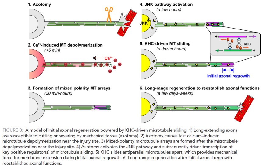
Fig. 8 from Lu, Lakonishok, & Gelfand (2015). License: Creative Commons 2.

The longest neuron that a human has is from the spinal cord to the tip of the toes. As a cell, it needs various proteins in various places. How is this transport done? Surely not by diffusion, the proteins would degrade or would arrive at inopportune membrane-moments (I just coined that). Molecular motors, on the other hand, are toiling proteins which haul huge cargoes for the benefit of the cell in an incredibly ingenious manner (they have feet and sticky soles and gears and so on). Notable motors are kinesin and dynein, the former brings stuff to the terminal buttons of the axon, the latter goes in the opposite direction, to the soma. They walk on a railway-like scaffold in a very funny manner, if you are to believe the simulations. Go on, I dare you, search kinesin or dynein animation on Google or YouTube and tell me then that biology is not funny.

And because no self-respectable scientist can work with the molecular motors without adding his/her contribution to the above-mentioned wealth of animations, the paper below comes with no less than 9 movies (as online supplemental material)! Lu et al. (2015) focused their attention on the role of kinesin in injured neurons. The authors dyed several types of proteins in fly neurons and then cultured the cells in a Petri dish. And then cut their axons with a glass needle. After that, they used a really fancy microscope (and a good microscopist, you should look at their pictures) to look at what happens. Which is this: the cut activates a c-Jun N-terminal kinase cascade (the cell’s response to stress), which leads to sliding of microtubules (part of cell’s cytoskeleton), which is com­pletely dependent on kinesin-1 heavy chain. This sliding initiates axonal regeneration (see picture).

I believe the kinesins and dyneins are the most charming, funny, and endearing proteins out there. Yes, I’m anthropomorphizing clumps of amino acids. I know, I’m a geek.

Reference: Lu W, Lakonishok M, & Gelfand VI (1 Apr 2015, Epub 5 Feb 2015). Kinesin-1–powered microtubule sliding initiates axonal regeneration in Drosophila cultured neurons. Molecular Biology of the Cell, 26(7):1296-307. doi: 10.1091/mbc.E14-10-1423. Article | FREE FULLTEXT PDF | Supplemental movies

Some youtube videos I mentioned before, quite accurate, too: best in show

by Neuronicus, 12 November 2015

The FIRSTS: Axon description (1865)

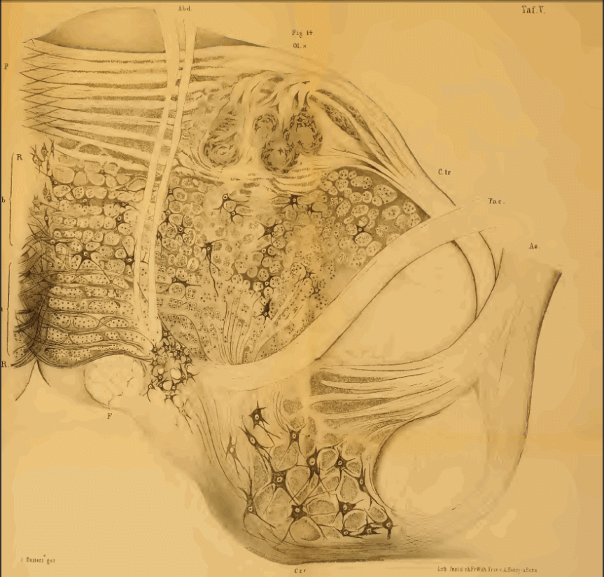
Human medulla oblongata sectioned at the level of the olivary nuclei. Drawing by Deiters (1965). Original caption: Fig. 15. Durchschnitt der medulla oblongata des Menschen in der Höhe der Olive (OL). R.R Raphe, Hyi). Nervus hypoglossus. Vag. Nervus vagus, deren Kerne in V und H liegen, aber in der Zeichnung nicht feiner ausgeführt sind. Den Haupttlißil der Figur nimmt die Formatio reticularis ein mit ihren zerstreuten Ganglienzellen, und die Olive mit den zu ihr hinzutretenden Fasern des Stratum zonale; C.c crura cerebelli ad medullam oblongatam; P Pyramidenstrang.
Human medulla oblongata sectioned at the level of the olivary nuclei. Drawing by Deiters (1865). Original caption: “Fig. 15. Durchschnitt der medulla oblongata des Menschen in der Höhe der Olive (OL). R.R Raphe, Hyi). Nervus hypoglossus. Vag. Nervus vagus, deren Kerne in V und H liegen, aber in der Zeichnung nicht feiner ausgeführt sind. Den Haupttlißil der Figur nimmt die Formatio reticularis ein mit ihren zerstreuten Ganglienzellen, und die Olive mit den zu ihr hinzutretenden Fasern des Stratum zonale; C.c crura cerebelli ad medullam oblongatam; P Pyramidenstrang”.

In 1863, using the microscope, a german neuroanatomist from the University of Bonn by the name of Otto Friedrich Karl Deiters describes in exquisite detail the branch-like processes of the neuron (i. e. dendrites) and the long, single “axis cylinder” (i.e. axon). Deiters’ nucleus is named after him (the place where a good portion of the cranial nerve VIII ends).

The book with the findings is published in German, posthumously (in 1865), with preface and under the editorial guidance of Max Schultze, another famous German anatomist. I got the information from Debanne et al. (2011), which is nice review on axon physiology (my German is kindda rusty due to lack of use). But I got my hands on the original German book (see link below) and, like a kid that doesn’t know how to read yet, all I could do was marvel at the absolutely stunning drawings by OFK Deiters. Which are truly and unequivocally beautiful. See for yourself.

Neurons with axons and dendrites. Drawings buy Deiters (1865.)
Neurons with axons and dendrites. Drawings by Deiters (1865.)

Reference: Debanne D, Campanac E, Bialowas A, Carlier E, & Alcaraz G (April 2011). Axon physiology. Physiological Reviews, 91(2):555-602. doi: 10.1152/physrev.00048.2009. Article | FREE FULLTEXT PDF

Original citation: Deiters OFK (1865). Untersuchungen über Gehirn und Rückenmark des Menschen und der Säugethiere. Ed. Max Schultze, Braunschweig: Vieweg, 1865. doi: 10.5962/bhl.title.15270. Book | PDF

By Neuronicus, 24 October 2015