The superiority illusion

Following up on my promise to cover a few papers about self-deception, the second in the series is about the superiority illusion, another cognitive bias (the first was about depressive realism).

Yamada et al. (2013) sought to uncover the origins of the ubiquitous belief that oneself is “superior to average people along various dimensions, such as intelligence, cognitive ability, and possession of desirable traits” (p. 4363). The sad statistical truth is that MOST people are average; that’s the whole definitions of ‘average’, really… But most people think they are superior to others, a.k.a. the ‘above-average effect’.

Twenty-four young males underwent resting-state fMRI and PET scanning. The first scanner is of the magnetic resonance type and tracks where you have most of the blood going in the brain at any particular moment. More blood flow to a region is interpreted as that region being active at that moment.

The word ‘functional’ means that the subject is performing a task while in the scanner and the resultant brain image is correspondent to what the brain is doing at that particular moment in time. On the other hand, ‘resting-state’ means that the individual did not do any task in the scanner, s/he just sat nice and still on the warm pads listening to the various clicks, clacks, bangs & beeps of the scanner. The subjects were instructed to rest with their eyes open. Good instruction, given than many subjects fall asleep in resting state MRI studies, even in the terrible racket that the coils make that sometimes can reach 125 Db. Let me explain: an MRI is a machine that generates a huge magnetic field (60,000 times stronger than Earth’s!) by shooting rapid pulses of electricity through a coiled wire, called gradient coil. These pulses of electricity or, in other words, the rapid on-off switchings of the electrical current make the gradient coil vibrate very loudly.

A PET scanner functions on a different principle. The subject receives a shot of a radioactive substance (called tracer) and the machine tracks its movement through the subject’s body. In this experiment’s case, the tracer was raclopride, a D2 dopamine receptor antagonist.

The behavioral data (meaning the answers to the questionnaires) showed that, curiously, the superiority illusion belief was not correlated with anxiety or self-esteem scores, but, not curiously, it was negatively correlated with helplessness, a measure of depression. Makes sense, especially from the view of depressive realism.

The imaging data suggests that dopamine binding to its striatal D2 receptors attenuate the functional connectivity between the left sensoriomotor striatum (SMST, a.k.a postcommissural putamen) and the dorsal anterior cingulate cortex (daCC). And this state of affairs gives rise to the superiority illusion (see Fig. 1).

125 superiority - Copy
Fig. 1. The superiority illusion arises from the suppression of the dorsal anterior cingulate cortex (daCC) – putamen functional connection by the dopamine coming from the substantia nigra/ ventral tegmental area complex (SN/VTA) and binding to its D2 striatal receptors. Credits: brain diagram: Wikipedia, other brain structures and connections: Neuronicus, data: Yamada et al. (2013, doi: 10.1073/pnas.1221681110). Overall: Public Domain

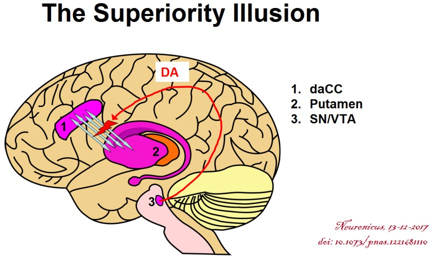
This was a frustrating paper. I cannot tell if it has methodological issues or is just poorly written. For instance, I have to assume that the dACC they’re talking about is bilateral and not ipsilateral to their SMST, meaning left. As a non-native English speaker myself I guess I should cut the authors a break for consistently misspelling ‘commissure’ or for other grammatical errors for fear of being accused of hypocrisy, but here you have it: it bugged me. Besides, mine is a blog and theirs is a published peer-reviewed paper. (Full Disclosure: I do get editorial help from native English speakers when I publish for real and, except for a few personal style quirks, I fully incorporate their suggestions). So a little editorial help would have gotten a long way to make the reading more pleasant. What else? Ah, the results are not clearly explained anywhere, it looks like the authors rely on obviousness, a bad move if you want to be understood by people slightly outside your field. From the first figure it looks like only 22 subjects out of 24 showed superiority illusion but the authors included 24 in the imaging analyses, or so it seems. The subjects were 23.5 +/- 4.4 years, meaning that not all subjects had the frontal regions of the brain fully developed: there are clear anatomical and functional differences between a 19 year old and a 27 year old.

I’m not saying it is a bad paper because I have covered bad papers; I’m saying it was frustrating to read it and it took me a while to figure out some things. Honestly, I shouldn’t even have covered it, but I spent some precious time going through it and its supplementals, what with me not being an imaging dude, so I said the hell with it, I’ll finish it; so here you have it :).

By Neuronicus, 13 December 2017

REFERENCE: Yamada M, Uddin LQ, Takahashi H, Kimura Y, Takahata K, Kousa R, Ikoma Y, Eguchi Y, Takano H, Ito H, Higuchi M, Suhara T (12 Mar 2013). Superiority illusion arises from resting-state brain networks modulated by dopamine. Proceedings of the National Academy of Sciences of the United States of America, 110(11):4363-4367. doi: 10.1073/pnas.1221681110. ARTICLE | FREE FULLTEXT PDF 

Amusia and stroke

Although a complete musical anti-talent myself, that doesn’t prohibit me from fully enjoying the works of the masters in the art. When my family is out of earshot, I even bellow – because it cannot be called music – from the top of my lungs alongside the most famous tenors ever recorded. A couple of days ago I loaded one of my most eclectic playlists. While remembering my younger days as an Iron Maiden concert goer (I never said I listen only to classical music :D) and screaming the “Fear of the Dark” chorus, I wondered what’s new on the front of music processing in the brain.

And I found an interesting recent paper about amusia. Amusia is, as those of you with ancient Greek proclivities might have surmised, a deficit in the perception of music, mainly the pitch but sometimes rhythm and other aspects of music. A small percentage of the population is born with it, but a whooping 35 to 69% of stroke survivors exhibit the disorder.

So Sihvonen et al. (2016) decided to take a closer look at this phenomenon with the help of 77 stroke patients. These patients had an MRI scan within the first 3 weeks following stroke and another one 6 months poststroke. They also completed a behavioral test for amusia within the first 3 weeks following stroke and again 3 months later. For reasons undisclosed, and thus raising my eyebrows, the behavioral assessment was not performed at 6 months poststroke, nor an MRI at the 3 months follow-up. It would be nice to have had behavioral assessment with brain images at the same time because a lot can happen in weeks, let alone months after a stroke.

Nevertheless, the authors used a novel way to look at the brain pictures, called voxel-based lesion-symptom mapping (VLSM). Well, is not really novel, it’s been around for 15 years or so. Basically, to ascertain the function of a brain region, researchers either get people with a specific brain lesion and then look for a behavioral deficit or get a symptom and then they look for a brain lesion. Both approaches have distinct advantages but also disadvantages (see Bates et al., 2003). To overcome the disadvantages of these methods, enter the scene VLSM, which is a mathematical/statistical gimmick that allows you to explore the relationship between brain and function without forming preconceived ideas, i.e. without forcing dichotomous categories. They also looked at voxel-based morphometry (VBM), which a fancy way of saying they looked to see if the grey and white matter differ over time in the brains of their subjects.

After much analyses, Sihvonen et al. (2016) conclude that the damage to the right hemisphere is more likely conducive to amusia, as opposed to aphasia which is due mainly to damage to the left hemisphere. More specifically,

“damage to the right temporal areas, insula, and putamen forms the crucial neural substrate for acquired amusia after stroke. Persistent amusia is associated with further [grey matter] atrophy in the right superior temporal gyrus (STG) and middle temporal gyrus (MTG), locating more anteriorly for rhythm amusia and more posteriorly for pitch amusia.”

The more we know, the better chances we have to improve treatments for people.

104-copy

unless you’re left-handed, then things are reversed.

References:

1. Sihvonen AJ, Ripollés P, Leo V, Rodríguez-Fornells A, Soinila S, & Särkämö T. (24 Aug 2016). Neural Basis of Acquired Amusia and Its Recovery after Stroke. Journal of Neuroscience, 36(34):8872-8881. PMID: 27559169, DOI: 10.1523/JNEUROSCI.0709-16.2016. ARTICLE  | FULLTEXT PDF

2.Bates E, Wilson SM, Saygin AP, Dick F, Sereno MI, Knight RT, & Dronkers NF (May 2003). Voxel-based lesion-symptom mapping. Nature Neuroscience, 6(5):448-50. PMID: 12704393, DOI: 10.1038/nn1050. ARTICLE

By Neuronicus, 9 November 2016

Save

The good and the bad of abstinence

The nucleus accumbens, a key region involved in reward processing and addiction. Credit: Zou et al. (2015)

Consumption of addictive drugs changes your brain and these changes underlie the consequent dependence. It is very difficult to quit, and certainly it is not a matter of lack of will power that the majority of drug users have such a great difficulty in trying to quit. But what happens to the brains of the lucky few who managed to kick the addiction out the window? Are their brains reverting to their pre-addiction states? As the paper below shows, sadly, no. But there is hope.

Zhou et al. ( 2015) used resting state fMRI (which means scanning your brain without requiring you to perform any task) to investigate the brains of 30 healthy controls and 30 heroin-addicts who were abstinent from the drug for more than 3 years. Specifically, they looked to see if the connectivity of certain brain regions involved in addiction is different after such a long abstinence time.

The bad news is that the abstinents still had some abnormal connectivity (for specialists: “stronger functional connectivity between the nucleus accumbens and the right ventromedial prefrontal cortex and relatively weaker connectivity between the nucleus accumbens and the left putamen, left precuneus, and supplementary motor area” p. 1697).

The good news: the longer the abstinence time, the greater the strength of the connection between nucleus accumbens and putamen (first structure involved in reward processing, the second in habit-learning), suggesting a partial neural recovery.

The study has some limitations: technical (did not control for heartbeat and respiration), methodological (most of the heroin abstinents were smokers, another addiction) and theoretical (one brain area does not support only one function, so its connectivity shouldn’t be over interpreted). With the caveat that the connectivity differences observed do not in any way point to a cause and effect relationship – that is we don’t know if these differences existed before the first heroin intake and caused it or they appeared after as a result of drug consumption, I think the paper is still is worth reading.

Reference: Zou F, Wu X, Zhai T, Lei Y, Shao Y, Jin X, Tan S, Wu B, Wang L, Yang Z (November 2015, Epub 17 Aug 2015). Abnormal resting-state functional connectivity of the nucleus accumbens in multi-year abstinent heroin addicts. Journal of Neuroscience Research, 93(11):1693-702. doi: 10.1002/jnr.23608. Article | FREE PDF

By Neuronicus, 9 October 2015