Who invented optogenetics?

Wayne State University. Ever heard of it? Probably not. How about Zhuo-Hua Pan? No? No bell ringing? Let’s try a different approach: ever heard of Stanford University? Why, yes, it’s one of the most prestigious and famous universities in the world. And now the last question: do you know who Karl Deisseroth is? If you’re not a neuroscientist, probably not. But if you are, then you would know him as the father of optogenetics.

Optogenetics is the newest tool in the biology kit that allows you to control the way a cell behaves by shining a light on it (that’s the opto part). Prior to that, the cell in question must be made to express a protein that is sensitive to light (i.e. rhodopsin) either by injecting a virus or breeding genetically modified animals that express that protein (that’s the genetics part).

If you’re watching the Nobel Prizes for Medicine, then you would also be familiar with Deisseroth’s name as he may be awarded the Nobel soon for inventing optogenetics. Only that, strictly speaking, he did not. Or, to be fair and precise at the same time, he did, but he was not the first one. Dr. Pan from Wayne State University was. And he got scooped.98.png

The story is at length imparted to us by Anna Vlasits in STAT and republished in Scientific American. In short, Dr. Pan, an obscure name in an obscure university from an ill-famed city (Detroit), does research for years in an unglamorous field of retina and blindness. He figured, quite reasonably, that restoring the proteins which sense light in the human eye (i.e. photoreceptor proteins) could restore vision in the congenitally blind. The problem is that human photoreceptor proteins are very complicated and efforts to introduce them into retinas of blind people have proven unsuccessful. But, in 2003, a paper was published showing how an algae protein that senses light, called channelrhodopsin (ChR), can be expressed into mammalian cells without loss of function.

So, in 2004, Pan got a colleague from Salus University (if Wayne State University is a medium-sized research university, then Salus is a really tiny, tiny little place) to engineer a ChR into a virus which Pan then injected in rodent retinal neurons, in vivo. After 3-4 weeks he obtained the expression of the protein and the expression was stable for at least 1 year, showing that the virus works nicely. Then his group did a bunch of electrophysiological recordings (whole cell patch-clamp and voltage clamp) to see if shining light on those neurons makes them fire. It did. Then, they wanted to see if ChR is for sure responsible for this firing and not some other proteins so they increased the intensity of the blue light that their ChR is known to sense and observed that the cell responded with increased firing. Now that they saw the ChR works in normal rodents, next they expressed the ChR by virally infecting mice who were congenitally blind and repeated their experiments. The electrophysiological experiments showed that it worked. But you see with your brain, not with your retina, so the researchers looked to see if these cells that express ChR project from the retina to the brain and they found their axons in lateral geniculate and superior colliculus, two major brain areas important for vision. Then, they recorded from these areas and the brain responded when blue light, but not yellow or other colors, was shone on the retina. The brain of congenitally blind mice without ChR does not respond regardless of the type of light shone on their retinas. But does that mean the mouse was able to see? That remains to be seen (har har) in future experiments. But the Pan group did demonstrate – without question or doubt – that they can control neurons by light.

All in all, a groundbreaking paper. So the Pan group was not off the mark when they submitted it to Nature on November 25, 2004. As Anna Vlasits reports in the Exclusive, Nature told Pan to submit to a more specialized journal, like Nature Neuroscience, which then rejected it. Pan submitted then to the Journal of Neuroscience, which also rejected it. He submitted it then to Neuron on November 29, 2005, which finally accepted it on February 23, 2006. Got published on April 6, 2006. Deisseroth’s paper was submitted to Nature Neuroscience on May 12, 2005, accepted on July, and published on August 14, 2005… His group infected rat hippocampal neurons cultured in a Petri dish with a virus carrying the ChR and then they did some electrophysiological recordings on those neurons while shining lights of different wavelengths on them, showing that these cells can be controlled by light.

There’s more on the saga with patent filings and a conference where Pan showed the ChR data in May 2005 and so on, you can read all about it in Scientific American. The magazine is just hinting to what I will say outright, loud and clear: Pan didn’t get published because of his and his institution’s lack of fame. Deisseroth did because of the opposite. That’s all. This is not about squabbles about whose work is more elegant, who presented his work as a scientific discovery or a technical report or whose title is more catchy, whose language is more boisterous or native English-speaker or luck or anything like that. It is about bias and, why not?, let’s call a spade a spade, discrimination. Nature and Journal of Neuroscience are not caught doing this for the first time. Not by a long shot. The problem is that they are still doing it, that is: discriminating against scientific work presented to them based on the name of the authors and their institutions.

Personally, so I don’t get comments along the lines of the fox and the grapes, I have worked at both high profile and low profile institutions. And I have seen the difference not in the work, but in the reception.

That’s my piece for today.

Source:  STAT, Scientific American.

References:

1) Bi A, Cui J, Ma YP, Olshevskaya E, Pu M, Dizhoor AM, & Pan ZH (6 April 2006). Ectopic expression of a microbial-type rhodopsin restores visual responses in mice with photoreceptor degeneration. Neuron, 50(1): 23-33. PMID: 16600853. PMCID: PMC1459045. DOI: 10.1016/j.neuron.2006.02.026. ARTICLE | FREE FULLTEXT PDF

2) Boyden ES, Zhang F, Bamberg E, Nagel G, & Deisseroth K. (Sep 2005, Epub 2005 Aug 14). Millisecond-timescale, genetically targeted optical control of neural activity. Nature Neuroscience, 8(9):1263-1268. PMID: 16116447. DOI: 10.1038/nn1525. doi:10.1038/nn1525. ARTICLE 

By Neuronicus, 11 September 2016

Save

Save

Save

Save

You were not my first choice either!

lego
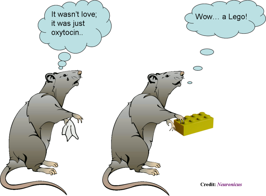
Sexually receptive female mice prefer a Lego brick over a male if their prefrontal oxytocin neurons are silenced.

Over the past five years or so, dopamine stepped down from the role of the “love molecule” in favor of oxytocin, a hormone previously known mostly for its crucial role in pregnancy, labor, delivery, lactation, and breastfeeding. Since some interesting discoveries in monogamous vs. polygamous voles (a type of rodent) pointing to oxytocin as essential for bonding, many studies implicated the chemical in all sorts of behaviors, from autistic to trusting, from generosity to wound healing.

Nakajima, Görlich, & Heintz (2015) add to that body of knowledge by finding that only a small group of cells in the medial prefrontal cortex express oxytocin receptors: a subpopulation of somatostatin cortical interneurons. Moreover, these neurons are gender dimorphic, meaning they differ from male to female: the female ones have twice as many action potentials upon application of oxytocin as compared to male’s.

And here is the more interesting part:
– Females in the sexually receptive phase of their estrus whose oxytocin neurons were silenced preferred to interact with a Lego brick over a male mouse (which, as you might have guessed, in not what they typically choose).
– Females that were not in their sexually receptive phase when their oxytocin neurons were silenced still preferred to interact with a mouse (male or female) over the Lego brick.
– Silencing of other neurons had no effect on their choice.
– Silencing had no effect on the males.

Hm… there are such things out there as oxytocin inter-nasal sprays… How soon do you think until the homeopaths, naturopaths, and other charlatans market oxytocin as a potent aphrodisiac? And it will take some deaths until the slow machine of beaurocracy turns its wheels and tightens the regulations on the accessibility to the hormone. Until then… as the cartoons say, don’t try this at home! Go buy some flowers or something for your intended one… it would work better, trust me on this.

Reference: Nakajima M, Görlich A, & Heintz N (9 October 2014). Oxytocin modulates female sociosexual behavior through a specific class of prefrontal cortical interneurons. Cell. 159(2): 295–305. doi:10.1016/j.cell.2014.09.020. Article | FREE FULLTEXT PDF

By Neuronicus, 23 October 2015

Giving up? Your parvalbumin neurons may have something to do with it

Cartoon from http://i393.photobucket.com/albums/pp20/saisi24/dontgivedup.jpg, licensing unknown
Cartoon from Photobucket, licensing unknown.

One of the most ecologically-valid rodent models of depression is the learned helplessness paradigm. You get a rat or a mouse and you confine it in a cage with an electrified grid. Then you apply mild foot shocks at random intervals and of random duration for an hour (which is one session). The mouse initially tries to escape, but there is no escape; the whole floor is electrified. After a couple of sessions, the mouse doesn’t try to escape anymore; it gives up. Even when you put the mouse in a cage with an open door, so it can flee to no-pain freedom, it doesn’t attempt to do so. The interpretation is that the mouse has learned that it cannot control the environment, no matter what he does, he’s helpless, so why bother? Hence the name of the behavioral paradigm: learned helplessness.

All antidepressants on the market have been tested at one point or another against this paradigm; if the drug got the mouse to try to escape more, then the drug passed the test.

Just like in the higher vertebrate realm, there are a few animals who keep trying to escape longer than the others, before they too finally give up; we call these resilient.

Perova, Delevich, & Li (2015) looked at a type of neuron that may have something to do with the capacity of some of the mice to be resilient; the parvalbumin interneurons (PAI) from the medial prefrontal cortex (mPFC). These neurons produce GABA, the major inhibitory neurotransmitter in the brain, and modulates the activity of the nearby neurons. Thanks to the ability to genetically engineer mice to have a certain kind of cell fluoresce, the researchers were able to identify and subsequently record from and manipulate the function of the PAIs. These PAIs’ response to stimulation was weaker in helpless animals compared to resilient or controls. Also, inactivation of the PAI via a designer virus promotes helplessness.

Reference: Perova Z, Delevich K, & Li B (18 Feb 2015). Depression of Excitatory Synapses onto Parvalbumin Interneurons in the Medial Prefrontal Cortex in Susceptibility to Stress. The Journal of Neuroscience, 35(7):3201–3206. doi: 10.1523/JNEUROSCI.2670-14.2015. Article | FREE FULLTEXT PDF

By Neuronicus, 21 October 2015

Viruses are as alive as crystals

Crystal of ultrapure bismuth. Photo credit: Intangir (public domain)
Crystal of ultrapure bismuth. Credit: Intangir. License: PD

The biology hype of the week is the notion that viruses are alive. Well, the true answer to that is… maybe. But that’s not catchy enough for headlines, is it?

Let’s start, as usual, with the source. Nasir & Caetano-Anollés (2015) published a paper where they did a lot of computer sniffing in existing proteomics databases to find out that viruses express a few dozen unique protein folds and they share several hundred more with cells. In other words, some of the viral proteins are unique. Using this information and some neat math they managed to calculate an evolutionary tree, that is, they classified the viruses via genetic relatedness to themselves and living organisms. That’s the strictu sensu of the term “tree of life”. From this taxonomy exercise, the authors speculate about when and how viruses might have appeared. They concluded that viruses appeared as RNA chunks spat out of cells. To give you a little background, there are two main hypotheses about the origin of viruses: appeared before cells as free floating RNA, or they were pieces of RNA that have been kicked out of a living cell, so after the evolution of cells. All well and good, I’m not going to open that can of worms, which hypothesis is more supported from data and so on.

Now, and this is the contentious part, verbatim:

“Here, we put forth the bold conjecture of a universal tree of life (uToL) that describes the evolution of cellular and viral proteomes. […]. Thus, viruses should be considered “living” organisms that simply survive by means of an atypical reproduction method that requires infecting a cell” (p. 18).

It’s their opinion. Not a fact. Not – and this is important – a direct consequence of their awesome taxonomy exercise. For a formal definition, a conjecture is an opinion or conclusion formed on the basis of incomplete information (Oxford Dictionary).

Don’t get me wrong, I think this is a neat paper, and, frankly, I don’t have a horse in this race: I don’t care whether viruses are alive or not. But I do care to distinguish between fact and opinion, an intellectual exercise that seem to have eluded the science websites and science popularization zines and e-zines like EurekaAlert, IFLS, ScienceAlert, Gizmodo, Daily Mail, Wired, Popular Science, R & D Magazine, Laboratory Equipment, and many others, who ran titles saying “Viruses are alive” in just so many words. Note that even the authors themselves put the word “living” between quotes. Know the difference between opinion (that is, we think that because x makes y, maybe x is blue) and a scientific fact (x makes y, y makes z, therefore x must make z and we know that not only because it’s logical, but also because no other wretched letter wants to make z, believe us we tried, we made sure z is where we put it, because we put dye on it, we measured it, chopped it, looked at it with 5 different instruments, and we cannot make z without x though a poor grad student tried and tried in vain, we even modeled z, and yes, it is made by x, otherwise known as we eliminated all other testable bloody possibilities that we could think of. Unless q makes z in humans, but we can’t measure that. Or r makes z under Jovian gravity, but we didn’t get a grant for that…. get my drift what a scientific fact is?).

Now, rant is not over. The authors argue that viruses have a sort of metabolism and they replicate, so they meet the requirements for being classified as alive. For the sake of the argument, I can posit that either these two conditions are not enough for something to be considered alive, or we have then to conclude that cave crystals are also alive. Crystals only grow in the appropriate environment of a saturated solution and bits shattered off from them go into an inert mode waiting for appropriate conditions. Crystal growth can even be looked at as a sort of metabolism. So, if they are willing to characterize crystal growth as somewhere on the continuous scale of life, viruses can be there as well. Using their analogy, the living, metabolically active form for crystals is when they are growing in a saturated solution; and bits breaking off or the bulk waiting for solution conditions to change is just an atypical reproductive scheme. It even gets more interesting with some of the more modern more complicated crystal growth theories with preassembly into nanocrystals, editing or incorporating defects, etc. Ok, I’m getting tired and I made my point anyway. Rant over. Happy debating!

Reference: Nasir, A. & Caetano-Anollés, G. (25 September 2015). A phylogenomic data-driven exploration of viral origins and evolution. Science Advances, 1(8): 1-24. DOI: 10.1126/sciadv.1500527. Article | FREE PDF

by Neuronicus, 29 September 2015

Save

Obscure protein restores memory decline

In the not-too-distant-future, your grandma may give you a run for your money on your video games. Photo credit: http://www.funtoosh.com/pictures/
In the not-too-distant-future, your grandma may give you a run for your money on your video games. Photo credit: Funtoosh

Aging comes with all sorts of maladies, but one of the most frustrating is the feeling that you are not as sharp as you used to be. Cognitive decline has been previously linked, at least in part, to a dysregulation in the neuronal calcium homeostasis in the hippocampus, which is a brain region essential for learning and memory. One player that keeps in check the proper balance of calcium use is the protein FKBP1b, and, not surprisingly, its amounts are reduced in aging rats and Alzheimer’s suffering patients.

FKBP1b overexpression in hippocampal neurons reversed spatial memory deficits in aged rats. Fig. 3 (partial) from Gant, J. C., Chen, K. C., Kadish, I., Blalock, E. M., Thibault, O., Porter, N. M., Landfield, P. W. (29 July 2015). Reversal of Aging-Related Neuronal Ca2+ Dysregulation and Cognitive Impairment by Delivery of a Transgene Encoding FK506-Binding Protein 12.6/1b to the Hippocampus. The Journal of Neuroscience, 35(30):10878 –10887. doi: 10.1523/JNEUROSCI.1248-15.2015.
FKBP1b overexpression in hippocampal neurons reversed spatial memory deficits in aged rats. Fig. 3 (partial) from Gant et al. (2015): doi: 10.1523/JNEUROSCI.1248-15.2015.

Gant et al. (2015) sought to increase the expression of the FKBP1b protein in the hippocampus, in the hopes that its increase would result in better calcium homeostasis and, as a result, better memory performance in aging rats. They built a virus that carried the gene for making the FKBP1b protein and they injected this directly in the hippocampus. After they waited 5-6 weeks for the gene to be expressed, they tested the rats in the Morris water maze, a test for spatial memory. The old rats that received the injection performed as well as the young rats, and far better than the old rats who didn’t get the injection. Then the researchers made sure that the injection is the one responsible for the results, by checking the levels of the FKBP1b protein in the hippocampus (increased, as per specs), by recording from those neurons (they were awesome), and by imaging the calcium to make sure the balance was restored (ditto).

Reference: Gant, J. C., Chen, K. C., Kadish, I., Blalock, E. M., Thibault, O., Porter, N. M., Landfield, P. W. (29 July 2015). Reversal of Aging-Related Neuronal Ca2+ Dysregulation and Cognitive Impairment by Delivery of a Transgene Encoding FK506-Binding Protein 12.6/1b to the Hippocampus. The Journal of Neuroscience, 35(30):10878 –10887. doi: 10.1523/JNEUROSCI.1248-15.2015. Article + FREE PDF + Journal of Neuroscience cover