The FIRSTS: Lack of happy events in depression (2003)

My last post focused on depression and it reminded me of something that I keep telling my students and they all react with disbelief. Well, I tell them a lot of things to which they react with disbelief, to be sure, but this one I keep thinking it should not generate such incredulity. The thing is: depressed people perceive the same amount of negative events happening to them as healthy people, but far fewer positive ones. This seems to be counter-intuitive to non-professionals, who believe depressed people are just generally sadder than average and that’s why they see the half-empty side of the glass of life.

So I dug out the original paper who found this… finding. It’s not as old as you might think. Peeters et al. (2003) paid $30/capita to 86 people, 46 of which were diagnosed with Major Depressive Disorder and seeking treatment in a community mental health center or outpatient clinic (this is in Netherlands). None were taking antidepressants or any other drugs, except low-level anxiolytics. Each participant was given a wristwatch that beeped 10 times a day at semi-random intervals of approximately 90 min. When the watch beeped, the subjects had to complete a form within maximum 25 min answering questions about their mood, currents events, and their appraisal of those events. The experiment took 6 days, including weekend.

The results? Contrary to popular belief, people with depression “did not report more frequent negative events, although they did report fewer positive events and appraised both types of events as more stressful” (p. 208). In other words, depressed people are not seeing half-empty glasses all the time; instead, they don’t see the half-full glasses. Note that they regarded both negative and positive events as stressful. We circle back to the ‘stress is the root of all evil‘ thing.

I would have liked to see if the decrease in positive affect and perceived happy events correlates with increased sadness. The authors say that “negative events were appraised as more unpleasant, more important, and more stressful by the depressed than by the healthy participants ” (p. 206), but, curiously, the  mood was assessed with ratings on the feeling anxious, irritated, restless, tense, guilty, irritable, easily distracted, and agitate, and not a single item on depression-iconic feelings: sad, empty, hopeless, worthless.

Nevertheless, it’s a good psychological study with in depth statistical analyses. I also found thought-provoking this paragraph: “The literature on mood changes in daily life is dominated by studies of daily hassles. The current results indicate that daily uplifts are also important determinants of mood, in both depressed and healthy people” (p. 209).

152 depression and lack of happy events - Copy

REFERENCE: Peeters F, Nicolson NA, Berkhof J, Delespaul P, & deVries M. (May 2003). Effects of daily events on mood states in major depressive disorder. Journal of Abnormal Psychology, 112(2):203-11. PMID: 12784829, DOI: 10.1037/0021-843X.112.2.203. ARTICLE

By Neuronicus, 4 May 2019

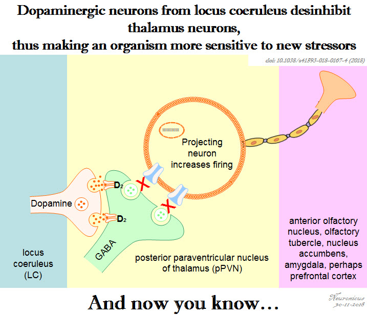
Pic of the day: Dopamine from a non-dopamine place

147 lc da ppvn - Copy

Reference: Beas BS, Wright BJ, Skirzewski M, Leng Y, Hyun JH, Koita O, Ringelberg N, Kwon HB, Buonanno A, & Penzo MA (Jul 2018, Epub 18 Jun 2018). The locus coeruleus drives disinhibition in the midline thalamus via a dopaminergic mechanism. Nature Neuroscience,21(7):963-973. PMID: 29915192, PMCID: PMC6035776 [Available on 2018-12-18], DOI:10.1038/s41593-018-0167-4. ARTICLE

The Mom Brain

Recently, I read an opinion titled When I Became A Mother, Feminism Let Me Down. The gist of it was that some (not all!) feminists, while empowering women and girls to be anything they want to be and to do anything a man or a boy does, they fail in uplifting the motherhood aspect of a woman’s life, should she choose to become a mother. In other words, even (or especially, in some cases) feminists look down on the women who chose to switch from a paid job and professional career to an unpaid stay-at-home mom career, as if being a mother is somehow beneath what a woman can be and can achieve. As if raising the next generation of humans to be rational, informed, well-behaved social actors instead of ignorant brutal egomaniacs is a trifling matter, not to be compared with the responsibilities and struggles of a CEO position.

Patriarchy notwithstanding, a woman can do anything a man can. And more. The ‘more’ refers to, naturally, motherhood. Evidently, fatherhood is also a thing. But the changes that happen in a mother’s brain and body during pregnancy, breastfeeding, and postpartum periods are significantly more profound than whatever happens to the most loving and caring and involved father.

Kim (2016) bundled some of these changes in a nice review, showing how these drastic and dramatic alterations actually have an adaptive function, preparing the mother for parenting. Equally important, some of the brain plasticity is permanent. The body might spring back into shape if the mother is young or puts into it a devilishly large amount of effort, but some brain changes are there to stay. Not all, though.

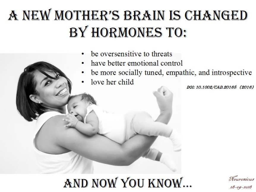
One of the most pervasive findings in motherhood studies is that hormones whose production is increased during pregnancy and postpartum, like oxytocin and dopamine, sensitize the fear circuit in the brain. During the second trimester of pregnancy and particularly during the third, expectant mothers start to be hypervigilent and hypersensitive to threats and to angry faces. A higher anxiety state is characterized, among other things, by preferentially scanning for threats and other bad stuff. Threats mean anything from the improbable tiger to the 1 in a million chance for the baby to be dropped by grandma to the slightly warmer forehead or the weirdly colored poopy diaper. The sensitization of the fear circuit, out of which the amygdala is an essential part, is adaptive because it makes the mother more likely to not miss or ignore her baby’s cry, thus attending to his or her needs. Also, attention to potential threats is conducive to a better protection of the helpless infant from real dangers. This hypersensitivity usually lasts 6 to 12 months after childbirth, but it can last lifetime in females already predisposed to anxiety or exposed to more stressful events than average.

Many new mothers worry if they will be able to love their child as they don’t feel this all-consuming love other women rave about pre- or during pregnancy. Rest assured ladies, nature has your back. And your baby’s. Because as soon as you give birth, dopamine and oxytocin flood the body and the brain and in so doing they modify the reward motivational circuit, making new mothers literally obsessed with their newborn. The method of giving birth is inconsequential, as no differences in attachment have been noted (this is from a different study). Do not mess with mother’s love! It’s hardwired.

Another change happens to the brain structures underlying social information processing, like the insula or fusiform gyrus, making mothers more adept at self-motoring, reflection, and empathy. Which is a rapid transformation, without which a mother may be less accurate in understanding the needs, mental state, and social cues of the very undeveloped ball of snot and barf that is the human infant (I said that affectionately, I promise).

In order to deal with all these internal changes and the external pressures of being a new mom the brain has to put up some coping mechanisms. (Did you know, non-parents, that for the first months of their newborn lives, the mothers who breastfeed must do so at least every 4 hours? Can you imagine how berserk with sleep deprivation you would be after 4 months without a single night of full sleep but only catnaps?). Some would be surprised to find out – not mothers, though, I’m sure – that “new mothers exhibit enhanced neural activation in the emotion regulation circuit including the anterior cingulate cortex, and the medial and lateral prefrontal cortex” (p. 50). Which means that new moms are actually better at controlling their emotions, particularly at regulating negative emotional reactions. Shocking, eh?

140 mom brain1 - Copy

Finally, it appears that very few parts of the brain are spared from this overhaul as the entire brain of the mother is first reduced in size and then it grows back, reorganized. Yeah, isn’t that weird? During pregnancy the brain shrinks, being at its lowest during childbirth and then starts to grow again, reaching its pre-pregnancy size 6 months after childbirth! And when it’s back, it’s different. The brain parts heavily involved in parenting, like the amygdala involved in the anxiety, the insula and superior temporal gyrus involved in social information processing and the anterior cingulate gyrus involved in emotional regulation, all these show increased gray matter volume. And many other brain structures that I didn’t list. One brain structure is rarely involved only in one thing so the question is (well, one of them) what else is changed about the mothers, in addition to their increased ability to parent?

I need to add a note here: the changes that Kim (2016) talks about are averaged. That means some women get changed more, some less. There is variability in plasticity, which should be a pleonasm. There is also variability in the human population, as any mother attending a school parents’ night-out can attest. Some mothers are paranoid with fear and overprotective, others are more laissez faire when it comes to eating from the floor.

But SOME changes do occur in all mothers’ brains and bodies. For example, all new mothers exhibit a heightened attention to threats and subsequent raised levels of anxiety. But when does heightened attention to threats become debilitating anxiety? Thanks to more understanding and tolerance about these changes, more and more women feel more comfortable reporting negative feelings after childbirth so that now we know that postpartum depression, which happens to 60 – 80% of mothers, is a serious matter. A serious matter that needs serious attention from both professionals and the immediate social circle of the mother, both for her sake as well as her infant’s. Don’t get me wrong, we – both males and females – still have a long way ahead of us to scientifically understand and to socially accept the mother brain, but these studies are a great start. They acknowledge what all mothers know: that they are different after childbirth than the way they were before. Now we have to figure out how are they different and what can we do to make everyone’s lives better.

Kim (2016) is an OK review, a real easy read, I recommend it to the non-specialists wholeheartedly; you just have to skip the name of the brain parts and the rest is pretty clear. It is also a very short review, which will help with reader fatigue. The caveat of that is that it doesn’t include a whole lotta studies, nor does it go in detail on the implications of what the handful cited have found, but you’ll get the gist of it. There is a vastly more thorough literature if one would include animal studies that the author, curiously, did not include. I know that a mouse is not a chimp is not a human, but all three of us are mammals, and social mammals at that. Surely, there is enough biological overlap so extrapolations are warranted, even if partially. Nevertheless, it’s a good start for those who want to know a bit about the changes motherhood does to the brain, behavior, thoughts, and feelings.

Corroborated with what I already know about the neuroscience of maternity, my favourite takeaway is this: new moms are not crazy. They can’t help most of these changes. It’s biology, you see. So go easy on new moms. Moms, also go easy on yourselves and know that, whether they want to share or not, the other moms probably go through the same stuff. The other moms are doing better than you are either. You’re not alone. And if that overactive threat circuit gives you problems, i.e. you feel overwhelmed, it’s OK to ask for help. And if you don’t get it, ask for it again and again until you do. That takes courage, that’s empowerment.

P. S. The paper doesn’t look like it’s peer-reviewed. Yes, I know the peer-reviewing publication system is flawed, I’ve been on the receiving end of it myself, but it’s been drilled into my skull that it’s important, flawed as it is, so I thought to mention it.

REFERENCE: Kim, P. (Sept. 2016). Human Maternal Brain Plasticity: Adaptation to Parenting, New Directions for Child and Adolescent Development, (153): 47–58. PMCID: PMC5667351, doi: 10.1002/cad.20168. ARTICLE | FREE FULLTEXT PDF

By Neuronicus, 28 September 2018

The Benefits of Vacation

My prolonged Internet absence from the last month or so was due to a prolonged vacation. In Europe. Which I loved. Both the vacation and the Europe. Y’all, people, young and old, listen to me: do not neglect vacations for they strengthen the body, nourish the soul, and embolden the spirit.

More pragmatically, vacations lower the stress level. Yes, even the stressful vacations lower the stress level, because the acute stress effects of “My room is not ready yet” / “Jimmy puked in the car” / “Airline lost my luggage” are temporary and physiologically different from the chronic stress effects of “I’ll lose my job if I don’t meet these deadlines” / “I hate my job but I can’t quit because I need health insurance” / “I’m worried for my child’s safety” / “My kids will suffer if I get a divorce” / “I can’t make the rent this month”.

Chronic stress results in a whole slew of real nasties, like cognitive, learning, and memory impairments, behavioral changes, issues with impulse control, immune system problems, weight gain, cardiovascular disease and so on and so on and so on. Even death. As I told my students countless of times, chronic stress to the body is as real and physical as a punch in the stomach but far more dangerous. So take a vacation as often as you can. Even a few days of total disconnect help tremendously.

There are literally thousands of peer-reviewed papers out there that describe the ways in which stress produces all those bad things, but not so many papers about the effects of vacations. I suspect this is due to the inherent difficulty in accounting for the countless environmental variables that can influence one’s vacation and its outcomes, whereas identifying and characterizing stressors is much easier. In other words, lack of experimental control leads to paucity of good data. Nevertheless, from this paucity, Chen & Petrick (2013) carefully selected 98 papers from both academic and nonacademic publications about the benefits of travel vacations.

These are my take-home bullet-points:

  • vacation effects last no more than a month
  • vacations reduce both the subjective perception of stress and the objective measurement of it (salivary cortisol)
  • people feel happier after taking a vacation
  • there are some people who do not relax in a vacation, presumably because they cannot ‘detach’ themselves from the stressors in their everyday life (long story here why some people can’t let go of problems)
  • vacations lower the occurrence of cardiovascular disease
  • vacations decrease work-related stress, work absenteeism, & work burnout
  • vacations increase job performance
  • the more you do on a vacation the better you feel, particularly if you’re older
  • you benefit more if you do new things or go to new places instead of just staying home
  • vacations increase overall life satisfaction

Happy vacationing!

137 - Copy

REFERENCE: Chen, C-C & Petrick, JF (Nov. 2013, Epub 17 Jul. 2013). Health and Wellness Benefits of Travel Experiences: A Literature Review, Journal of Travel Research, 52(6):709-719. doi: 10.1177/0047287513496477. ARTICLE | FULLTEXT PDF via ResearchGate.

By Neuronicus, 20 July 2018

The FIRSTS: The roots of depressive realism (1979)

There is a rumor stating that depressed people see the world more realistically and the rest of us are – to put it bluntly – deluded optimists. A friend of mine asked me if this is true. It took me a while to find the origins of this claim, but after I found it and figured out that the literature has a term for the phenomenon (‘depressive realism’), I realized that there is a whole plethora of studies on the subject. So the next following posts will be centered, more or less, on the idea of self-deception.

It was 1979 when Alloy & Abramson published a paper who’s title contained the phrase ‘Sadder but Wiser’, even if it was followed by a question mark. The experiments they conducted are simple, but the theoretical implications are large.

The authors divided several dozens of male and female undergraduate students into a depressed group and a non-depressed group based on their Beck Depression Inventory scores (a widely used and validated questionnaire for self-assessing depression). Each subject “made one of two possible responses (pressing a button or not pressing a button) and received one of two possible outcomes (a green light or no green light)” (p. 447). Various conditions presented the subjects with various degrees of control over what the button does, from 0 to 100%. After the experiments, the subjects were asked to estimate their control over the green light, how many times the light came on regardless of their behavior, what’s the percentage of trials on which the green light came on when they pressed or didn’t press the button, respectively, and how did they feel. In some experiments, the subjects were wining or losing money when the green light came on.

Verbatim, the findings were that:

“Depressed students’ judgments of contingency were surprisingly accurate in all four experiments. Nondepressed students, on the other hand, overestimated the degree of contingency between their responses and outcomes when noncontingent outcomes were frequent and/or desired and underestimated the degree of contingency when contingent outcomes were undesired” (p. 441).

In plain English, it means that if you are not depressed, when you have some control and bad things are happening, you believe you have no control. And when you have no control but good things are happening, then you believe you have control. If you are depressed, it does not matter, you judge your level of control accurately, regardless of the valence of the outcome.

Such illusion of control is a defensive mechanism that surely must have adaptive value by, for example, allowing the non-depressed to bypass a sense of guilt when things don’t work out and increase self-esteem when they do. This is fascinating, particularly since it is corroborated by findings that people receiving gambling wins or life successes like landing a good job, rewards that at least in one case are demonstrably attributable to chance, believe, nonetheless, that it is due to some personal attributes that make them special, that makes them deserving of such rewards. (I don’t remember the reference of this one so don’t quote me on it. If I find it, I’ll post it, it’s something about self-entitlement, I think). That is not to say that life successes are not largely attributable to the individual; they are. But, statistically speaking, there must be some that are due to chance alone, and yet most people feel like they are the direct agents for changes in luck.

Another interesting point is that Alloy & Abramson also tried to figure out how exactly their subjects reasoned when they asserted their level of control through some clever post-experiment questioners. Long story short (the paper is 45 pages long), the illusion of control shown by nondepressed subjects in the no control condition was the result of incorrect logic, that is, faulty reasoning.

In summary, the distilled down version of depressive realism that non-depressed people see the world through rose-colored glasses is correct only in certain circumstances. Because only in particular conditions this illusion of control applies and that is overestimation of control only when good things are happening and underestimation of control when bad things are happening. But, by and large, it does seem that depression clears the fog a bit.

Of course, it has been over 40 years since the publication of this paper and of course it has its flaws. Many replications and replications with caveats and meta-analyses and reviews and opinions and alternative hypotheses have been confirmed and infirmed and then confirmed again with alterations, so there is still a debate out there about the causes/ functions/ ubiquity/ circumstantiality of the depressive realism effect. One thing seems to be constant though: the effect exists.

I will leave you with the ponders of Alloy & Abramson (1979):

“A crucial question is whether depression itself leads people to be “realistic” or whether realistic people are more vulnerable to depression than other people” (p. 480).

124 - Copy

REFERENCE: Alloy LB, & Abramson LY (Dec. 1979). Judgment of contingency in depressed and nondepressed students: sadder but wiser? Journal of Experimental Psychology: General, 108(4): 441-485. PMID: 528910. http://dx.doi.org/10.1037/0096-3445.108.4.441. ARTICLE | FULLTEXT PDF via ResearchGate

By Neuronicus, 30 November 2017

Painful Pain Paper

There has been much hype over the new paper published in the latest Nature issue which claims to have discovered an opioid analgesic that doesn’t have most of the side effects of morphine. If the claim holds, the authors may have found the Holy Grail of pain research chased by too many for too long (besides being worth billions of dollars to its discoverers).

The drug, called PZM21, was discovered using structure-based drug design. This means that instead of taking a drug that works, say morphine, and then tweaking its molecular structure in various ways and see if the resultant drugs work, you take the target of the drug, say mu-opioid receptors, and design a drug that fits in that slot. The search and design are done initially with sophisticated software and there are many millions of virtual candidates. So it takes a lot of work and ingenuity to select but a few drugs that will be synthesized and tested in live animals.

Manglik et al. (2016) did just that and they came up with PZM21 which, compared to morphine, is:

1) selective for the mu-opioid receptors (i.e. it doesn’t bind to anything else)
2) produces no respiratory depression (maybe a touch on the opposite side)
3) doesn’t affect locomotion
4) produces less constipation
5) produces long-lasting affective analgesia
6) and has less addictive liability

The Holy Grail, right? Weeell, I have some serious issues with number 5 and, to some extent, number 6 on this list.

Normally, I wouldn’t dissect a paper so thoroughly because, if there is one thing I learned by the end of GradSchool and PostDoc, is that there is no perfect paper out there. Consequently, anyone with scientific training can find issues with absolutely anything published. I once challenged someone to bring me any loved and cherished paper and I would tear it apart; it’s much easier to criticize than to come up with solutions. Probably that’s why everybody hates Reviewer No. 2…

But, for extraordinary claims, you need extraordinary evidence. And the evidence simply does not support the 5 and maybe 6 above.

Let’s start with pain. The authors used 3 tests: hotplate (drop a mouse on a hot plate for 10 sec and see what it does), tail-flick (give an electric shock to the tail and see how fast the mouse flicks its tail) and formalin (inject an inflammatory painful substance in the mouse paw and see what the animal does). They used 3 doses of PZM21 in the hotplate test (10, 20, and 40 mg/Kg), 2 doses in the tail-flick test (10 and 20), and 1 dose in the formalin test (20). Why? If you start with a dose-response in a test and want to convince me it works in the other tests, then do a dose-response for those too, so I have something to compare. These tests have been extensively used in pain research and the standard drug used is morphine. Therefore, the literature is clear on how different doses of morphine work in these tests. I need your dose-responses for your new drug to be able to see how it measures up, since you claim it is “more efficacious than morphine”. If you don’t want to convince me there is a dose-response effect, that’s fine too, I’ll frown a little, but it’s your choice. However, then choose a dose and stick with it! Otherwise I cannot compare the behaviors across tests, rendering one or the other test meaningless. If you’re wondering, they used only one dose of morphine in all the tests, except the hotplate, where they used two.

Another thing also related to doses. The authors found something really odd: PZM21 works (meaning produces analgesia) in the hotplate, but not the tail-flick tests. This is truly amazing because no opiate I know of can make such a clear-cut distinction between those two tests. Buuuuut, and here is a big ‘BUT” they did not test their highest dose (40mg/kg) in the tail-flick test! Why? I’ll tell you how, because I am oh sooo familiar with this argument. It goes like this:

Reviewer: Why didn’t you use the same doses in all your 3 pain tests?

Author: The middle and highest doses have similar effects in the hotplate test, ok? So it doesn’t matter which one of these doses I’ll use in the tail-flick test.

Reviewer: Yeah, right, but, you have no proof that the effects of the two doses are indistinguishable because you don’t report any stats on them! Besides, even so, that argument applies only when a) you have ceiling effects (not the case here, your morphine hit it, at any rate) and b) the drug has the expected effects on both tests and thus you have some logical rationale behind it. Which is not the case here, again: your point is that the drug DOESN’T produce analgesia in the tail-flick test and yet you don’t wanna try its HIGHEST dose… REJECT AND RESUBMIT! Awesome drug discovery, by the way!

So how come the paper passed the reviewers?! Perhaps the fact that two of the reviewers are long term publishing co-authors from the same University had something to do with it, you know, same views predisposes them to the same biases and so on… But can you do that? I mean, have reviewers for Nature from the same department for the same paper?

Alrighty then… let’s move on to the stats. Or rather not. Because there aren’t any for the hotplate or tail-flick! Now, I know all about the “freedom from the tyranny of p” movement (that is: report only the means, standard errors of mean, and confidence intervals and let the reader judge the data) and about the fact that the average scientist today needs to know 100-fold more stats that his predecessors 20 years ago (although some biologists and chemists seem to be excused from this, things either turn color or not, either are there or not etc.) or about the fact that you cannot get away with only one experiment published these days, but you need a lot of them so you have to do a lot of corrections to your stats so you don’t fall into the Type 1 error. I know all about that, but just like the case with the doses, choose one way or another and stick to it. Because there are ANOVAs ran for the formalin test, the respiration, constipation, locomotion, and conditioned place preference tests, but none for the hotplate or tailflick! I am also aware that to be published in Science or Nature you have to strip your work and wordings to the bare minimum because the insane wordcount limits, but you have free rein in the Supplementals. And I combed through those and there are no stats there either. Nor are there any power analyses… So, what’s going on here? Remember, the authors didn’t test the highest dose on the tail-flick test because – presumably – the highest and intermediary doses have indistinguishable effects, but where is the stats to prove it?

And now the thing that really really bothered me: the claim that PZM21 takes away the affective dimension of pain but not the sensory. Pain is a complex experience that, depending on your favourite pain researcher, has at least 2 dimensions: the sensory (also called ‘reflexive’ because it is the immediate response to the noxious stimulation that makes you retract by reflex the limb from whatever produces the tissue damage) and the affective (also called ‘motivational’ because it makes the pain unpleasant and motivates you to get away from whatever caused it and seek alleviation and recovery). The first aspect of pain, the sensory, is relatively easy to measure, since you look at the limb withdrawal (or tail, in the case of animals with prolonged spinal column). By contrast, the affective aspect is very hard to measure. In humans, you can ask them how unpleasant it is (and even those reports are unreliable), but how do you do it with animals? Well, you go back to humans and see what they do. Humans scream “Ouch!” or swear when they get hurt (so you can measure vocalizations in animals) or humans avoid places in which they got hurt because they remember the unpleasant pain (so you do a test called Conditioned Place Avoidance for animals, although if you got a drug that shows positive results in this test, like morphine, you don’t know if you blocked the memory of unpleasantness or the feeling of unpleasantness itself, but that’s a different can of worms). The authors did not use any of these tests, yet they claim that PZM21 takes away the unpleasantness of pain, i.e. is an affective analgesic!

What they did was this: they looked at the behaviors the animal did on the hotplate and divided them in two categories: reflexive (the lifting of the paw) and affective (the licking of the paw and the jumping). Now, there are several issues with this dichotomy, I’m not even going to go there; I’ll just say that there are prominent pain researchers that will scream from the top of their lungs that the so-called affective behaviors from the hotplate test cannot be indexes of pain affect, because the pain affect requires forebrain structures and yet these behaviors persist in the decerebrated rodent, including the jumping. Anyway, leaving the theoretical debate about what those behaviors they measured really mean aside, there still is the problem of the jumpers: namely, the authors excluded from the analysis the mice who tried to jump out of the hotplate test in the evaluation of the potency of PZM21, but then they left them in when comparing the two types of analgesia because it’s a sign of escaping, an emotionally-valenced behavior! Isn’t this the same test?! Seriously? Why are you using two different groups of mice and leaving the impression that is only one? And oh, yeah, they used only the middle dose for the affective evaluation, when they used all three doses for potency…. And I’m not even gonna ask why they used the highest dose in the formalin test… but only for the normal mice, the knockouts in the same test got the middle dose! So we’re back comparing pears with apples again!

Next (and last, I promise, this rant is way too long already), the non-addictive claim. The authors used the Conditioned Place Paradigm, an old and reliable method to test drug likeability. The idea is that you have a box with 2 chambers, X and Y. Give the animal saline in chamber X and let it stay there for some time. Next day, you give the animal the drug and confine it in chamber Y. Do this a few times and on the test day you let the animal explore both chambers. If it stays more in chamber Y then it liked the drug, much like humans behave by seeking a place in which they felt good and avoiding places in which they felt bad. All well and good, only that is standard practice in this test to counter-balance the days and the chambers! I don’t know about the chambers, because they don’t say, but the days were not counterbalanced. I know, it’s a petty little thing for me to bring that up, but remember the saying about extraordinary claims… so I expect flawless methods. I would have also liked to see a way more convincing test for addictive liability like self-administration, but that will be done later, if the drug holds, I hope. Thankfully, unlike the affective analgesia claims, the authors have been more restrained in their verbiage about addiction, much to their credit (and I have a nasty suspicion as to why).

I do sincerely think the drug shows decent promise as a painkiller. Kudos for discovering it! But, seriously, fellows, the behavioral portion of the paper could use some improvements.

Ok, rant over.

EDIT (Aug 25, 2016): I forgot to mention something, and that is the competing financial interests declared for this paper: some of its authors already filed a provisional patent for PZM21 or are already founders or consultants for Epiodyne (a company that that wants to develop novel analgesics). Normally, that wouldn’t worry me unduly, people are allowed to make a buck from their discoveries (although is billions in this case and we can get into that capitalism-old debate whether is moral to make billions on the suffering of other people, but that’s a different story). Anyway, combine the financial interests with the poor behavioral tests and you get a very shoddy thing indeed.

Reference: Manglik A, Lin H, Aryal DK, McCorvy JD, Dengler D, Corder G, Levit A, Kling RC, Bernat V, Hübner H, Huang XP, Sassano MF, Giguère PM, Löber S, Da Duan, Scherrer G, Kobilka BK, Gmeiner P, Roth BL, & Shoichet BK (Epub 17 Aug 2016). Structure-based discovery of opioid analgesics with reduced side effects. Nature, 1-6. PMID: 27533032, DOI: 10.1038/nature19112. ARTICLE 

By Neuronicus, 21 August 2016

Stress can kill you and that’s no metaphor

The term ‘heartbreak’ is used as a metaphor to describe the intense feeling of loss, sometimes also called emotional pain. But what if the metaphor has roots into something more tangible than a feeling, that of the actual muscular organ giving signs of failure?

Although there have been previous reports that found stress causes cardiovascular problems, including myocardial infarction, Graff et al. (2016) conducted the largest study to date that investigated this link: they had almost 1 million subjects. That’s right, 1 million people (well, actually 974 732). Out of these, almost 20% of them had a partner who died between 1995 and 2014. The chosen stressor was the loss of a loved one because “the loss of a partner is considered one of the most severely stressful life events and is likely to affect most people, independently of coping mechanisms” (p. 1-2). The authors looked at Danish hospital records for people who were diagnosed with atrial fibrillation (AF) for the first time and correlated that data with bereavement information. AF increases the risk of death due to stroke or heart failure.

The people who underwent loss had an increased risk to develop AF for 1 year after the loss. The risk was more pronounced in the first 8-14 days after the loss, the bereaved people having a 90% higher risk of developing AF than non-bereaved people. By the end of the first month the risk had declined, but still was a whooping 41% higher than the average. Only 1 year after the loss the risk of developing AF was similar to that of non-bereaved people.

The risk was even higher in young people or if the death of the partner was unexpected. The authors also looked to see if other variables play a role in the risk, like gender, civil status, education, diabetes, or cardiovascular medication and none influenced the results.

I suspect the number of people that have heart problems after major stress is actually a lot higher because of the under-reporting bias. In other words, not everybody who feels their heart aching would go to the hospital, particularly in the first couple of weeks after losing a loved one.

As for the mechanism, there is some data pointing to some stress hormones (like adrenaline or cortisol) which can damage the heart. Other substances released in abundance during stress and likely to act in concert with the stress hormones are proinflamatory cytokines which also can lead to arrhythmias.

85heart - Copy

Reference: Graff S, Fenger-Grøn M, Christensen B, Søndergaard Pedersen H, Christensen J, Li J, & Vestergaard M (2016). Long-term risk of atrial fibrillation after the death of a partner. Open Heart, 3: e000367. doi:10.1136/openhrt-2015-000367. Article  | FREE FULTEXT PDF

By Neuronicus, 16 April 2016

Mechanisms of stress resilience

71 stress - CopyLast year a new peer-reviewed journal called Neurobiology of Stress made its debut. The journal is published by Elsevier, who, in an uncharacteristic move, has provided Open Access for its first three issues. So hurry up and download the papers.
The very first issue is centered around the idea of resilience. That is, exposed to the same stressors, some people are more likely to develop stress-induced diseases, whereas others seem to be immune to the serious effects of stress.

Much research has been carried out to uncover the effects of chronic stress or of an exposure to a single severe stressor, which vary from cardiovascular disorders, obesity, irritable bowel syndrome, immune system dysfunctions to posttraumatic stress disorder, generalized anxiety, specific phobias, or depression. By comparison, there is little, but significant data on resilience: the ability to NOT develop those nasty stress-induced disorders. Without doubt, one reason for this scarcity is the difficulty in finding such rare subjects in our extremely stressful society. Therefore most of the papers in this issue focus on animal models.

Nevertheless, there is enough data on resilience to lead to no less that twenty reviews on the subject. It was difficult to choose one as most are very interesting, tackling various aspects of resilience, from sex differences to prenatal exposure to stress, from epigenetic to neurochemical modifications, from social inequalities to neurogenesis and so on.

So I chose for today a more general review of Pfau & Russo (2015), entitled “Peripheral and central mechanisms of stress resilience”. After it introduces the reader to four animal models of resilience, the paper looks at the neruoendocrine responses to stress and identifies some possible chemical mediators of resilience (like certain hormones), then at the immune responses to stress (bad, bad cytokines), and finally at the brain responses to stress (surprisingly, not focusing on amygdala, hypothalamus or hippocampus, but on the dopamine system originating from ventral tegmental area).

I catalogue the review as a medium difficulty read because it requires a certain amount of knowledge of the stress field beforehand. But do check out the other ones in the issue, too!

Reference: Pfau ML & Russo SJ (1 Jan 2015). Peripheral and central mechanisms of stress resilience. Neurobiology of Stress, 1:66-79. PMID: 25506605, PMCID: PMC4260357, DOI: 10.1016/j.ynstr.2014.09.004. Article | FREE FULLTEXT PDF

By Neuronicus, 24 January 2016

Terrorist attacks increase the male fetal loss

effelThe odds of having a baby boy decreases after terrorist attacks, natural or man-made disasters, or economical depression. There are several studies worldwide that support this finding. This is somewhat counter-intuitive, because there are anecdotal accounts that report an increase in male births after a war, presumably to make up for the lost men.

Bruckner et al. (2010) wanted to see if this decrease in the odds of a male births, also called the secondary sex ratio, is due to a failure to conceive male babies or the male fetuses die in the womb before birth. They looked at the public databases from 1996-2002 fetal deaths and births from the U.S. National Center for Health Statistics.

The results showed that in the months following the September 11, 2001 terrorist attacks the deaths of male fetuses older that 20 weeks increased significantly. The authors make reference to the communal bereavement hypothesis, which stipulates that stress increases in persons not directly affected by a tragedy. Although the effects of stress on pregnant females is well documented, why the male fetuses seem to be more susceptible to mother’s stress is unknown.

I chose to feature this paper because of the recent Paris atrocities.

Reference: Bruckner TA, Catalano R, & Ahern J. (25 May 2010). Male fetal loss in the U.S. following the terrorist attacks of September 11, 2001. BMC Public Health.;10:273. doi: 10.1186/1471-2458-10-273. Article | FREE FULLTEXT PDF

By Neuronicus, 15 November 2015

How long does it take for environmental enrichment to show effects?

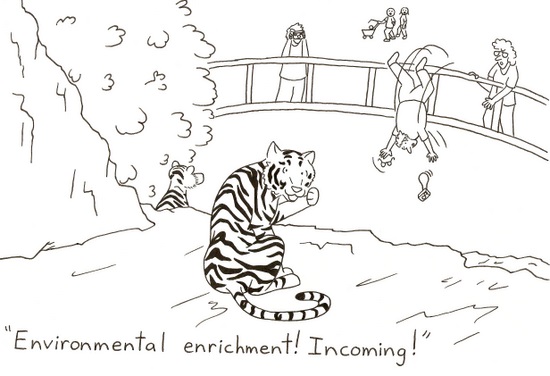
From funnyvet.
From funnyvet.

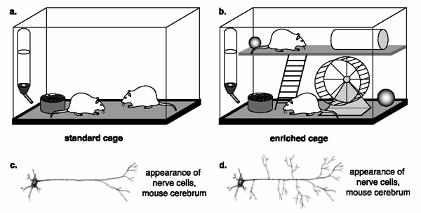
Environmental enrichment is a powerful way to give a boost to neurogenesis and alleviate some anxiety and depression symptoms. For the laboratory rodents, who spend their lives in cages with water and food access, environmental enrichment can refer to as little as a toy or two or as much as large room colonies with different size tubes, different levels to explore, nesting materials, plenty of toys with various shapes, textures, and colors, exercise wheels, and even the occasional fruit or peanut butter snack. But for how long does a mouse need to be exposed to enrichment to show cognitive and emotional improvement?

Leger et al. (2015) ran several anxiety, depression, and long-term memory tests in mice who have been exposed to environmental enrichment for 24 h, 1, 3, or 5 weeks. Although 24 h exposure was enough to improve memory, only after 3-week exposure some anxiety behaviors were attenuated. No effect on depressive behaviors or coticosterone levels, which may be due to that particular strain of mouse (several other studies found that environmental enrichment ameliorates depressive symptoms in other mice strains and rats). The 3-week exposure also increased the levels of serotonin in the frontal cortex. Only after 5-eweek exposure there was a significant survival rate of the hippocampal new cells. Of note, these were normal mice, i.e. they were not suffering from any disorder prior to exposure.

Mice raised in an impoverished environment (a) show less dendrite growth (c) than do mice raised in an enriched environment (b, d). Copyright: BSCS.

The findings give us a nice timeline for environmental enrichment to show its desired effects. But… if there are differences in the timeline and effects of environmental enrichment exposure from mouse strain to mouse strain, then what can we say for humans? Probably not much, unfortunately. As the ad nauseam overused phrase goes at the end of so many papers, ‘more research is needed to elucidate this problem’.

Reference: Leger M, Paizanis E, Dzahini K, Quiedeville A, Bouet V, Cassel JC, Freret T, Schumann-Bard P, & Boulouard M. (Nov 2015, Epub 5 Jun 2014). Environmental Enrichment Duration Differentially Affects Behavior and Neuroplasticity in Adult Mice. Cerebral Cortex, 25(11):4048-61. doi: 10.1093/cercor/bhu119. Article | FREE PDF

By Neuronicus, 1 November 2015

Fat & afraid or slim & brave (Leptin and anxiety in ventral tegmental area)

A comparison of a mouse unable to produce leptin thus resulting in obesity (left) and a normal mouse (right). Courtesy of Wikipedia. License: PD

Leptin is a small molecule produced mostly by the adipose tissue, whose absence is the cause of morbid obesity in the genetically engineered ob/ob mice. Here is a paper that gives us another reason to love this hormone.

Liu, Guo, & Lu (2015) build upon their previous work of investigating the leptin action(s) in the ventral tegmental area of the brain (VTA), a region that houses dopamine neurons and widely implicated in pleasure and drug addiction (among other things). They did a series of very straightforward experiments in which the either infused leptin directly into the mouse VTA or deleted the leptin receptors in this region (by using a virus in genetically engineered mice). Then they tested the mice on three different anxiety tests.

The results: leptin decreases anxiety; absence of leptin receptors increases anxiety. Simple and to the point. And also makes sense, given that leptin receptors are mostly located on the VTA neurons that project to the central amygdala, a region involved in fear and anxiety (curiously, the authors cite the amygdala papers, but do not comment on the leptin-VTA-dopamine-amygdala connection). For the specialists, I would say that they are a little liberal with their VTA hit assessment (they are mostly targeting the posterior VTA) and their GFP (green fluorescent protein) is sparsely expressed.

Reference: Liu J, Guo M, & Lu XY (Epub ahead of print 5 Oct 2015). Leptin/LepRb in the Ventral Tegmental Area Mediates Anxiety-Related Behaviors. International Journal of Neuropsychopharmacology, 1–11. doi:10.1093/ijnp/pyv115. Article | FREE PDF

By Neuronicus, 28 October 2015

The F in memory

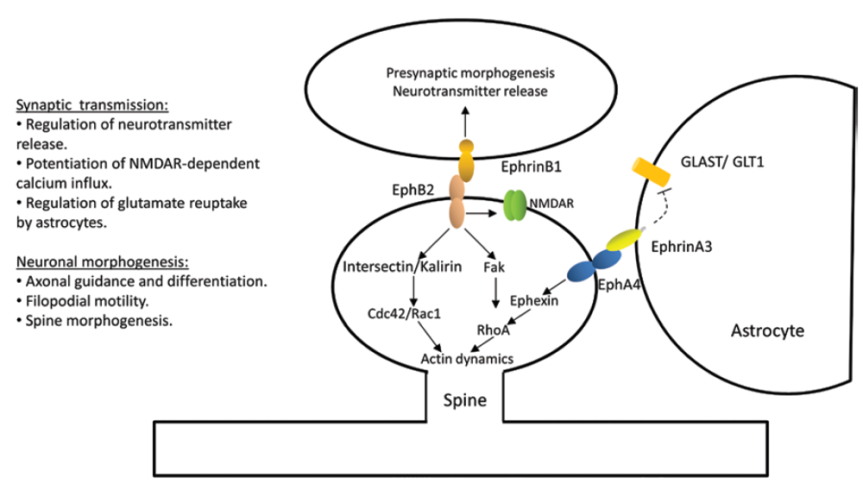
"Figure 2. Ephs and ephrins mediate molecular events that may be involved in memory formation. Evidence shows that memory formation involves alterations of presynaptic neurotransmitter release, activation of glutamate receptors, and neuronal morphogenesis. Eph receptors regulate synaptic transmission by regulating synaptic release, glutamate reuptake from the synapse (via astrocytes), and glutamate receptor conductance and trafficking. Ephs and ephrins also regulate neuronal morphogenesis of axons and dendritic spines through controlling the actin cytoskeleton structure and dynamics" (Dines & Lamprecht, 2015, p. 3).
“Figure 2. Ephs and ephrins mediate molecular events that may be involved in memory formation. Evidence shows that memory formation involves alterations of presynaptic neurotransmitter release, activation of glutamate receptors, and neuronal morphogenesis. Eph receptors regulate synaptic transmission by regulating synaptic release, glutamate reuptake from the synapse (via astrocytes), and glutamate receptor conductance and trafficking. Ephs and ephrins also regulate neuronal morphogenesis of axons and dendritic spines through controlling the actin cytoskeleton structure and dynamics” (Dines & Lamprecht, 2015, p. 3).

When thinking about long-term memory formation, most people immediately picture glutamate synapses. Dines & Lamprecht (2015) review the role of a family of little known players, but with big roles in learning and long-term memory consolidation: the ephs and the ephrines.

Ephs (the name comes from erythropoietin-producing human hepatocellular, the cancer line from which the first member was isolated) are transmembranal tyrosine kinase receptors. Ephrines (Eph receptor interacting protein) bind to them. Ephrines are also membrane-bound proteins, which means that in order for the aforementioned binding to happen, cells must touch each other, or at least be in a very very cozy vicinity. They are expressed in many regions of the brain like hippocampus, amygdala, or cortex.

The authors show that “interruption of Ephs/ephrins mediated functions is sufficient for disruption of memory formation” (p. 7) by reviewing a great deal of genetic, pharmacologic, and electrophysiological studies employing a variety of behavioral tasks, from spatial memory to fear conditioning. The final sections of the review focus on the involvement of ephs/ephrins in Alzheimer’s and anxiety disorders, suggesting that drugs that reverse the impairment on eph/ephrin signaling in these brain diseases may lead to an eventual cure.

Reference: Dines M & Lamprecht R (8 Oct 2015, Epub 13 Sept 2015). The Role of Ephs and Ephrins in Memory Formation. International Journal of Neuropsychopharmacology, 1-14. doi:10.1093/ijnp/pyv106. Article | FREE FULLTEXT PDF

By Neuronicus, 26 October 2015

Nettles are good for you in more ways than one

Urtica Urens (the small nettle). Photo by H. Zell, released under CC BY-SA 3.0. Courtesy of Wikipedia.

Many cultures, specially East-European and North-African, use nettles in their cuisine, as soups, creams, or teas. Nettles’ taste resemble spinach. Now Doukkali et al. (2015) discovered a new use for the plant.

The authors harvested Urtica urens from north Morocco, dried the plants, and prepared a methanolic extract (see p. 2 for the procedure. Don’t drink methanol!). Then, they gave the extract in 3 different doses to mice and assessed its effects in two anxiety and one locomotor test against saline (the control) and diazepam (Valium), a powerful anxiolytic from the benzodiazepine class. Like diazepam, the plant extract had anxiolytic properties, but unlike diazepam, it did not induce any locomotor effects. And this is where the big thing is: ALL benzos on the market have significant side effects in the form of drowsiness, impaired coordination, sedation and so on. Having an anxiolytic without motor impairment would be wonderful.

This is a short, simple to read paper, clearly written, and covers some classic aspects of new drug discovery (like dose-response and lethal dose assessment). The reasons why I think it did not make it to one of the big journals is the small sample size, the relatively moderate effect, and the lack of identifying the active compound (there are virtually no straight-forward behavioral studies published in Nature or Science any more; you’ve got to have the molecules, or proteins, or cells, or what-have-yous as proof that you mean hard-science business).

Or, the fact that it does not have any graphs, all data is presented in tables, which I personally enjoy, as it is oh so easy to manipulate with a graph; and the fancier looking the image, the better chances few people get it anyway. Give me tables with standard deviation any day, as I suspect is the position of the authors of the paper too. But, for the visually inclined, I made a Fig. with some of their data, took only 15 minutes in Excel.

Fig. 1. The effect of saline (S), diazepam (D), and nettle extract (N) on the light-dark test (left) and hole board test (right). Data from Doukkali et al. (2015) , graph by Neuronicus.

Or, not to put a too fine point to it, the authors are from Morocco, so they don’t come shrouded in the A-list universities glamour. In any case, the next obvious step is to isolate the active compound and replicate its anxiolytic effects on other tests and other species.

Reference: Doukkali Z, Taghzouti K, Bouidida EL, Nadjmouddine M, Cherrah Y, & Alaoui K. (24 April 2015). Evaluation of anxiolytic activity of methanolic extract of Urtica urens in a mice model. Behavioral and Brain Functions, 11(19): 1-5. doi: 10.1186/s12993-015-0063-y. Article | FREE PDF

By Neuronicus, 16 October 2015

Cell phones give you hallucinations

A young businessman in a suit screaming at a cell phone. By: Benjamin Miller. License FSP Standard FreeStockPhotos.biz
Photo by Benjamin Miller. License: FSP Standard FreeStockPhotos.biz

Medical doctors (MD) are overworked, particularly when they are hatchlings (i.e. Medical School students) and fledglings (interns and residents). So overworked, that in many countries is routine to have 80-hour weeks and 30-hour shifts as residents and interns. This is a concern as it has been shown that sleep deprivation impairs learning (which is the whole point of residency) and increases the number of medical mistakes (the lack of which is the whole point of their profession).

Lin et al. (2013) show that it can do more than that. Couple internship and cell phones and you get… hallucinations. That’s right. The authors asked 73 medical interns to complete some tests before their internship, then every third, sixth, and twelfth months of their internship, and after the internship. The questionnaires were on anxiety, depression, personality, and cell phone habits and hallucinations. That is: the sensation that your cell phone is vibrating or ringing when, in fact, it is not (which fully corresponds to the definition of hallucination). And here is what they found:

 Before internship, 78% of MDs experienced phantom vibration and 27% experienced phantom ringing.
 During their 1-year internship, about 85 to 95% of MDs experienced phantom vibration and phantom ringing.
 After the internship when the MDs did no work for two weeks, 50% still had these hallucinations.

Composite figure from Lin et al. (2015) showing the interns' depression (above) and anxiety (below) scores before, during, and after internship. The differences are statistically significant.
Fig. 1. Composite figure from Lin et al. (2015) showing the interns’ depression (above) and anxiety (below) scores before, during, and after internship. The differences are statistically significant.

The MDs’ depression and anxiety were also elevated more during the internship than before or after (see Fig. 1), but there was no correlation between the hallucinations and the depression and anxiety scores.

These findings are disturbing on so many levels… Should we be worried that prolonged exposure to cell phones can produce hallucinations? Or that o good portion of the MDs have hallucinations before going to internship? Or that 90% the people in charge with your life or your child’s life are so overworked that are hallucinating on a regular basis? Fine, fine, believing that your phone is ringing or vibrating may not be such a big deal of a hallucination, compared with, let’s say, “the voices told me to give you a lethal dose of morphine”, but as a neuroscientist I beg the question: is there a common mechanism between these two types of hallucinations and, if so, what ELSE is the MD hallucinating about while reassuring you that your CAT scan is normal? Or, forget about the hallucinations, should we worry that your MD is probably more depressed and anxious than you? Or, the “good” news, that the medical interns provide “a model of stress-induced psychotic symptoms” better that previous models, as the authors put it (p. 5)? I really wish there was more research on positive things (… that was a pun; hallucinations are a positive schizophrenic symptom, look it up 🙂 ).

Reference: Lin YH, Lin SH, Li P, Huang WL, & Chen CY. (10 June 2013). Prevalent hallucinations during medical internships: phantom vibration and ringing syndromes. PLoS One, 8(6): e65152. doi: 10.1371/journal.pone.0065152. Article | FREE PDF | First time the phenomenon was documented in press

By Neuronicus, 14 October 2015

Stressed out? Blame your amygdalae

amygdala
Clipart: Royalty free from http://www.cliparthut.com. Text: Neuronicus.

Sooner or later, everyone is exposed to high amounts of stress, whether it is in the form losing someone dear, financial insecurity, or health problems and so on. Most of us manage to bounce right up and continue with our lives, but there is a considerable segment of the population who do not and develop all sorts of problems, from autoimmune disorders to severe depression and anxiety. What makes those people more susceptible to stress? And, more importantly, can we do something about it (yeah, besides making the world a less stressful place)?

Swartz et al. (2015) scanned the brain of 753 healthy young adults (18-22 yrs) while performing a widely used paradigm that elicits amygdalar activation (brain structure, see pic): the subjects had to match a face appearing in the upper part of the screen with one of the faces in the lower part of the screen. The faces looked fearful, angry, surprised, or neutral and amygdalae are robustly activated when matching the fearful face. Then the authors had the participants fill out questionnaires regarding their life events and perceived stress level every 3 months over a period of 2 years (they say 4 years everywhere else in the paper minus Methods & Results, which are the sections that count if one wants to replicate; maybe this is only half of the study and they intend to follow-up to 4 years?).

The higher your baseline amygdalar activation, the higher the risk to develop anxiety disorders later on if expossed to life stressors. Yellow = amygdala. Photo credit: https://www.youtube.com/watch?v=JD44PbAOTy8, presumably copyrighted to Duke University.

The finding of the study is this: baseline amygdalar activation can predict who will develop anxiety later on. In other words, if your natural, healthy, non-stressed self has a an overactive amygdala, you will develop some anxiety disorder later on if exposed to stressors (and who isn’t?). The good news is that knowing this, the owner of the super-sensitive amygdalae, even if s/he may not be able to protect her/himself from stressors, at least can engage in some preventative therapy or counseling to be better equipped with adaptive coping mechanisms when the bad things come. Probably we could all benefit from being “better equipped with adaptive coping mechanisms”, feisty amygdalae or not. Oh, well…

Reference: Swartz, J.R., Knodt, A.R., Radtke, S.R., & Hariri, A.R. (2015). A neural biomarker of psychological vulnerability to future life stress. Neuron, 85, 505-511. doi: 10.1016/j.neuron.2014.12.055. Article | PDF | Video

By Neuronicus, 12 October 2015

Stress can get you fat. And then kill you.

stress meSome people lose weight under stressful conditions and some gain weight. How does that play into the risk for the cardiovascular disease and subsequent mortality? Medical doctors keep warning us that fat people are at risk for diabetes and heart disease. Turns out that being a little on the heavy side might actually not be that bad. It all depends on what kind of fat and where it is.

The paper featured today reviews a series of interesting articles with surprising results. Peters & McEwen (2015) identify three distinct phenotypes:

1) The good stress leads to well-proportionate body shape. People who live in safe environments, they do well socioeconomically, they have good self-esteem, and they have a fulfilling social and family life. They experience low levels of stress, they are well proportionate, and have a low mortality rate due to cardiovascular disease. Might as well call these ones the lucky ones.

2) The tolerable stress leads to corpulent-but-narrow-waisted body shape. People who experience stress but in order to cope with it they supply the brain with more energy by eating more. So they become more corpulent, gaining subcutaneous fat, but their cardiovascular mortality risk remains low.

3) The toxic stress leads to lean-but-wide-waisted body shape. People who experience prolonged stress exposure to uncertain socioeconomic conditions, poor work, or family life. They have low self-esteem, often associated with depressive periods. They are or become lean, but they accumulate large visceral fat deposits (as opposed to subcutaneous), and their cardiovascular mortality risk is the highest. They also are at risk for other physical and mental disorders. The phenotype 3 people have a wider waist relative to their body mass index and height.

Source: Peters & McEwen (2015, p.144)

Thus, the authors propose that instead or along with the body mass index, another metric should be used to identify the ones in dire need of help: the body shape index. Also, the review outlines the mechanisms responsible for these findings.

So next time you see a not so well-proportionate person, smile. Maybe even offer to help or chat; you don’t know what they’re going through.

Reference: Peters, A. & McEwen, B. S. (September 2015, Epub 3 July 2015). Stress habituation, body shape and cardiovascular mortality. Neuroscience Biobehavioral Reviews, 56:139-50. doi: 10.1016/j.neubiorev.2015.07.001. Article | FREE PDF

By Neuronicus, 5 October 2015