Aging and its 11 hippocampal genes

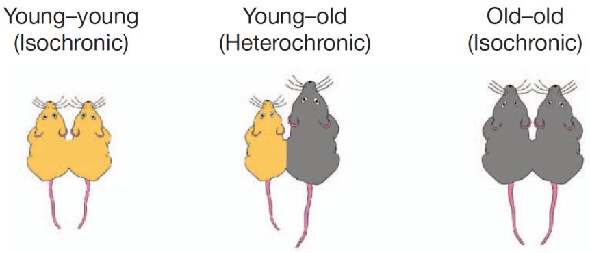
Aging is being quite extensively studied these days and here is another advance in the field. Pardo et al. (2017) looked at what happens in the hippocampus of 2-months old (young) and 28-months old (old) female rats. Hippocampus is a seahorse shaped structure no more than 7 cm in length and 4 g in weight situated at the level of your temples, deep in the brain, and absolutely necessary for memory.

First the researchers tested the rats in a classical maze test (Barnes maze) designed to assess their spatial memory performance. Not surprisingly, the old performed worse than the young.

Then, they dissected the hippocampi and looked at neurogenesis and they saw that the young rats had more newborn neurons than the old. Also, the old rats had more reactive microglia, a sign of inflammation. Microglia are small cells in the brain that are not neurons but serve very important functions.

After that, the researchers looked at the hippocampal transcriptome, meaning they looked at what proteins are being expressed there (I know, transcription is not translation, but the general assumption of transcriptome studies is that the amount of protein X corresponds to the amount of the RNA X). They found 210 genes that were differentially expressed in the old, 81 were upregulated and 129 were downregulated. Most of these genes are to be found in human too, 170 to be exact.

But after looking at male versus female data, at human and mouse aging data, the authors came up with 11 genes that are de-regulated (7 up- and 4 down-) in the aging hippocampus, regardless of species or gender. These genes are involved in the immune response to inflammation. More detailed, immune system activates microglia, which stays activated and this “prolonged microglial activation leads to the release of pro-inflammatory cytokines that exacerbate neuroinflammation, contributing to neuronal loss and impairment of cognitive function” (p. 17). Moreover, these 11 genes have been associated with neurodegenerative diseases and brain cancers.

112hc-copy

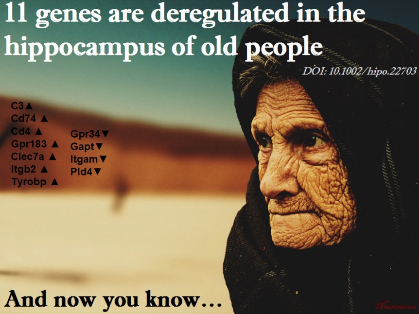
These are the 11 genes: C3 (up), Cd74  (up), Cd4 (up), Gpr183 (up), Clec7a (up), Gpr34 (down), Gapt (down), Itgam (down), Itgb2 (up), Tyrobp (up), Pld4 (down).”Up” and “down” indicate the direction of deregulation: upregulation or downregulation.

I wish the above sentence was as explicitly stated in the paper as I wrote it so I don’t have to comb through their supplemental Excel files to figure it out. Other than that, good paper, good work. Gets us closer to unraveling and maybe undoing some of the burdens of aging, because, as the actress Bette Davis said, “growing old isn’t for the sissies”.

Reference: Pardo J, Abba MC, Lacunza E, Francelle L, Morel GR, Outeiro TF, Goya RG. (13 Jan 2017, Epub ahead of print). Identification of a conserved gene signature associated with an exacerbated inflammatory environment in the hippocampus of aging rats. Hippocampus, doi: 10.1002/hipo.22703. ARTICLE

By Neuronicus, 25 January 2017

Save

Save

CCL11 found in aged but not young blood inhibits adult neurogenesis

vil - Copy
Portion of Fig. 1 from Villeda et al. (2011, doi: 10.1038/nature10357) describing the parabiosis procedure. Basically, under complete anesthesia, the peritoneal membranes and the skins of the two mice were sutured together. The young mice were 3–4 months (yellow) and old mice were 18–20 months old (grey).

My last post was about parabiosis and its sparse revival as a technique in physiology experiments. Parabiosis is the surgical procedure that joins two living animals allowing them to share their circulatory systems. Here is an interesting paper that used the method to tackle blood’s contribution to neurogenesis.

Adult neurogenesis, that is the birth of new neurons in the adult brain, declines with age. This neurogenesis has been observed in some, but not all brain regions, called neurogenic niches.

Because these niches occur in blood-rich areas of the brain, Villeda et al. (2011) wondered if, in addition with the traditional factors required for neurogenesis like enrichment or running, blood factors may also have something to do with neurogenesis. The authors made a young and an old mouse to share their blood via parabiosis (see pic.).

Five weeks after the parabiosis procedure, the young mouse had decreased neurogenesis and the old mouse had increased neurogenesis compared to age-matched controls. To make sure their results are due to something in the blood, they injected plasma from an old mouse into a young mouse and that also resulted in reduced neurogenesis. Moreover, the reduced neurogenesis was correlated with impaired learning as shown by electrophysiological recordings from the hippocampus and from behavioral fear conditioning.

So what in the blood does it? The authors looked at 66 proteins found in the blood (I don’t know the blood make-up, so I can’t tell if 66 is a lot or not ) and noticed that 6 of these had increased levels in the blood of ageing mice whether linked by parabiosis or not. Out of these six, the authors focus on CCL11 (unclear to me why that one, my bet is that they tried the others too but didn’t have enough data). CCLL11 is a small signaling protein involved in allergies. So the authors injected it into young mice and Lo and Behold! there was decreased neurogenesis in their hippocampus. Maybe the vampires were onto something, whadda ya know? Just kidding… don’t go around sucking young people’s blood!

This paper covers a lot of work and, correspondingly, has no less than 23 authors and almost 20 Mb of supplemental documents! The story it tells is very interesting and as complete as it gets, covering many aspects of the problems investigated and many techniques to address those problems. Good read.

Reference: Villeda SA, Luo J, Mosher KI, Zou B, Britschgi M, Bieri G, Stan TM, Fainberg N, Ding Z, Eggel A, Lucin KM, Czirr E, Park JS, Couillard-Després S, Aigner L, Li G, Peskind ER, Kaye JA, Quinn JF, Galasko DR, Xie XS, Rando TA, Wyss-Coray T. (31 Aug 2011). The ageing systemic milieu negatively regulates neurogenesis and cognitive function. Nature. 477(7362):90-94. doi: 10.1038/nature10357. Article | FREE Fulltext PDF

By Neuronicus, 6 January 2016

The FIRSTS: Adult neurogenesis (1962)

New neurons in the granular layer of the hippocampus. Fig. 30 from Altman & Das (1965).

I am starting a new category today: the Firsts. It will feature articles that showed something really interesting for the first time. Yes, all articles show something for the first time, that’s why they are published. But I have noticed either a lack of acknowledgment (“it is known that x”) or a disregard for the old papers (“doesn’t count if it’s before, say, 2001”), particularly among the new generation of scientists. So I will feature both the really big ones (e.g., first proof of adult neurogenesis) or the more obscure, but nonetheless, first in their field (e.g., first synthesis of morphine).

Today, first proof of adult neurogenesis. Altman (1962) wanted to see the kinetics of glial proliferation after brain injury. Glial cells are the other type of cells in the brain and they outnumber the neurons 10 to 1. Altman lesioned the rat lateral geniculate nucleus (a portion of the thalamus that deals primarily with vision) and then injected the rats with thymidine-H3, a dye that labels the newly formed cells. In addition to the expected glial proliferation, he also observed (by microscope and careful histology) that some neurons were also stained with the dye, which means that they were born after the injection. The new neurons were in many regions of the brain (so not only those associated with the lesioned area), including the cortical areas.

Altman followed up and three years later published the first comprehensive study of postnatal (not adult) neurogenesis in dendate gyrus of the hippocampus.

References:

  1. Altman, J. (30 March 1962). Are New Neurons Formed in the Brains of Adult Mammals?. Science, 135 (3509): 1127-1128. DOI: 10.1126/science.135.3509.1127. Article | PDF
  2. Altman, J, & Das, G. D. (June 1965). Autoradiographic and histological evidence of postnatal hippocampal neurogenesis in rats. The Journal of Comparative Neurology, 124 (3): 319 –335. DOI: 10.1002/cne.901240303. Article | PDF

by Neuronicus, 30 September 2015