Interview with Jason D. Shepherd, PhD

During the first week of the publication, a Cell paper that I covered a couple of weeks ago has received a lot of attention from media outlets, like The Atlantic, Scicasts and Neuroscience News/University of Utah Press Release. It is not my intention to duplicate here their wonderfully done summaries and interviews; rather to provide answers to some geeky questions arisen from the minds of nerdy scientists like me.

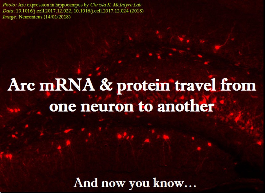
Dr. Shepherd, you are the corresponding author of a paper published on Jan. 11 in Cell about a protein heavily involved in memory formation, called Arc. Your team and another team from University of Massachusetts, who published in the same issue of Cell, simultaneously discovered that Arc looks like and behaves like a virus. The protein “infects” nearby cells, in this case neurons, with instructions of how to make more of itself, i.e. it shuttles its own mRNA from one cell to another.

Neuronicus: Why is this discovery so important?

​Jason D. Shepherd: I think there’s a couple of big implications of this work:

  1. ​The so called “junk” DNA in our genomes that come from viruses and transposable elements actually provide source material for new genes. Arc isn’t the first example, but it’s the first prominent brain gene to have these kinds of origins.

  2. This is the first demonstration that cellular proteins are capable of assembling into capsid-like structures. This is a completely new way of thinking about communication between cells.

  3. We think there may be other genes that can also form capsids, suggesting this method of signaling is fairly common in organisms.

N: 2) When you and your colleagues compared Arc’s genetic sequence across species you concluded Arc comes from a virus that infected four-legged animals some time ago. A little time later the virus infected the flies too. When did these events occur?

​JDS: So we think the origins are from a retrotransposon not a virus. These are DNA sequences or elements that “jump” into the host genome. Think of them as primitive viruses. Indeed, these elements are thought to be the ancestors of retroviruses like HIV. The mammalian Arc gene seems to have originated ~400 million years ago, the fly about 150 million years ago. ​

N: 3) So, if Arc has been so successfully repurposed by the tetrapod and fly ancestors to add memory formation, what does that mean for the animals and insects before the infection? I understand that we move now in the realm of speculation, but who better to speculate on these things than the people who work on Arc? The question is: did these pre-infection creatures have bad and short memories? The alternate view would be that they had similar memory abilities due to a different mechanism that was replaced by Arc. Which one do you think is more likely?

​JDS: Good question. It’s certainly the case that memory capacity improved in tetrapods, but unclear if Arc is the sole reason. I suspect that Arc confers some unique aspects to brains, otherwise it would not have been so conserved after the initial insertion event, but I also think there are probably other Arc-like genes in other organisms that do not have Arc. I will also note that we are not even sure, yet, that the fly Arc is important for fly memory/learning.

N: 4) Remaining in the realm of speculation, if this intercellular mRNA transport proves to be ubiquitous for a variety of mRNAs, what does that say of the transcriptome of a cell at any given time? From a practical point of view, a cell is what is made off, meaning the ensemble of all its enzymes and proteins and so on, collectively termed transcriptome. So if a cell can just alter its neighbor’s transcriptome array, does that mean that it’s possible to alter also its function? Even more outrageously speculative, perhaps even its type? Can we make cancer cells commit suicide by shooting Arc capsules of mRNA at them?

​JDS: Yes! Cool ideas. I think this is quite likely, that these signaling extracellular vesicles can dramatically alter the state of a cell. We are obviously looking into this. ​

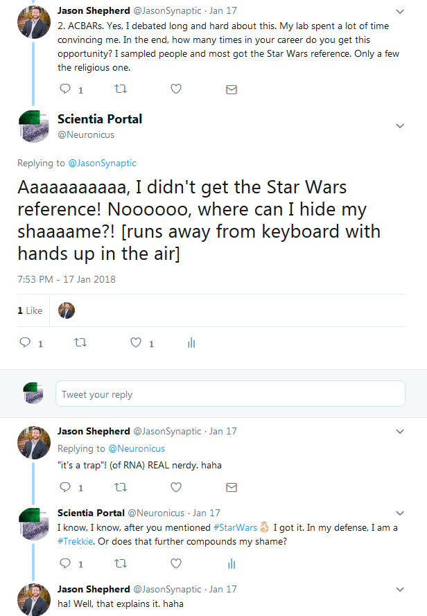
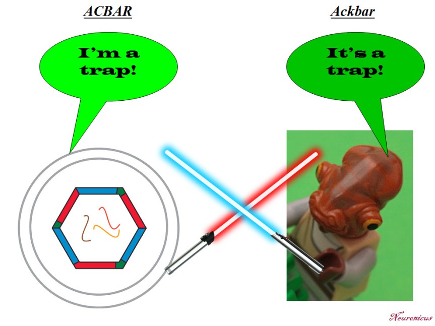
N: 5) Finally, in the paper, the Arc capsules containing mRNA are referred to as ACBAR (Arc Capsid Bearing Any RNA). At first I thought it was a reference to “Allahu akbar” which is Arabic for ‘God is greatest’, the allusion being “ACBAR! Our exosome is the greatest!” or “Arc Acbar! Our Arc is the greatest!”. Is this where the naming is coming from?

​JDS: No no. As I said on twitter, my lab came up with this acronym because we are all Star Wars nerds and the classic “It’s a trap!” line from general Ackbar seemed apt for something that was trapping RNA. ​

Below is the Twitter exchange Dr. Shepherd refers to:

129shepherd - Copy

Dr. Shepherd, thank you for your time! And congratulations on a well done paper and a well told story. Your Methods section is absolutely great; anybody can follow the instructions and replicate your data. Somebody in your lab must have kept great records. Congratulations again!

129_starwars - Copy
The ACBAR graphic is from the Cell’s abstract (©2017 Elsevier Inc.) but since it’s for comedic purposes, I’d say is fair use. Same for the Lego Ackbar.

By Neuronicus, 28 January 2018

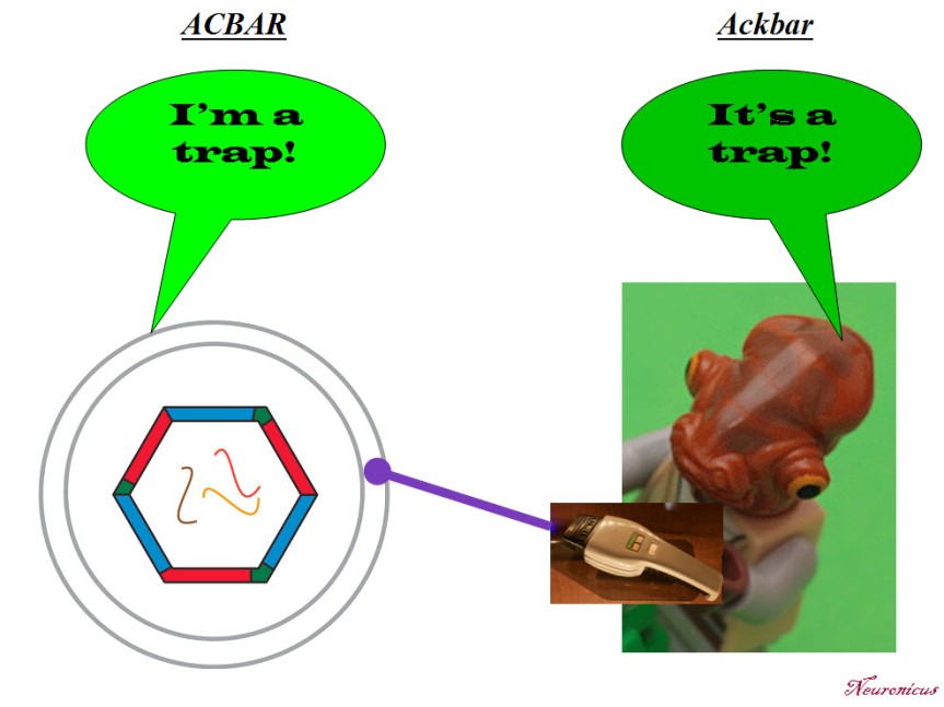
P. S. Since I have obviously managed to annoy the #StarWars universe and twitterverse because I depicted General Ackbar using a Jedi sword when he’s not a Jedi, I thought only fair to annoy the other half of the world, the #trekkies. So here you go:

129_startrek - Copy.jpg


Arc: mRNA & protein from one neuron to another

EDIT 1 [Jan 17, 2018]: I promised four days ago that I will post this, while it was still hot, but my Internet was down, thanks to the only behemoth provider in USA. And rated the worst company in the Nation, too. You definitely know by now about whom I’m talking about. Grrrr…  Anyway, here is the paper:

As promised, today’s paper talks about mRNA transfer between neurons.

Pastuzyn et al. (2018) looked at the gene Arc in neurons because they thought its Gag sequence looks suspiciously similar to some retroviruses. Could it be possible that it also behaves like a virus?

Arc is heavily involved in the immune system, is essential for the formation of long-term memories, and is involved in all sorts of diseases, like schizophrenia and Alzheimer’s, among other things.

Pastuzyn et al. (2018) is a relatively long and dense paper, albeit well written. So, I thought that this time, instead of giving you a summary of their research it would be better to give you the authors’ story directly in their own words written as subtitles in the Results section (bold letters – the authors words, normal font – mine). Warning: this is a much more jargon-dense blog post than my previous one on the same topic and, because it is so much material, I will not explain every term.

  • Fly and Tetrapod (us) Arc Genes Independently Originated from Distinct Lineages of Ty3/gypsy Retrotransposons, the phylogenomic analyses tell us, meaning the authors have done a lot of computer-assisted comparisons of similar forms of the gene in hundreds of species.
  • Arc Proteins Self-Assemble into Virus-like Capsids. Arc likes to oligomerize spontaneously (dimers and trimers). The oligomers resemble virus-like capsids, similar to HIV.
  • Arc Binds and Encapsulates RNA. Although it loves its own RNA about 10 times more than other RNAs, it’s a promiscuous protein (doesn’t care which RNA as long as it follows the rules of stoichiometry). Arc capsids encapsulate both the Arc protein (maybe other proteins too?), its mRNA, and whatever mRNA happened to be in the vicinity at the time of encapsulation. Arc capsids are able to protect the mRNA from RNAases.
  • Arc Capsid Assembly Requires RNA. If there is no RNA around, the capsids are few and poorly formed.
  • Arc Protein and Arc mRNA Are Released by Neurons in Extracellular Vesicles. Arc capsid packages Arc protein & Arc mRNA into extracellular vesicles (EV). The size of these EVs is < 100nm, putting them in the exosome category. This exosome, which the authors gave the unfortunate name of ACBAR (Arc Capsid Bearing Any RNA), is being expelled from cortical neurons in an activity-dependent manner. In other words, when neurons are stimulated, they release ACBARs.
  • Arc Mediates Intercellular Transfer of mRNA in Extracellular Vesicles. ACBARs dock to the host cell and then undergo clathrin-dependent endocytosis, meaning they expel their cargo in the host cell. The levels of Arc protein and Arc mRNA peaks in a host hippocampal cell in four hours from incubation. The ACBARs tend to congregate around donor cell’s dendrites.
  • Transferred Arc mRNA Can Undergo Activity-Dependent Translation. Activating the group 1 metabotropic glutamate receptor (mGluR1/5) by application of the agonist DHPG induces a significant increase of the amount of Arc protein in the host neurons.

This is a veritable tour de force paper. The Results section has 7 sub-sections, each with multiple experiments to dot every i and cross every t. I’m eyeballing about 40 experiments. It is true that there are 13 authors on the paper from different institutions – yeay for collaboration! – but c’mon! Is this what you need to get in Cell these days? Apparently so. Don’t get me wrong, this is an outstanding paper. But in the end it is still only one paper, which means only one first author. The rest are there for the ride because for a tenure track application nobody cares about your papers in CNS (Cell, Nature, Science = The Central Nervous System of the scientific community, har, har) if you’re not the first author. It looks like the increasing amount of work you need to be published in top tier journals these days is becoming a pet peeve of mine as I keep mentioning it (for example, here).

My pet peeves aside, Pastuzyn et al. (2018) is an excellent paper that opens interesting practical (drug delivery) and theoretical (biological repurpose of ancient invaders) gates. Kudos!

128-1 - Copy

REFERENCE: Pastuzyn ED, Day CE, Kearns RB, Kyrke-Smith M, Taibi AV, McCormick J, Yoder N, Belnap DM, Erlendsson S, Morado DR, Briggs JAG, Feschotte C, & Shepherd JD. (11 Jan 2018). The Neuronal Gene Arc Encodes a Repurposed Retrotransposon Gag Protein that Mediates Intercellular RNA Transfer. Cell, 172(1-2):275-288.e18. PMID: 29328916. doi: 10.1016/j.cell.2017.12.024. ARTICLE | FULLTEXT PDF via ResearchGate

P.S. I said that ACBAR is an unfortunate acronym because I don’t know about you but I for one wouldn’t want my discovery to be linked either with a religion or with terrorist cries, even if that link is done only by a small fraction of the population. Although I can totally see the naming-by-committee going: “ACBAR! Our exosome is the greatest! Yeay!” or “Arc Acbar! Our Arc is the greatest. Double yeay!”. On a second thought, it’s kindda nerdy geeky neat. I still wouldn’t have done it though…

By Neuronicus, 14 January 2018

EDIT 2 [Jan 22, 2018]: There is another paper that discovered that Arc forms capsids that encapsulate RNA and then shuttles it across the neuromuscular junction in Drosophila (fly). To their credit, Cell published both these papers back-to-back so no researcher gets scooped of their discovery. From what I can see, the discovery really happened simultaneously, so I modified my infopic to reflect that (both papers were submitted in January 2017, received in revised version on August 15, 2017 and published in the same issue on January 11, 2018). Here is the reference to the other article:

Ashley J, Cordy B, Lucia D, Fradkin LG, Budnik V, & Thomson T (11 Jan 2018). Retrovirus-like Gag Protein Arc1 Binds RNA and Traffics across Synaptic Boutons, Cell. 172(1-2): 262-274.e11. PMID: 29328915. doi: 10.1016/j.cell.2017.12.022. ARTICLE

EDIT 3 [Jan 29, 2018]: Dr. Shepherd, the last author of the paper I featured, was kind enough to answer a few of my questions about the implications of his and his team’s findings, answers which you will find here.

By Neuronicus, 22 January 2018

Old chimpanzees get Alzheimer’s pathology

Alzheimer’s Disease (AD) is the most common type of dementia with a progression that can span decades. Its prevalence is increasing steadily, particularly in the western countries and Australia. So some researchers speculated that this particular disease might be specific to humans. For various reasons, either genetic, social, or environmental.

A fresh e-pub brings new evidence that Alzheimer’s might plague other primates as well. Edler et al. (2017) studied the brains of 20 old chimpanzees (Pan troglodytes) for a whole slew of Alzheimer’s pathology markers. More specifically, they looked for these markers in brain regions commonly affected by AD, like the prefrontal cortex, the midtemporal gyrus, and the hippocampus.

Alzheimer’s markers, like Tau and Aβ lesions, were present in the chimpanzees in an age-dependent manner. In other words, the older the chimp, the more severe the pathology.

Interestingly, all 20 animals displayed some form of Alzheimer’s pathology. This finding points to another speculation in the field which is: dementia is just part of normal aging. Meaning we would all get it, eventually, if we would live long enough; some people age younger and some age older, as it were. This hypothesis, however, is not favored by most researchers not the least because is currently unfalsifiable. The longest living humans do not show signs of dementia so how long is long enough, exactly? But, as the authors suggest, “Aβ deposition may be part of the normal aging process in chimpanzees” (p. 24).

Unfortunately, “the chimpanzees in this study did not participate in formal behavioral or cognitive testing” (p. 6). So we cannot say if the animals had AD. They had the pathological markers, yes, but we don’t know if they exhibited the disease as is not uncommon to find these markers in humans who did not display any behavioral or cognitive symptoms (Driscoll et al., 2006). In other words, one might have tau deposits but no dementia symptoms. Hence the title of my post: “Old chimpanzees get Alzheimer’s pathology” and not “Old chimpanzees get Alzheimer’s Disease”

Good paper, good methods and stats. And very useful because “chimpanzees share 100% sequence homology and all six tau isoforms with humans” (p. 4), meaning we have now a closer to us model of the disease so we can study it more, even if primate research has taken significant blows these days due to some highly vocal but thoroughly misguided groups. Anyway, the more we know about AD the closer we are of getting rid of it, hopefully. And, soon enough, the aforementioned misguided groups shall have to face old age too with all its indignities and my guess is that in a couple of decades or so there will be fresh money poured into aging diseases research, primates be damned.

121-chimps get Alz - Copy

REFERENCE: Edler MK, Sherwood CC, Meindl RS, Hopkins WD, Ely JJ, Erwin JM, Mufson EJ, Hof PR, & Raghanti MA. (EPUB July 31, 2017). Aged chimpanzees exhibit pathologic hallmarks of Alzheimer’s disease. Neurobiology of Aging, PII: S0197-4580(17)30239-7, DOI: http://dx.doi.org/10.1016/j.neurobiolaging.2017.07.006. ABSTRACT  | Kent State University press release

By Neuronicus, 23 August 2017

Save

Save

Aging and its 11 hippocampal genes

Aging is being quite extensively studied these days and here is another advance in the field. Pardo et al. (2017) looked at what happens in the hippocampus of 2-months old (young) and 28-months old (old) female rats. Hippocampus is a seahorse shaped structure no more than 7 cm in length and 4 g in weight situated at the level of your temples, deep in the brain, and absolutely necessary for memory.

First the researchers tested the rats in a classical maze test (Barnes maze) designed to assess their spatial memory performance. Not surprisingly, the old performed worse than the young.

Then, they dissected the hippocampi and looked at neurogenesis and they saw that the young rats had more newborn neurons than the old. Also, the old rats had more reactive microglia, a sign of inflammation. Microglia are small cells in the brain that are not neurons but serve very important functions.

After that, the researchers looked at the hippocampal transcriptome, meaning they looked at what proteins are being expressed there (I know, transcription is not translation, but the general assumption of transcriptome studies is that the amount of protein X corresponds to the amount of the RNA X). They found 210 genes that were differentially expressed in the old, 81 were upregulated and 129 were downregulated. Most of these genes are to be found in human too, 170 to be exact.

But after looking at male versus female data, at human and mouse aging data, the authors came up with 11 genes that are de-regulated (7 up- and 4 down-) in the aging hippocampus, regardless of species or gender. These genes are involved in the immune response to inflammation. More detailed, immune system activates microglia, which stays activated and this “prolonged microglial activation leads to the release of pro-inflammatory cytokines that exacerbate neuroinflammation, contributing to neuronal loss and impairment of cognitive function” (p. 17). Moreover, these 11 genes have been associated with neurodegenerative diseases and brain cancers.

112hc-copy

These are the 11 genes: C3 (up), Cd74  (up), Cd4 (up), Gpr183 (up), Clec7a (up), Gpr34 (down), Gapt (down), Itgam (down), Itgb2 (up), Tyrobp (up), Pld4 (down).”Up” and “down” indicate the direction of deregulation: upregulation or downregulation.

I wish the above sentence was as explicitly stated in the paper as I wrote it so I don’t have to comb through their supplemental Excel files to figure it out. Other than that, good paper, good work. Gets us closer to unraveling and maybe undoing some of the burdens of aging, because, as the actress Bette Davis said, “growing old isn’t for the sissies”.

Reference: Pardo J, Abba MC, Lacunza E, Francelle L, Morel GR, Outeiro TF, Goya RG. (13 Jan 2017, Epub ahead of print). Identification of a conserved gene signature associated with an exacerbated inflammatory environment in the hippocampus of aging rats. Hippocampus, doi: 10.1002/hipo.22703. ARTICLE

By Neuronicus, 25 January 2017

Save

Save

Fructose bad effects reversed by DHA, an omega-3 fatty acid

Despite alarm signals raised by various groups and organizations regarding the dangers of the presence of sugars – particularly fructose derived from corn syrup – in almost every food in the markets, only in the past decade there has been some serious evidence against high consumption of fructose.

A bitter-sweet (sic!) paper comes from Meng et al. (2016) who, in addition to showing some bad things that fructose does to brain and body, it also shows some rescue from its deleterious effects by DHA (docosahexaenoic acid), an omega-3 fatty acid.

The authors had 3 groups of rodents: one group got fructose in their water for 6 weeks, another group got fructose and DHA, and another group got their normal chow. The amount of fructose was calculated to be ecologically valid, meaning that they fed the animals the equivalent of 1 litre soda bottle per day (130 g of sugar for a 60 Kg human).

The rats that got fructose had worse learning and memory performance at a maze test compared to the other two groups.

The rats that got fructose had altered gene expression in two brain areas: hypothalamus (involved in metabolism) and hippocampus (involved in learning and memory) compared to the other two groups.

The rats that got fructose had bad metabolic changes that are precursors for Type 2 diabetes, obesity and other metabolic disorders (high blood glucose, triglycerides, insulin, and insulin resistance index) compared to the other two groups.

86 - Copy.jpg

The genetic analyses that the researchers did (sequencing the RNA and analyzing the DNA methylation) revealed a whole slew of the genes that had been affected by the fructose treatment. So, they did some computer work that involved Bayesian modeling  and gene library searching and they selected two genes (Bgn and Fmod) out of almost a thousand possible candidates who seemed to be the drivers of these changes. Then, they engineered mice that lacked these genes. The resultant mice had the same metabolic changes as the rats that got fructose, but… their learning and memory was even better than that of the normals? I must have missed something here. EDIT: Well… yes and no. Please read the comment below from the Principal Investigator of the study.

It is an ok paper, done by the collaboration of 7 laboratories from 3 countries. But there are a few things that bother me, as a neuroscientist, about it. First is the behavior of the genetic knock-outs. Do they really learn faster? The behavioral results are not addressed in the discussion. Granted, a genetic knockout deletes that gene everywhere in the brain and in the body, whereas the genetic alterations induced by fructose are almost certainly location-specific.

Which brings me to the second bother: nowhere in the paper (including the supplemental materials, yeas, I went through those) are any brain pictures or diagrams or anything that can tell us which nuclei of the hypothalamus the samples came from. Hypothalamus is a relatively small structure with drastically different functional nuclei very close to one another. For example, the medial preoptic nucleus that deals with sexual hormones is just above the suprachiasmatic nucleus that deals with circadian rhythms and near the preoptic is the anterior nucleus that deals mainly with thermoregulation. The nuclei that deal with hunger and satiety (the lateral and the ventromedial nucleus, respectively) are located in different parts of the hypothalamus. In short, it would matter very much where they got their samples from because the transcriptome and methylome would differ substantially from nucleus to nucleus. Hippocampus is not so complicated as that, but it also has areas with specialized functions. So maybe they messed up the identification of the two genes Bgn and Fmod as drivers of the changes; after all, they found almost 1 000 genes altered by fructose. And that mess-up might have been derived by their blind hypothalamic and hippocampal sampling. EDIT: They didn’t mess up,  per se. Turns out there were technical difficulties of extracting enough nucleic acids from specific parts of hypothalamus for analyses. I told you them nuclei are small…

Anyway, the good news comes from the first experiment, where DHA reverses the bad effects of fructose. Yeay! As a side note, the fructose from corn syrup is metabolized differently than the fructose from fruits. So you are far better off consuming the equivalent amount on fructose from a litre of soda in fruits. And DHA comes either manufactured from algae or extracted from cold-water oceanic fish oils (but not farmed fish, apparently).

If anybody that read the paper has some info that can help clarify my “bothers”, please do so in the Comment section below. The other media outlets covering this paper do not mention anything about the knockouts. Thanks! EDIT: The last author of the paper, Dr. Yang, was very kind and clarified a bit of my “bothers” in the Comments section. Thanks again!

Reference: Meng Q, Ying Z, Noble E, Zhao Y, Agrawal R, Mikhail A, Zhuang Y, Tyagi E, Zhang Q, Lee J-H, Morselli M, Orozco L, Guo W, Kilts TM, Zhu J, Zhang B, Pellegrini M, Xiao X, Young MF, Gomez-Pinilla F, Yang X (2016). Systems Nutrigenomics Reveals Brain Gene Networks Linking Metabolic and Brain Disorders. EBioMedicine, doi: 10.1016/j.ebiom.2016.04.008. Article | FREE fulltext PDF | Supplementals | Science Daily cover | NeuroscienceNews cover

By Neuronicus, 24 April 2016

A third way of neuronal communication

electrical fields - Copy
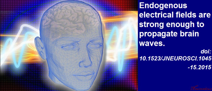
Every neuroscience or biology textbook will tell you that neurons communicate with one another in two ways: via chemical synapses (one neuron releases some substances that change the membrane potential of the receiving neuron) or via electrical synapses (neurons physically share some membrane proteins that allows the electrical impulse to go from one neuron to another). (Nota bene: for the taxonomic nitpickers, I decided that the other non-conventional forms of communication, like diffusion, fall in one or the other of the two categories above).

Now Qiu et al. (2015) have some evidence that there may be another way of neuronal chatting. Long ago (in 1924), Hans Berger observed that neurons have rhythmic and spontaneous electrical activity. The rhythmic activity of an entire population of neurons is called a wave and can be detected with rudimentary tools such as EEG. Qiu et al. reasoned that the speed of such a traveling wave is too slow to be explained by conventional neuronal communication (of which we know quite a lot).

So the researchers ran some computer simulations to test if diffuse electrical fields can explain the speed of a wave, instead of the conventional fast communications. In other words, can local, weak, slow endogenous fields sweep the brain by recruiting nearby neurons? The computer simulations said it is possible, with a very slow speed of 10 cm/s.

Then they recorded the electrical activity of mouse hippocampus isolated in a Petri dish. When the researchers blocked the electrical field there was a decrease in the speed of the wave, meaning the field is required for the wave speed observed.

To be fair, this is not a new idea; even in the early eighties these fields were suggested, because the wave persisted even when the other two ways of communicating have been blocked. And in the early noughts it was shown that a neural network is much more sensitive to electrical field manipulation than individual neurons. What makes this paper interesting is that the authors show that the endogenous electrical fields are not too weak to underlie the wave, as previously thought. The work has implications for the study of epilepsy, sleep, and memory.

Reference: Qiu C, Shivacharan RS, Zhang M, & Durand DM (2 December 2015). Can Neural Activity Propagate by Endogenous Electrical Field?, Journal of Neuroscience, 35(48): 15800-15811; doi: 10.1523/JNEUROSCI.1045-15.2015 Article | Full Text via Research Gate | ScienceAlert cover

By Neuronicus, 11 February 2016

CCL11 found in aged but not young blood inhibits adult neurogenesis

vil - Copy
Portion of Fig. 1 from Villeda et al. (2011, doi: 10.1038/nature10357) describing the parabiosis procedure. Basically, under complete anesthesia, the peritoneal membranes and the skins of the two mice were sutured together. The young mice were 3–4 months (yellow) and old mice were 18–20 months old (grey).

My last post was about parabiosis and its sparse revival as a technique in physiology experiments. Parabiosis is the surgical procedure that joins two living animals allowing them to share their circulatory systems. Here is an interesting paper that used the method to tackle blood’s contribution to neurogenesis.

Adult neurogenesis, that is the birth of new neurons in the adult brain, declines with age. This neurogenesis has been observed in some, but not all brain regions, called neurogenic niches.

Because these niches occur in blood-rich areas of the brain, Villeda et al. (2011) wondered if, in addition with the traditional factors required for neurogenesis like enrichment or running, blood factors may also have something to do with neurogenesis. The authors made a young and an old mouse to share their blood via parabiosis (see pic.).

Five weeks after the parabiosis procedure, the young mouse had decreased neurogenesis and the old mouse had increased neurogenesis compared to age-matched controls. To make sure their results are due to something in the blood, they injected plasma from an old mouse into a young mouse and that also resulted in reduced neurogenesis. Moreover, the reduced neurogenesis was correlated with impaired learning as shown by electrophysiological recordings from the hippocampus and from behavioral fear conditioning.

So what in the blood does it? The authors looked at 66 proteins found in the blood (I don’t know the blood make-up, so I can’t tell if 66 is a lot or not ) and noticed that 6 of these had increased levels in the blood of ageing mice whether linked by parabiosis or not. Out of these six, the authors focus on CCL11 (unclear to me why that one, my bet is that they tried the others too but didn’t have enough data). CCLL11 is a small signaling protein involved in allergies. So the authors injected it into young mice and Lo and Behold! there was decreased neurogenesis in their hippocampus. Maybe the vampires were onto something, whadda ya know? Just kidding… don’t go around sucking young people’s blood!

This paper covers a lot of work and, correspondingly, has no less than 23 authors and almost 20 Mb of supplemental documents! The story it tells is very interesting and as complete as it gets, covering many aspects of the problems investigated and many techniques to address those problems. Good read.

Reference: Villeda SA, Luo J, Mosher KI, Zou B, Britschgi M, Bieri G, Stan TM, Fainberg N, Ding Z, Eggel A, Lucin KM, Czirr E, Park JS, Couillard-Després S, Aigner L, Li G, Peskind ER, Kaye JA, Quinn JF, Galasko DR, Xie XS, Rando TA, Wyss-Coray T. (31 Aug 2011). The ageing systemic milieu negatively regulates neurogenesis and cognitive function. Nature. 477(7362):90-94. doi: 10.1038/nature10357. Article | FREE Fulltext PDF

By Neuronicus, 6 January 2016

How long does it take for environmental enrichment to show effects?

From funnyvet.
From funnyvet.

Environmental enrichment is a powerful way to give a boost to neurogenesis and alleviate some anxiety and depression symptoms. For the laboratory rodents, who spend their lives in cages with water and food access, environmental enrichment can refer to as little as a toy or two or as much as large room colonies with different size tubes, different levels to explore, nesting materials, plenty of toys with various shapes, textures, and colors, exercise wheels, and even the occasional fruit or peanut butter snack. But for how long does a mouse need to be exposed to enrichment to show cognitive and emotional improvement?

Leger et al. (2015) ran several anxiety, depression, and long-term memory tests in mice who have been exposed to environmental enrichment for 24 h, 1, 3, or 5 weeks. Although 24 h exposure was enough to improve memory, only after 3-week exposure some anxiety behaviors were attenuated. No effect on depressive behaviors or coticosterone levels, which may be due to that particular strain of mouse (several other studies found that environmental enrichment ameliorates depressive symptoms in other mice strains and rats). The 3-week exposure also increased the levels of serotonin in the frontal cortex. Only after 5-eweek exposure there was a significant survival rate of the hippocampal new cells. Of note, these were normal mice, i.e. they were not suffering from any disorder prior to exposure.

Mice raised in an impoverished environment (a) show less dendrite growth (c) than do mice raised in an enriched environment (b, d). Copyright: BSCS.

The findings give us a nice timeline for environmental enrichment to show its desired effects. But… if there are differences in the timeline and effects of environmental enrichment exposure from mouse strain to mouse strain, then what can we say for humans? Probably not much, unfortunately. As the ad nauseam overused phrase goes at the end of so many papers, ‘more research is needed to elucidate this problem’.

Reference: Leger M, Paizanis E, Dzahini K, Quiedeville A, Bouet V, Cassel JC, Freret T, Schumann-Bard P, & Boulouard M. (Nov 2015, Epub 5 Jun 2014). Environmental Enrichment Duration Differentially Affects Behavior and Neuroplasticity in Adult Mice. Cerebral Cortex, 25(11):4048-61. doi: 10.1093/cercor/bhu119. Article | FREE PDF

By Neuronicus, 1 November 2015

The F in memory

"Figure 2. Ephs and ephrins mediate molecular events that may be involved in memory formation. Evidence shows that memory formation involves alterations of presynaptic neurotransmitter release, activation of glutamate receptors, and neuronal morphogenesis. Eph receptors regulate synaptic transmission by regulating synaptic release, glutamate reuptake from the synapse (via astrocytes), and glutamate receptor conductance and trafficking. Ephs and ephrins also regulate neuronal morphogenesis of axons and dendritic spines through controlling the actin cytoskeleton structure and dynamics" (Dines & Lamprecht, 2015, p. 3).
“Figure 2. Ephs and ephrins mediate molecular events that may be involved in memory formation. Evidence shows that memory formation involves alterations of presynaptic neurotransmitter release, activation of glutamate receptors, and neuronal morphogenesis. Eph receptors regulate synaptic transmission by regulating synaptic release, glutamate reuptake from the synapse (via astrocytes), and glutamate receptor conductance and trafficking. Ephs and ephrins also regulate neuronal morphogenesis of axons and dendritic spines through controlling the actin cytoskeleton structure and dynamics” (Dines & Lamprecht, 2015, p. 3).

When thinking about long-term memory formation, most people immediately picture glutamate synapses. Dines & Lamprecht (2015) review the role of a family of little known players, but with big roles in learning and long-term memory consolidation: the ephs and the ephrines.

Ephs (the name comes from erythropoietin-producing human hepatocellular, the cancer line from which the first member was isolated) are transmembranal tyrosine kinase receptors. Ephrines (Eph receptor interacting protein) bind to them. Ephrines are also membrane-bound proteins, which means that in order for the aforementioned binding to happen, cells must touch each other, or at least be in a very very cozy vicinity. They are expressed in many regions of the brain like hippocampus, amygdala, or cortex.

The authors show that “interruption of Ephs/ephrins mediated functions is sufficient for disruption of memory formation” (p. 7) by reviewing a great deal of genetic, pharmacologic, and electrophysiological studies employing a variety of behavioral tasks, from spatial memory to fear conditioning. The final sections of the review focus on the involvement of ephs/ephrins in Alzheimer’s and anxiety disorders, suggesting that drugs that reverse the impairment on eph/ephrin signaling in these brain diseases may lead to an eventual cure.

Reference: Dines M & Lamprecht R (8 Oct 2015, Epub 13 Sept 2015). The Role of Ephs and Ephrins in Memory Formation. International Journal of Neuropsychopharmacology, 1-14. doi:10.1093/ijnp/pyv106. Article | FREE FULLTEXT PDF

By Neuronicus, 26 October 2015

I can watch you learning

Human Stereotaxic System. Photo credit: The Mind Project

Recording directly form the healthy living human brain has always been a coveted goal of many neuroscientists, thus bypassing the limitations of non-invasive techniques or animal work. But, understandably, nobody would seek or grant approval for inserting an electrode in the healthy living human brain, on moral and ethical grounds. The next best thing is to insert an electrode into the not so healthy living human brain.

Ison, Quiroga, & Fried (2015) got lucky and gained access to 14 patients with intractable epilepsy that had electrodes implanted in their brain to find where the seizure focus is (for possible surgical resection later on). Using these electrodes, they recorded the activity of single neurons within the medial temporal lobe (MTL, a brain area paramount for learning) while the patients performed some simple association tasks. First, they presented images of places, people, and animals to the patients to see “which (if any) of the recorded neurons responded to a picture” (p. 220). When they got a neuron responding to something, they rushed out, did some data and image processing, and after an hour they started the experiment. Which was showing the patient the picture to which the neuron responded to (e.g. Stimulus 1 = patient’s daughter) overimposed on a background that the neuron did not respond to (e.g. Stimulus 2 = the Eiffel tower). After one single trial (although there was some variability), the patients learned the associations (i.e. Stimulus 3 = daughter in front of Eiffel tower) and this learning was mirrored by how the neuron responded. Namely, the neuron increased its activity by 200% to 400% (counted in spikes per second) when shown the previously un-responded to image alone (i.e. Stimulus 2).

Excerpt from Fig. 5 from Ison, Quiroga, & Fried (2015).
Excerpt from Fig. 5 from Ison, Quiroga, & Fried (2015). “Average normalized neural activity (black squares) and behavioral responses (green circles) to the non-preferred stimulus as a function of trial number. Data were aligned to the learning time (relative trial number 0)”, i.e. when they showed the composite image between Stimulus 1 and Stimulus 2. “Note that the neural activity follows the sudden increase in behavioral learning”.

The authors recorded from over 600 neurons from various MTL regions, out of which 51 responded to a Stimulus 1. From these, only half learned, that is, they increased their activity when Stimulus 2 was shown. For the picky specialist, the cells were both Type 1 and Type 2 neurons, located 6 in the hippocampus, 4 in the entorhinal cortex, 11 in the parahippocampal cortex, and 1 in the amygdala. And the authors controlled for familiarity, attentional demands, and other extraneous variables (with some very fancy and hard to follow stats, I might add).

The paper settles an old psychology dispute. Do we learn an association gradually or at once? In other words, do we learn gradually that A and B occur together, or do we learn that the first time we are shown A and B together and the next trials serve just to refine and consolidate the new knowledge? Ison, Quiroga, & Fried (2015) data show that learning happens at once, in an all-or-none fashion.

Reference: Ison, M. J., Quian Quiroga, R., & Fried, I. (1 July 2015). Rapid Encoding of New Memories by Individual Neurons in the Human Brain. Neuron, 87(1): 220-30. doi: 10.1016/j.neuron.2015.06.016. Article | FREE PDF

By Neuronicus, 4 October 2015

The FIRSTS: Adult neurogenesis (1962)

New neurons in the granular layer of the hippocampus. Fig. 30 from Altman & Das (1965).

I am starting a new category today: the Firsts. It will feature articles that showed something really interesting for the first time. Yes, all articles show something for the first time, that’s why they are published. But I have noticed either a lack of acknowledgment (“it is known that x”) or a disregard for the old papers (“doesn’t count if it’s before, say, 2001”), particularly among the new generation of scientists. So I will feature both the really big ones (e.g., first proof of adult neurogenesis) or the more obscure, but nonetheless, first in their field (e.g., first synthesis of morphine).

Today, first proof of adult neurogenesis. Altman (1962) wanted to see the kinetics of glial proliferation after brain injury. Glial cells are the other type of cells in the brain and they outnumber the neurons 10 to 1. Altman lesioned the rat lateral geniculate nucleus (a portion of the thalamus that deals primarily with vision) and then injected the rats with thymidine-H3, a dye that labels the newly formed cells. In addition to the expected glial proliferation, he also observed (by microscope and careful histology) that some neurons were also stained with the dye, which means that they were born after the injection. The new neurons were in many regions of the brain (so not only those associated with the lesioned area), including the cortical areas.

Altman followed up and three years later published the first comprehensive study of postnatal (not adult) neurogenesis in dendate gyrus of the hippocampus.

References:

  1. Altman, J. (30 March 1962). Are New Neurons Formed in the Brains of Adult Mammals?. Science, 135 (3509): 1127-1128. DOI: 10.1126/science.135.3509.1127. Article | PDF
  2. Altman, J, & Das, G. D. (June 1965). Autoradiographic and histological evidence of postnatal hippocampal neurogenesis in rats. The Journal of Comparative Neurology, 124 (3): 319 –335. DOI: 10.1002/cne.901240303. Article | PDF

by Neuronicus, 30 September 2015

Obscure protein restores memory decline

In the not-too-distant-future, your grandma may give you a run for your money on your video games. Photo credit: http://www.funtoosh.com/pictures/
In the not-too-distant-future, your grandma may give you a run for your money on your video games. Photo credit: Funtoosh

Aging comes with all sorts of maladies, but one of the most frustrating is the feeling that you are not as sharp as you used to be. Cognitive decline has been previously linked, at least in part, to a dysregulation in the neuronal calcium homeostasis in the hippocampus, which is a brain region essential for learning and memory. One player that keeps in check the proper balance of calcium use is the protein FKBP1b, and, not surprisingly, its amounts are reduced in aging rats and Alzheimer’s suffering patients.

FKBP1b overexpression in hippocampal neurons reversed spatial memory deficits in aged rats. Fig. 3 (partial) from Gant, J. C., Chen, K. C., Kadish, I., Blalock, E. M., Thibault, O., Porter, N. M., Landfield, P. W. (29 July 2015). Reversal of Aging-Related Neuronal Ca2+ Dysregulation and Cognitive Impairment by Delivery of a Transgene Encoding FK506-Binding Protein 12.6/1b to the Hippocampus. The Journal of Neuroscience, 35(30):10878 –10887. doi: 10.1523/JNEUROSCI.1248-15.2015.
FKBP1b overexpression in hippocampal neurons reversed spatial memory deficits in aged rats. Fig. 3 (partial) from Gant et al. (2015): doi: 10.1523/JNEUROSCI.1248-15.2015.

Gant et al. (2015) sought to increase the expression of the FKBP1b protein in the hippocampus, in the hopes that its increase would result in better calcium homeostasis and, as a result, better memory performance in aging rats. They built a virus that carried the gene for making the FKBP1b protein and they injected this directly in the hippocampus. After they waited 5-6 weeks for the gene to be expressed, they tested the rats in the Morris water maze, a test for spatial memory. The old rats that received the injection performed as well as the young rats, and far better than the old rats who didn’t get the injection. Then the researchers made sure that the injection is the one responsible for the results, by checking the levels of the FKBP1b protein in the hippocampus (increased, as per specs), by recording from those neurons (they were awesome), and by imaging the calcium to make sure the balance was restored (ditto).

Reference: Gant, J. C., Chen, K. C., Kadish, I., Blalock, E. M., Thibault, O., Porter, N. M., Landfield, P. W. (29 July 2015). Reversal of Aging-Related Neuronal Ca2+ Dysregulation and Cognitive Impairment by Delivery of a Transgene Encoding FK506-Binding Protein 12.6/1b to the Hippocampus. The Journal of Neuroscience, 35(30):10878 –10887. doi: 10.1523/JNEUROSCI.1248-15.2015. Article + FREE PDF + Journal of Neuroscience cover