Tomato transcriptome

As most children, growing up I showed little appreciation for what I had, coveting instead what I did not. Now I realize how fortunate I have been to have grown up half the time in a metropolis and the other half at the countryside. At the farm. A subsistence farm, although I truly loathe the term because we were not just subsisting but thriving off the land, as we planted and harvested a bit of everything and we had a specimen or four of almost all the farm animals, from bipeds to quadrupeds.

I got on this memory lane after reading the paper of Shinozaki et al. (2018) on tomatoes. It was a difficult read for me as it was punctured by many term definition lookups since botany evolved quite steeply since the last time I checked, about 25 years or so.

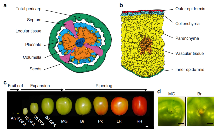
Briefly, the scientists grew tomato plants in a greenhouse at Cornell, NY. They harvested the fruit from 60 plants about 5 to 50 days after the flower was at its peak (DPA, days post anthesis) following this chart:

  • Expanding [fruit] stage (harvested at 5, 10, 20, or 30 DPA)
  • Mature Green stage (full-size green fruit, ≈ 39 DPA),
  • Breaker stage (definite break in color from green to tannish-yellow with less than 10% of the surface, ≈ 42 DPA),
  • Pink stage (50% pink or red color, ≈ 44 DPA),
  • Light red stage (100% light red, ≈ 46 DPA),
  • Red ripe stage (full red for 8 days, ≈ 50 DPA).

(simplified from the Methods section, p. 10, see pic)
130-shinozaki - Copy
Fig. 1 (partial from Shinozaki et al., 2018). A tissue/cell-based transcript profiling of developing tomato fruit. a Traced image of six targeted fruit tissues. Shaded areas of the total pericarp and the placenta were not harvested. b Traced image of five pericarp cells. c Representative pictures of harvested fruit spanning ten developmental stages. d Representative pictures of the stylar end of MG and Br stage fruit. DPA, days post anthesis; MG, mature green; Br, breaker; Pk, pink; LR, light red; RR, red ripe. Credit: DOI: 10.1038/s41467-017-02782-9. License: CC BY 4.0 IL.

Immediately after harvesting, the tomato was scanned with a micro-computed tomograph (micro-CT) to generate a 3D image of the fruit, including its internal structures. Then, the fruit was dissected by hand or laser, depending of its size, divided into various tissue types and then preserved either via snap-freezing in liquid nitrogen or standard tissue fixation for light or transmission electron microscopy. Finally, the researchers used kits to extract and analyze the RNA from their samples. And, last but not least, a lot of math & stats.

This is what I got out of it:

  1. A total of 24,660 genes were uniquely expressed in various tomato cell types and at various stages of development.
  2. The tomato ripens from within, meaning from the interior to the exterior and not the other way around.
  3. The ripening seems to be a continuous process, starting before the ‘Breaker’ stage.
  4. The ripening signals originate in the locular tissues (the goo around the seeds; it’s possible that the seeds themselves send the signals to the locular tissue to start the ripening process).
  5. The flesh of the fruit is only one part of the tomato and the most investigated, but the other types of tissue are also important. For example, some genes responsible for aroma and flavor (CTOMT1, TOMLOXC) are predominantly or even exclusively expressed in the flesh, but some genes that improve the nutritional value (SlGAD3) are expressed mostly in the placenta.
  6. The fruit can do photosynthesis, probably for the benefit of its seeds.
  7. Each developmental stage is characterized by a distinct transcriptome profile (by inference, also a distinct proteomic profile, although not necessarily in exact correspondence)
  8. Botany, like any serious science, is complicated.

Ah, I have been vindicated. By science, nonetheless! You see, in my pursuit to recapture the tomato taste of my childhood I sample various homegrown exemplars of Solanum lycopersicum derived both from more or less failed personal attempts with pots on the balcony and from various farmer’s market vendors. While I can understand – though not approve of – the industrial scale agro-growers’ practice to pick the tomatoes green, unripe and then artificially injecting them with ethylene to prolong shelf life, I completely fail to understand the picking them up when green by the sellers in the farmer’s markets. I had many surreal conversations with such vendors (I cannot call them farmers for the life of me) who more than once attempted to reassure me that 1) Everybody’s picking tomatoes green off the vine because that’s how it’s done and 2) Ripening happens on the window sill. In vain have I tried to explain the difference between ripen and rotten; in vain have I pointed out that color is only one indicator of ripening; in vain did I explain that during ripening on the vine the plant delivers certain substances to the fruit that lead to changes in the flesh composition to make it more nutritious for the future seedling, process that the aforesaid window sill does nor partake in. Alas, ultimately, my arguments (and my family’s last 400 years of farming experience) hit the wall of “I am growing tomatoes for three years now and I know what I’m doing. Are you buying or not?” As you might imagine, I end up going home frustrated and yet staring at some exorbitantly expensive and looking as sad as I feel greenish tomatoes.

For me, this is what Shinozaki et al. (2018) validated: Ripening is a complex process that involves a lot of physiological changes in the fruit, not merely some extra production of ethylene that can be conveniently supplied externally by a syringe or rotting on the window sill. Of course, there is nowhere in the paper that Shinozaki et al. (2018) say that. What they do say is this: “The ripening program is revealed as comprising gradients of gene expression, initiating in internal tissues then radiating outward, and basipetally along a latitudinal axis. We also identify spatial variations in the patterns of epigenetic control superimposed on ripening gradients” (Abstract). Tomayto, tomahto…

Now we know that… simply put, I’m right. Sometimes is good to be right. I am old enough to prefer happiness and tranquility over rightness & righteousness, but still young enough that sometimes, just sometimes, it feels good to be right. Yes, the Shinozaki et al. (2018) paper exists only for my vindication in my farmer’s market squabbles and not for providing a huge comprehensive atlas on the tomato transcriptome, along with an awesome spatiotemporal map showing the place and time of the expression of genes responsible for fruit ripening, quality traits and so on.

Good job, Shinozaki et al. (2018)!

130-tomato - Copy

REFERENCE: Shinozaki Y, Nicolas P, Fernandez-Pozo N, Ma Q, Evanich DJ, Shi Y, Xu Y, Zheng Y, Snyder SI, Martin LBB, Ruiz-May E, Thannhauser TW, Chen K, Domozych DS, Catalá C, Fei Z, Mueller LA, Giovannoni JJ, & Rose JKC (25 Jan 2018). High-resolution spatiotemporal transcriptome mapping of tomato fruit development and ripening. Nature Communications, 9(1):364. PMID: 29371663, PMCID: PMC5785480, DOI: 10.1038/s41467-017-02782-9. ARTICLE | FREE FULLTEXT PDF | The Tomato Expression Atlas database

By Neuronicus, 7 February 2018

Aging and its 11 hippocampal genes

Aging is being quite extensively studied these days and here is another advance in the field. Pardo et al. (2017) looked at what happens in the hippocampus of 2-months old (young) and 28-months old (old) female rats. Hippocampus is a seahorse shaped structure no more than 7 cm in length and 4 g in weight situated at the level of your temples, deep in the brain, and absolutely necessary for memory.

First the researchers tested the rats in a classical maze test (Barnes maze) designed to assess their spatial memory performance. Not surprisingly, the old performed worse than the young.

Then, they dissected the hippocampi and looked at neurogenesis and they saw that the young rats had more newborn neurons than the old. Also, the old rats had more reactive microglia, a sign of inflammation. Microglia are small cells in the brain that are not neurons but serve very important functions.

After that, the researchers looked at the hippocampal transcriptome, meaning they looked at what proteins are being expressed there (I know, transcription is not translation, but the general assumption of transcriptome studies is that the amount of protein X corresponds to the amount of the RNA X). They found 210 genes that were differentially expressed in the old, 81 were upregulated and 129 were downregulated. Most of these genes are to be found in human too, 170 to be exact.

But after looking at male versus female data, at human and mouse aging data, the authors came up with 11 genes that are de-regulated (7 up- and 4 down-) in the aging hippocampus, regardless of species or gender. These genes are involved in the immune response to inflammation. More detailed, immune system activates microglia, which stays activated and this “prolonged microglial activation leads to the release of pro-inflammatory cytokines that exacerbate neuroinflammation, contributing to neuronal loss and impairment of cognitive function” (p. 17). Moreover, these 11 genes have been associated with neurodegenerative diseases and brain cancers.

112hc-copy

These are the 11 genes: C3 (up), Cd74  (up), Cd4 (up), Gpr183 (up), Clec7a (up), Gpr34 (down), Gapt (down), Itgam (down), Itgb2 (up), Tyrobp (up), Pld4 (down).”Up” and “down” indicate the direction of deregulation: upregulation or downregulation.

I wish the above sentence was as explicitly stated in the paper as I wrote it so I don’t have to comb through their supplemental Excel files to figure it out. Other than that, good paper, good work. Gets us closer to unraveling and maybe undoing some of the burdens of aging, because, as the actress Bette Davis said, “growing old isn’t for the sissies”.

Reference: Pardo J, Abba MC, Lacunza E, Francelle L, Morel GR, Outeiro TF, Goya RG. (13 Jan 2017, Epub ahead of print). Identification of a conserved gene signature associated with an exacerbated inflammatory environment in the hippocampus of aging rats. Hippocampus, doi: 10.1002/hipo.22703. ARTICLE

By Neuronicus, 25 January 2017

Save

Save

The FIRSTS: The rise and fall of Pokemon (2001-2005?)

90pok - Copy

Few people know that Pokemon refers not only to a game, but also to a gene. An oncogene, to be precise, with a rather strange story.

An oncogene is a gene that promotes cancer (from oncology). Conventionally, a gene name is written in lowercase italicized letters (pokemon), whereas the protein the gene makes is not italicized (POKEMON, Pokemon, or pokemon, depending on the species). Maeda et al. (2005) first established in a Petri dish that the Pokemon is required for the growth of malignant tumors. Then, through a series of classic molecular biology experiments, the scientists found out how exactly Pokemon acts to accomplish this (by suppressing the expression of anti-cancer genes). Next, they engineered mice with pokemon overexpressed and saw that the mice with a lot of Pokemon “developed aggressive tumours” (p. 282). Then the authors checked how is this gene behaving in human cancers and found out that “Pokemon is expressed at very high levels in a subset of human lymphomas” (p. 284).

And here is how the gene got its name, according to Pier Paolo Pandolfi, the leader of the research group. Bear with me because it’s complicated. [*Takes deep breath*]: PO in POK stands for POZ domain (poxvirus and zinc finger) and K in POK stands for Krüppel (zinc finger transcription factor) whereas EMON stands for erythroid myeloid ontogenic factor. POK-EMON. Simple, eh? Phew…

Truth be told, Pandolfi first named the gene pokemon at a conference in 2001 (Simonite, 2005). Then the name has been used by researchers at various scientific meetings and poster presentations.

But when the Maeda et al. paper was published in Nature in 2005 which discovered the mechanism through which the gene promotes cancer, a lot of people, scientists and journalists alike, in an attempt at humour, flooded the internet with eye-catching titles along the lines of “Pokemon causes cancer”, “Pokemon kills you” and the like. I mean, even the researchers themselves in the abstract of the paper state: “Pokemon is aberrantly overexpressed in human cancers”. In response, The Pokémon Company threatened to sue for trademark copyright infringement because they didn’t want the game to be associated with cancer, like the gene is, even if the researches said the name is an acronym (maybe they meant backronym?). In the end, the researchers changed the name of the pokemon gene to the far less enticing zbtb7.

As the question mark in the title of the post suggests, the pokeman gene may not be entirely dead yet because there are stubborn scientists that still use the name pokemon and not zbtb7. I hope they have the cash to take on Nintendo if they decide to sue after all.

Too bad the zbtb7 (a.k.a. pokemon) gene was not a beneficial gene… Because another group of researchers named their new-found gene in 2008 pikachurin and so far, Nintendo din not make any waves… That is, probably, because Pikachurin is a protein in the eye retina that is required for proper vision by speeding the electric signals. Zip zip zip Pikachurin goes…

References:

  1. Maeda T, Hobbs RM, Merghoub T, Guernah I, Zelent A, Cordon-Cardo C, Teruya-Feldstein J, & Pandolfi PP (20 Jan 2005). Role of the proto-oncogene Pokemon in cellular transformation and ARF repression. Nature, 433(7023):278-85. PMID: 15662416, DOI: 10.1038/nature03203. ARTICLE | FULLTEXT PDF at Univ. Barcelona
  2. Simonite T (15 Dec 2005). Pokémon blocks gene name. Nature, 438(7070):897. PMID: 16355177, DOI: 10.1038/438897a. ARTICLE 

By Neuronicus, 18 July 2016