The third eye

The pineal gland has held fascination since Descartes’ nefarious claim that it is the seat of the soul. There is no evidence of that; he said it might be where the soul resides because he thought the pineal gland was the only solitaire structure in the brain so it must be special. By ‘solitaire’ I mean that all other brain structures come in doublets: 2 amygdalae, 2 hippocampi, 2 thalami, 2 hemispheres etc. He was wrong about that as well, in that there are some other singletons in the brain besides the pineal, like the anterior or posterior commissure, the cerebellar vermis, some deep brainstem and medullary structures etc.

Descartes’ dualism was the only escape route the mystics at the time had from the demands for evidence by the budding natural philosophers later known as scientists. So when some scientists noted that some lizards have a third eye on top of their head connected to the pineal gland, the mystics and, later, the conspiracy theorists went nuts. Here, see, if the seat of the soul is linked with the third eye, then the awakening of this eye in people would surely result in heightened awareness, closeness to the Divinity, oneness with Universe and other similar rubbish that can otherwise easily and reliably be achieved by a good dollop of magic mushrooms. Cheaper, too.

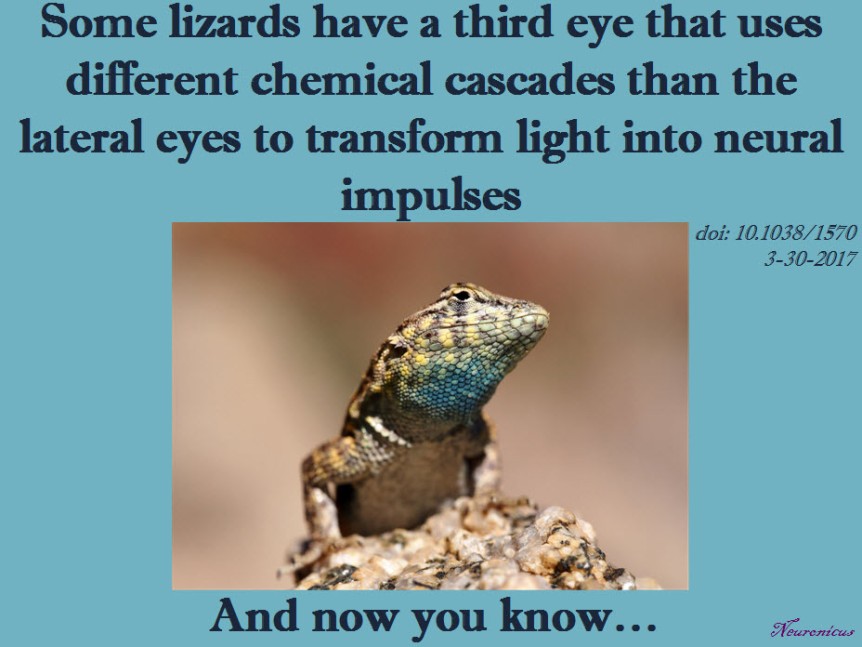
Back to the lizards. Yes, you read right: some lizards and frogs have a third eye. This eye is not exactly like the other two, but it has cells sensitive to light, even if they are not perceiving light in the same way the retinal cells from the lateral eyes are. It is located on the top of the skull, so sometimes it is called the parietal organ (because it is in-between the parietal skull bones, see pic).

Anolis_carolinensis_parietal_eye.CC BY-SA 3.0jpg
Dorsal view of the head of the adult Carolina anole (Anolis carolinensis) clearly showing the parietal eye (small gray/clear oval) at the top of its head. Photo by TheAlphaWolf. License: CC BY-SA 3.0, courtesy of Wikipedia.

It is believed to be a vestigial organ, meaning that primitive vertebrates might have had it as a matter of course but it disappeared in the more recently evolved animals. Importantly, birds and mammals don’t have it. Not at all, not a bit, not atrophied, not able to be “awakened” no matter what your favorite “lemme see your chakras” guru says. Go on, touch your top of the skull and see if you have some peeking soft tissue there. And no, the soft tissue that babies are born with right there on the top of the skull is not a third eye; it’s a fontanelle that allows for the rapid expansion of the brain during the first year of life.

The parietal organ’s anatomical connection to the pineal gland is not surprising at all for scientists because the pineal’s role in every single animal that has it is the regulation of some circadian rhythms by the production of melatonin. In humans, the eyes tell the pineal that is day or night and the pineal adjusts the melatonin production accordingly, i.e. less melatonin produced during the day and more during the night. Likewise, the lizards’ third eye’s main role is to provide information to the pineal about the ambient light for thermoregulatory purposes.

After this long introduction, here is the point: almost twenty years ago Xiong et al. (1998) looked at how this third eye perceives light. In the human eye, light hitting the rods and cones in the retina (reception) launches a biochemical cascade (transduction) that results in seeing (coding of the stimulus in the brain). Briefly, transduction goes thusly: the photon(s) cause(s) a special protein sensitive to light (e.g. rhodopsin) in the photoreceptor cells in the retina to split into its components (photobleaching), one of these components (11-cis-retinal) changes its conformation (photoisomerization), then activates a G-protein (transducin), which then activates the enzyme phosphodiesterase (PDE), which then destroys a nucleotide called cyclic guanosine monophosphate (cGMP), which results in the closing of the cell’s sodium ion channels, which leads to less neurotransmitter released (glutamate), which causes the nearby cells (bipolar cells) to release the same neurotransmitter, which now has the opposite effect, meaning it increases the firing rate of another set of cells (ganglion cells) and from there to the brain we go. Phew, visual transduction IS difficult. And this is the brief version.

It turns out that the third eye’s retina doesn’t have all the types of cells that the normal eyes have. Specifically, it misses the bipolar, horizontal and amacrine cells, having only ganglion and photoreception cells. So how goes the phototransduction in the third eye’s retina, if at all?

Xiong et al. (1998) isolated photoreceptor cells from the third eyes of the lizard Uta stansburiana. And then they did a bunch of electrophysiological recording on those cells under different illumination and chemical conditions.

They found that the phototransduction in the third eye is different from the lateral eyes in that when they expected to see hyperpolarization of the cell, they observed depolarization instead. Also, when they expected the PDE to break down cGMP they found that PDE is inhibited thereby increasing the amount of cGMP.  The fact that G-protein can inhibit PDE was totally unexpected and showed a novel way of cellular signaling. Moreover, they speculate that their results can make sense only if not one, but two G-proteins with opposite actions work in tandem.

A probably dumb technical question though: the human rhodopsin takes about 30 minutes to restore itself from photobleaching. Xiong et al. (1998) let the cells adapt to dark for 10 minutes before recordings. So I wonder if the results would have been slightly different if they allowed the cell more time to adapt? But I’m not an expert in retina science, you’ve seen how difficult it is, right? Maybe the lizard proteins are different or rhodopsin adaptation time has little or nothing to do with their experiments? After all, later research has shown that the third eye has its own unique opsins, like the green-sensitive parietopsin discovered by Su et al. (2006).

115 third eye - Copy

REFERENCE:  Xiong WH, Solessio EC, & Yau KW (Sep 1998). An unusual cGMP pathway underlying depolarizing light response of the vertebrate parietal-eye photoreceptor. Nature Neuroscience, 1(5): 359-365. PMID: 10196524, DOI: 10.1038/1570. ARTICLE

Additional bibliography: Su CY, Luo DG, Terakita A, Shichida Y, Liao HW, Kazmi MA, Sakmar TP, Yau KW (17 Mar 2006). Parietal-eye phototransduction components and their potential evolutionary implications. Science, 311(5767): 1617-1621. PMID: 16543463, DOI: 10.1126/science.1123802. ARTICLE

By Neuronicus, 30 March 2017

Save

Save

Save

Who invented optogenetics?

Wayne State University. Ever heard of it? Probably not. How about Zhuo-Hua Pan? No? No bell ringing? Let’s try a different approach: ever heard of Stanford University? Why, yes, it’s one of the most prestigious and famous universities in the world. And now the last question: do you know who Karl Deisseroth is? If you’re not a neuroscientist, probably not. But if you are, then you would know him as the father of optogenetics.

Optogenetics is the newest tool in the biology kit that allows you to control the way a cell behaves by shining a light on it (that’s the opto part). Prior to that, the cell in question must be made to express a protein that is sensitive to light (i.e. rhodopsin) either by injecting a virus or breeding genetically modified animals that express that protein (that’s the genetics part).

If you’re watching the Nobel Prizes for Medicine, then you would also be familiar with Deisseroth’s name as he may be awarded the Nobel soon for inventing optogenetics. Only that, strictly speaking, he did not. Or, to be fair and precise at the same time, he did, but he was not the first one. Dr. Pan from Wayne State University was. And he got scooped.98.png

The story is at length imparted to us by Anna Vlasits in STAT and republished in Scientific American. In short, Dr. Pan, an obscure name in an obscure university from an ill-famed city (Detroit), does research for years in an unglamorous field of retina and blindness. He figured, quite reasonably, that restoring the proteins which sense light in the human eye (i.e. photoreceptor proteins) could restore vision in the congenitally blind. The problem is that human photoreceptor proteins are very complicated and efforts to introduce them into retinas of blind people have proven unsuccessful. But, in 2003, a paper was published showing how an algae protein that senses light, called channelrhodopsin (ChR), can be expressed into mammalian cells without loss of function.

So, in 2004, Pan got a colleague from Salus University (if Wayne State University is a medium-sized research university, then Salus is a really tiny, tiny little place) to engineer a ChR into a virus which Pan then injected in rodent retinal neurons, in vivo. After 3-4 weeks he obtained the expression of the protein and the expression was stable for at least 1 year, showing that the virus works nicely. Then his group did a bunch of electrophysiological recordings (whole cell patch-clamp and voltage clamp) to see if shining light on those neurons makes them fire. It did. Then, they wanted to see if ChR is for sure responsible for this firing and not some other proteins so they increased the intensity of the blue light that their ChR is known to sense and observed that the cell responded with increased firing. Now that they saw the ChR works in normal rodents, next they expressed the ChR by virally infecting mice who were congenitally blind and repeated their experiments. The electrophysiological experiments showed that it worked. But you see with your brain, not with your retina, so the researchers looked to see if these cells that express ChR project from the retina to the brain and they found their axons in lateral geniculate and superior colliculus, two major brain areas important for vision. Then, they recorded from these areas and the brain responded when blue light, but not yellow or other colors, was shone on the retina. The brain of congenitally blind mice without ChR does not respond regardless of the type of light shone on their retinas. But does that mean the mouse was able to see? That remains to be seen (har har) in future experiments. But the Pan group did demonstrate – without question or doubt – that they can control neurons by light.

All in all, a groundbreaking paper. So the Pan group was not off the mark when they submitted it to Nature on November 25, 2004. As Anna Vlasits reports in the Exclusive, Nature told Pan to submit to a more specialized journal, like Nature Neuroscience, which then rejected it. Pan submitted then to the Journal of Neuroscience, which also rejected it. He submitted it then to Neuron on November 29, 2005, which finally accepted it on February 23, 2006. Got published on April 6, 2006. Deisseroth’s paper was submitted to Nature Neuroscience on May 12, 2005, accepted on July, and published on August 14, 2005… His group infected rat hippocampal neurons cultured in a Petri dish with a virus carrying the ChR and then they did some electrophysiological recordings on those neurons while shining lights of different wavelengths on them, showing that these cells can be controlled by light.

There’s more on the saga with patent filings and a conference where Pan showed the ChR data in May 2005 and so on, you can read all about it in Scientific American. The magazine is just hinting to what I will say outright, loud and clear: Pan didn’t get published because of his and his institution’s lack of fame. Deisseroth did because of the opposite. That’s all. This is not about squabbles about whose work is more elegant, who presented his work as a scientific discovery or a technical report or whose title is more catchy, whose language is more boisterous or native English-speaker or luck or anything like that. It is about bias and, why not?, let’s call a spade a spade, discrimination. Nature and Journal of Neuroscience are not caught doing this for the first time. Not by a long shot. The problem is that they are still doing it, that is: discriminating against scientific work presented to them based on the name of the authors and their institutions.

Personally, so I don’t get comments along the lines of the fox and the grapes, I have worked at both high profile and low profile institutions. And I have seen the difference not in the work, but in the reception.

That’s my piece for today.

Source:  STAT, Scientific American.

References:

1) Bi A, Cui J, Ma YP, Olshevskaya E, Pu M, Dizhoor AM, & Pan ZH (6 April 2006). Ectopic expression of a microbial-type rhodopsin restores visual responses in mice with photoreceptor degeneration. Neuron, 50(1): 23-33. PMID: 16600853. PMCID: PMC1459045. DOI: 10.1016/j.neuron.2006.02.026. ARTICLE | FREE FULLTEXT PDF

2) Boyden ES, Zhang F, Bamberg E, Nagel G, & Deisseroth K. (Sep 2005, Epub 2005 Aug 14). Millisecond-timescale, genetically targeted optical control of neural activity. Nature Neuroscience, 8(9):1263-1268. PMID: 16116447. DOI: 10.1038/nn1525. doi:10.1038/nn1525. ARTICLE 

By Neuronicus, 11 September 2016

Save

Save

Save

Save