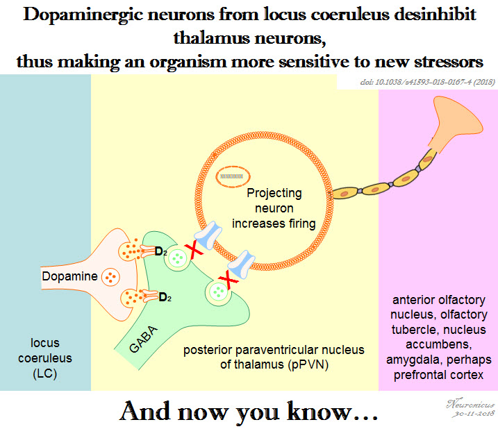
Pic of the day: Dopamine from a non-dopamine place

147 lc da ppvn - Copy

Reference: Beas BS, Wright BJ, Skirzewski M, Leng Y, Hyun JH, Koita O, Ringelberg N, Kwon HB, Buonanno A, & Penzo MA (Jul 2018, Epub 18 Jun 2018). The locus coeruleus drives disinhibition in the midline thalamus via a dopaminergic mechanism. Nature Neuroscience,21(7):963-973. PMID: 29915192, PMCID: PMC6035776 [Available on 2018-12-18], DOI:10.1038/s41593-018-0167-4. ARTICLE

Drink before sleep

Among the many humorous sayings, puns, and jokes that one inevitably encounters on any social medium account, one that was popular this year was about the similarity between putting a 2 year old to bed and putting your drunk friend to bed, which went like this: they both sing to themselves, request water, mumble and blabber incoherently, do some weird yoga posses, cry, hiccup, and then they pass out. The joke manages to steal a smile only if someone has been through both situations, otherwise it looses its appeal.

Being exposed to both situations, I thought that while the water request from the drunk friend is a response to the dehydrating effects of alcohol, the water request from the toddler is probably nothing more than a delaying tactic to postpone bedtime. Whether there may or may not be some truth to my assumption in the case of the toddler, here is a paper to show that there is definitely more to the water request than meets the eye.

Generally, thirst is generated by the hypothalamus when its neurons and neurons from organum vasculosum lamina terminalis (OVLT) in the brainstem sense that the blood is either too viscous (hypovolaemia) or too salty (hyperosmolality), both phenomena indicating a need for water. Ingesting water would bring these indices to homeostatic values.

More than a decade ago, researchers observed that rodents get a good gulp of water just before going to sleep. This surge was not motivated by thirst because the mice were not feverish, were not hungry and they did not have a too viscous or a too salty blood. So why do it then? If the rodents are restricted from drinking the water they get dehydrated, so obviously the behavior has function. But is not motivated by thirst, at least not the way we know it. Huh… The authors call this “anticipatory thirst”, because it keeps the animal from becoming dehydrated later on.

Since the behavior occurs with regularity, maybe the neurons that control circadian rhythms have something to do with it. So Gizowski et al. (2016) took a closer look at  the activity of clock neurons from the suprachiasmatic nucleus (SCN), a well known hypothalamic nucleus heavily involved in circadian rhythms. The authors did a lot of work on SCN and OVLT neurons: fluorescent labeling, c-fos expression, anatomical tracing, optogenetics, genetic knockouts, pharmacological manipulations, electrophysiological recordings, and behavioral experiments. All these to come to this conclusion:

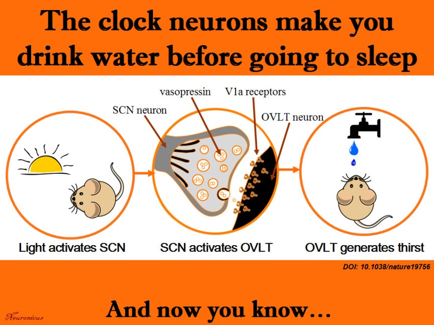
SCN neurons release vasopressin and that excites the OVLT neurons via V1a receptors. This is necessary and sufficient to make the animal drink the water, even if it’s not thirsty.

That’s a lot of techniques used in a lot of experiments for only three authors. Ten years ago, you needed only one, maybe two techniques to prove the same point. Either there have been a lot of students and technicians who did not get credit (there isn’t even an Acknowledgements section. EDIT: yes, there is, see the comments below or, if they’re missing, the P.S.) or these three authors are experts in all these techniques. In this day and age, I wouldn’t be surprised by either option. No wonder small universities have difficulty publishing in Big Name journals; they don’t have the resources to compete. And without publishing, no tenure… And without tenure, less research… And thus shall the gap widen.

Musings about workload aside, this is a great paper, shedding light on yet another mysterious behavior and elucidating the mechanism behind it. There’s still work to be done though, like answering how accurate is the SCN in predicting bedtime to activate the drinking behavior. Does it take its cues from light only? Does ambient temperature play a role and so on. This line of work can help people that work in shifts to prevent certain health problems. Their SCN is out of rhythm and that can influence deleteriously the activity of a whole slew of organs.

scn-h2o-copy
Summary of the doi: 10.1038/nature19756 findings. 1) The light is a cue for suprachiasmatic nulceus (SCN) that bedtime is near. 2) The SCN vasopressin neurons that project to organum vasculosum lamina terminalis (OVLT) are activated. 3) The OVLT generates the anticipatory thirst. 4) The animal drinks fluids.

Reference: Gizowski C, Zaelzer C, & Bourque CW (28 Sep 2016). Clock-driven vasopressin neurotransmission mediates anticipatory thirst prior to sleep. Nature, 537(7622): 685-688. PMID: 27680940. DOI: 10.1038/nature19756. ARTICLE

By Neuronicus, 5 October 2016

EDIT (12 Oct 2016): P.S. The blog comments are automatically deleted after a period of time. In case of this post that would be a pity because I have been fortunate to receive comments from at least one of the authors of the paper, the PI, Dr. Charles Bourque and, presumably under pseudonym, but I don’t know that for sure, also the first author, Claire Gizowski. So I will include here, in a post scriptum, the main idea of their comments. Here is an excerpt from Dr. Bourque’s comment:

“Let me state for the record that Claire accomplished pretty much ALL of the work in this paper (there is a description of who did what at the end of the paper). More importantly, there were no “unthanked” undergraduates, volunteers or other parties that contributed to this work.”

My hat, Ms. Gizowski. It is tipped. To you. Congratulations! With such an impressive work I am sure I will hear about you again and that pretty soon I will blog about Dr. Gizowski.

Who invented optogenetics?

Wayne State University. Ever heard of it? Probably not. How about Zhuo-Hua Pan? No? No bell ringing? Let’s try a different approach: ever heard of Stanford University? Why, yes, it’s one of the most prestigious and famous universities in the world. And now the last question: do you know who Karl Deisseroth is? If you’re not a neuroscientist, probably not. But if you are, then you would know him as the father of optogenetics.

Optogenetics is the newest tool in the biology kit that allows you to control the way a cell behaves by shining a light on it (that’s the opto part). Prior to that, the cell in question must be made to express a protein that is sensitive to light (i.e. rhodopsin) either by injecting a virus or breeding genetically modified animals that express that protein (that’s the genetics part).

If you’re watching the Nobel Prizes for Medicine, then you would also be familiar with Deisseroth’s name as he may be awarded the Nobel soon for inventing optogenetics. Only that, strictly speaking, he did not. Or, to be fair and precise at the same time, he did, but he was not the first one. Dr. Pan from Wayne State University was. And he got scooped.98.png

The story is at length imparted to us by Anna Vlasits in STAT and republished in Scientific American. In short, Dr. Pan, an obscure name in an obscure university from an ill-famed city (Detroit), does research for years in an unglamorous field of retina and blindness. He figured, quite reasonably, that restoring the proteins which sense light in the human eye (i.e. photoreceptor proteins) could restore vision in the congenitally blind. The problem is that human photoreceptor proteins are very complicated and efforts to introduce them into retinas of blind people have proven unsuccessful. But, in 2003, a paper was published showing how an algae protein that senses light, called channelrhodopsin (ChR), can be expressed into mammalian cells without loss of function.

So, in 2004, Pan got a colleague from Salus University (if Wayne State University is a medium-sized research university, then Salus is a really tiny, tiny little place) to engineer a ChR into a virus which Pan then injected in rodent retinal neurons, in vivo. After 3-4 weeks he obtained the expression of the protein and the expression was stable for at least 1 year, showing that the virus works nicely. Then his group did a bunch of electrophysiological recordings (whole cell patch-clamp and voltage clamp) to see if shining light on those neurons makes them fire. It did. Then, they wanted to see if ChR is for sure responsible for this firing and not some other proteins so they increased the intensity of the blue light that their ChR is known to sense and observed that the cell responded with increased firing. Now that they saw the ChR works in normal rodents, next they expressed the ChR by virally infecting mice who were congenitally blind and repeated their experiments. The electrophysiological experiments showed that it worked. But you see with your brain, not with your retina, so the researchers looked to see if these cells that express ChR project from the retina to the brain and they found their axons in lateral geniculate and superior colliculus, two major brain areas important for vision. Then, they recorded from these areas and the brain responded when blue light, but not yellow or other colors, was shone on the retina. The brain of congenitally blind mice without ChR does not respond regardless of the type of light shone on their retinas. But does that mean the mouse was able to see? That remains to be seen (har har) in future experiments. But the Pan group did demonstrate – without question or doubt – that they can control neurons by light.

All in all, a groundbreaking paper. So the Pan group was not off the mark when they submitted it to Nature on November 25, 2004. As Anna Vlasits reports in the Exclusive, Nature told Pan to submit to a more specialized journal, like Nature Neuroscience, which then rejected it. Pan submitted then to the Journal of Neuroscience, which also rejected it. He submitted it then to Neuron on November 29, 2005, which finally accepted it on February 23, 2006. Got published on April 6, 2006. Deisseroth’s paper was submitted to Nature Neuroscience on May 12, 2005, accepted on July, and published on August 14, 2005… His group infected rat hippocampal neurons cultured in a Petri dish with a virus carrying the ChR and then they did some electrophysiological recordings on those neurons while shining lights of different wavelengths on them, showing that these cells can be controlled by light.

There’s more on the saga with patent filings and a conference where Pan showed the ChR data in May 2005 and so on, you can read all about it in Scientific American. The magazine is just hinting to what I will say outright, loud and clear: Pan didn’t get published because of his and his institution’s lack of fame. Deisseroth did because of the opposite. That’s all. This is not about squabbles about whose work is more elegant, who presented his work as a scientific discovery or a technical report or whose title is more catchy, whose language is more boisterous or native English-speaker or luck or anything like that. It is about bias and, why not?, let’s call a spade a spade, discrimination. Nature and Journal of Neuroscience are not caught doing this for the first time. Not by a long shot. The problem is that they are still doing it, that is: discriminating against scientific work presented to them based on the name of the authors and their institutions.

Personally, so I don’t get comments along the lines of the fox and the grapes, I have worked at both high profile and low profile institutions. And I have seen the difference not in the work, but in the reception.

That’s my piece for today.

Source:  STAT, Scientific American.

References:

1) Bi A, Cui J, Ma YP, Olshevskaya E, Pu M, Dizhoor AM, & Pan ZH (6 April 2006). Ectopic expression of a microbial-type rhodopsin restores visual responses in mice with photoreceptor degeneration. Neuron, 50(1): 23-33. PMID: 16600853. PMCID: PMC1459045. DOI: 10.1016/j.neuron.2006.02.026. ARTICLE | FREE FULLTEXT PDF

2) Boyden ES, Zhang F, Bamberg E, Nagel G, & Deisseroth K. (Sep 2005, Epub 2005 Aug 14). Millisecond-timescale, genetically targeted optical control of neural activity. Nature Neuroscience, 8(9):1263-1268. PMID: 16116447. DOI: 10.1038/nn1525. doi:10.1038/nn1525. ARTICLE 

By Neuronicus, 11 September 2016

Save

Save

Save

Save
The song of a fly… the courtship of another

Drosophila melanogaster image illustrating sexual dimorphism and mating behavior. Credit: TheAlphaWolf (Wikimedia Commons)

Did you know that flies sing? True to the dictum that I just made up – ‘where is song, there is lust’ – it turns out not only that flies can sing, but they even have courtship songs! Granted, since they don’t have a larynx, the male flies sing by vibrating their wings in a certain way, which is unique to each fly species, and females listen with the feather-looking bit on top of their antennae, called arista. The behavior has generated enough research that a fairly hefty review about it has been published two years ago in Nature Reviews Neuroscience, pointing to a gene central to the male courtship circuitry and expressed only in the fly’s neurons, the fru gene (I bet it was called that way because when you make mutants you get fru/fru …).

Zhou et al. (2015) used a series of complicated experiments to successively activate or inhibit the neurons which express the fru gene, in order to identify the neural circuitry underlying hearing and processing the courtship songs. This circuitry is different in males and females, which makes sense since the serenading male expects different behaviors from his audience, depending on their sex; the listening males hurry to compete for the intended female and the females slow down and… listen carefully. Mind wondering: if I was the one serenading, wouldn’t I want to drive away the competitors, instead of drawing them in towards the object of my desire? Perhaps I want the competitors to also engage in courtship behavior so I can show off my wing vibrating prowess… Anyway, digression aside, in addition to figuring out which neuron does what, the authors managed to elicit courtship behavior in the listening males by optogenetically stimulating the 3rd and 4th order neurons in the newly identified circuit.

Besides being strangely interesting in itself, the research fills a gap in the understanding how courtship behavior is recognized, at least in fruit flies, which may be very useful information for other species as well, humans included.

Reference: Zhou, C., Franconville, R., Vaughan, A. G., Robinett, C. C., Jayaraman, V., & Baker, B. S. (21 September 2015). Central neural circuitry mediating courtship song perception in male Drosophila. Elife, 4:1-15. doi: 10.7554/eLife.08477. Article + FREE PDF

For the interested specialist, the MATLAB source code for analyzing calcium-imaging data can be found here.

By Neuronicus, 24 September 2015

Wireless mice, batteries not included (optrodes and electrodes in one wireless contraption)

A neuron is expressing the light-gated cation channel channelrhodopsin-2 (small green shapes), and is being illuminated by a focused beam of blue light (coming from the top). The opsins have opened, depolarizing or electrically activating the cell. The pulse of electricity spreads throughout the inside of the cell (white highlights), triggering the release of neurotransmitters to downstream cells. Credit: Ed Boyden and Massachusetts Institute of Technology McGovern Institute Courtesy: National Science Foundation
A neuron is expressing the light-gated cation channel channelrhodopsin-2 (small green shapes), and is being illuminated by a focused beam of blue light (coming from the top). The opsins have opened, depolarizing or electrically activating the cell. The pulse of electricity spreads throughout the inside of the cell (white highlights), triggering the release of neurotransmitters to downstream cells. Credit: Ed Boyden and Massachusetts Institute of Technology McGovern Institute. Courtesy: National Science Foundation

Optogenetics is the new (and very popular) kid on the block of neurotechniques arsenal. Before optogenetics, in order to manipulate how the neurons behave in a particular brain region, one had to do so electrically or chemically by inserting into that region an electrode or a tube, respectively. The trouble with that is that you never know exactly which neurons you are manipulating, given that any brain regions has many kinds of cells that project to many other regions and they are not nicely arranged in clumps but interspersed in a messy mesh. But with optogenetics, you can select exactly which neurons you want to manipulate. You take a mouse and you genetically engineer it to express in your favorite neurons a particular protein that is sensitive to a certain light wavelength (like a rhodopsin). Then, you shine the light in the mouse’s brain, turning on and off the neurons of your interest. The technique, besides having tremendous success in fundamental research, also shows promise for neuroprosthetics for some diseases, like Parkinson’s.

Despite rapid improvements of the technique due to increasing demand form the neuroscience community, it is still difficult to record from the freely-moving animal, mainly because the animal is attached to the fiber optic that delivers the light pulses. There have been some wireless attempts (some successful), but because the resultant contraption was too big and heavy for the animal, they have reduced options as compared to the wired devices. Gagnon-Turcotte et al. (2015) describe here a way to build a wireless headtsage that can deliver light pulses AND record the electrophysiological activity of the neurons. And, equally important, is cheap and can be build with off-the-shelf components.

Reference: Gagnon-Turcotte, G., Kisomi, A. A., Ameli, R., Camaro, C.-O. D., LeChasseur, Y., Néron, J.-L., , Bareil, P. B., Fortier, P., Bories, C., de Koninck, Y., & Gosselin, B. (9 September 2015). A Wireless Optogenetic Headstage with Multichannel Electrophysiological Recording Capability. Sensors, 15(9): 22776-22797; doi:10.3390/s150922776. Article + FREE PDF

By Neuronicus, 22 September 2015

Update (27 September 2015). The author Gabriel Gagnon-Turcotte wins the Mitacs 2015 prize for exceptional innovation for the development of a wireless system to study the brain