The pineal gland has held fascination since Descartes’ nefarious claim that it is the seat of the soul. There is no evidence of that; he said it might be where the soul resides because he thought the pineal gland was the only solitaire structure in the brain so it must be special. By ‘solitaire’ I mean that all other brain structures come in doublets: 2 amygdalae, 2 hippocampi, 2 thalami, 2 hemispheres etc. He was wrong about that as well, in that there are some other singletons in the brain besides the pineal, like the anterior or posterior commissure, the cerebellar vermis, some deep brainstem and medullary structures etc.
Descartes’ dualism was the only escape route the mystics at the time had from the demands for evidence by the budding natural philosophers later known as scientists. So when some scientists noted that some lizards have a third eye on top of their head connected to the pineal gland, the mystics and, later, the conspiracy theorists went nuts. Here, see, if the seat of the soul is linked with the third eye, then the awakening of this eye in people would surely result in heightened awareness, closeness to the Divinity, oneness with Universe and other similar rubbish that can otherwise easily and reliably be achieved by a good dollop of magic mushrooms. Cheaper, too.
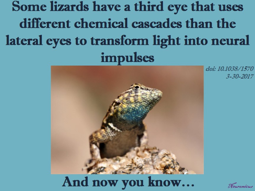
Back to the lizards. Yes, you read right: some lizards and frogs have a third eye. This eye is not exactly like the other two, but it has cells sensitive to light, even if they are not perceiving light in the same way the retinal cells from the lateral eyes are. It is located on the top of the skull, so sometimes it is called the parietal organ (because it is in-between the parietal skull bones, see pic).

It is believed to be a vestigial organ, meaning that primitive vertebrates might have had it as a matter of course but it disappeared in the more recently evolved animals. Importantly, birds and mammals don’t have it. Not at all, not a bit, not atrophied, not able to be “awakened” no matter what your favorite “lemme see your chakras” guru says. Go on, touch your top of the skull and see if you have some peeking soft tissue there. And no, the soft tissue that babies are born with right there on the top of the skull is not a third eye; it’s a fontanelle that allows for the rapid expansion of the brain during the first year of life.
The parietal organ’s anatomical connection to the pineal gland is not surprising at all for scientists because the pineal’s role in every single animal that has it is the regulation of some circadian rhythms by the production of melatonin. In humans, the eyes tell the pineal that is day or night and the pineal adjusts the melatonin production accordingly, i.e. less melatonin produced during the day and more during the night. Likewise, the lizards’ third eye’s main role is to provide information to the pineal about the ambient light for thermoregulatory purposes.
After this long introduction, here is the point: almost twenty years ago Xiong et al. (1998) looked at how this third eye perceives light. In the human eye, light hitting the rods and cones in the retina (reception) launches a biochemical cascade (transduction) that results in seeing (coding of the stimulus in the brain). Briefly, transduction goes thusly: the photon(s) cause(s) a special protein sensitive to light (e.g. rhodopsin) in the photoreceptor cells in the retina to split into its components (photobleaching), one of these components (11-cis-retinal) changes its conformation (photoisomerization), then activates a G-protein (transducin), which then activates the enzyme phosphodiesterase (PDE), which then destroys a nucleotide called cyclic guanosine monophosphate (cGMP), which results in the closing of the cell’s sodium ion channels, which leads to less neurotransmitter released (glutamate), which causes the nearby cells (bipolar cells) to release the same neurotransmitter, which now has the opposite effect, meaning it increases the firing rate of another set of cells (ganglion cells) and from there to the brain we go. Phew, visual transduction IS difficult. And this is the brief version.
It turns out that the third eye’s retina doesn’t have all the types of cells that the normal eyes have. Specifically, it misses the bipolar, horizontal and amacrine cells, having only ganglion and photoreception cells. So how goes the phototransduction in the third eye’s retina, if at all?
Xiong et al. (1998) isolated photoreceptor cells from the third eyes of the lizard Uta stansburiana. And then they did a bunch of electrophysiological recording on those cells under different illumination and chemical conditions.
They found that the phototransduction in the third eye is different from the lateral eyes in that when they expected to see hyperpolarization of the cell, they observed depolarization instead. Also, when they expected the PDE to break down cGMP they found that PDE is inhibited thereby increasing the amount of cGMP. The fact that G-protein can inhibit PDE was totally unexpected and showed a novel way of cellular signaling. Moreover, they speculate that their results can make sense only if not one, but two G-proteins with opposite actions work in tandem.
A probably dumb technical question though: the human rhodopsin takes about 30 minutes to restore itself from photobleaching. Xiong et al. (1998) let the cells adapt to dark for 10 minutes before recordings. So I wonder if the results would have been slightly different if they allowed the cell more time to adapt? But I’m not an expert in retina science, you’ve seen how difficult it is, right? Maybe the lizard proteins are different or rhodopsin adaptation time has little or nothing to do with their experiments? After all, later research has shown that the third eye has its own unique opsins, like the green-sensitive parietopsin discovered by Su et al. (2006).

REFERENCE: Xiong WH, Solessio EC, & Yau KW (Sep 1998). An unusual cGMP pathway underlying depolarizing light response of the vertebrate parietal-eye photoreceptor. Nature Neuroscience, 1(5): 359-365. PMID: 10196524, DOI: 10.1038/1570. ARTICLE
Additional bibliography: Su CY, Luo DG, Terakita A, Shichida Y, Liao HW, Kazmi MA, Sakmar TP, Yau KW (17 Mar 2006). Parietal-eye phototransduction components and their potential evolutionary implications. Science, 311(5767): 1617-1621. PMID: 16543463, DOI: 10.1126/science.1123802. ARTICLE
By Neuronicus, 30 March 2017


Very interesting, thanks!
LikeLiked by 2 people