Love and the immune system

Valentine’s day is a day when we celebrate romantic love (well, some of us tend to) long before the famous greeting card company Hallmark was established. Fittingly, I found the perfect paper to cover for this occasion.

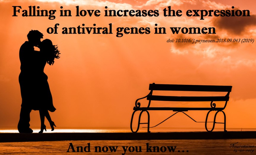
In the past couple of decades it became clear to scientists that there is no such thing as a mental experience that doesn’t have corresponding physical changes. Why should falling in love be any different? Several groups have already found that levels of some chemicals (oxytocin, cortisol, testosterone, nerve growth factor, etc.) change when we fall in love. There might be other changes as well. So Murray et al. (2019) decided to dive right into it and check how the immune system responds to love, if at all.

For two years, the researchers looked at certain markers in the immune system of 47 women aged 20 or so. They drew blood when the women reported to be “not in love (but in a new romantic relationship), newly in love, and out-of-love” (p. 6). Then they sent their samples to their university’s Core to toil over microarrays. Microarray techniques can be quickly summarized thusly: get a bunch of molecules of interest, in this case bits of single-stranded DNA, and stick them on a silicon plate or a glass slide in a specific order. Then you run your sample over it and what sticks, sticks, what not, not. Remember that DNA loves to be double stranded, so any single strand will stick to their counterpart, called complementary DNA. You put some fluorescent dye on your genes of interest and voilà, here you have an array of genes expressed in a certain type of tissue in a certain condition.

Talking about microarrays got me a bit on memory lane. When fMRI started to be a “must” in neuroscience, there followed a period when the science “market” was flooded by “salad” papers. We called them that because there were so many parts of the brain reported as “lit up” in a certain task that it made a veritable “salad of brain parts” out of which it was very difficult to figure out what’s going on. I swear that now that the fMRI field matured a bit and learned how to correct for multiple comparisons as well as to use some other fancy stats, the place of honor in the vegetable mix analogy has been relinquished to the ‘-omics’ studies. In other words, a big portion of the whole-genome or transcriptome studies became “salad” studies: too many things show up as statistically significant to make head or tail of it.

However, Murray et al. (2019) made a valiant – and successful – effort to figure out what those up- or down- regulated 61 gene transcripts in the immune system cells of 17 women falling in love actually mean. There’s quite a bit I am leaving out but, in a nutshell, love upregulated (that is “increased”) the expressions of genes involved in the innate immunity to viruses, presumably to facilitate sexual reproduction, the authors say.

The paper is well written and the authors graciously remind us that there are some limitations to the study. Nevertheless, this is another fine addition to the unbelievably fast growing body of knowledge regarding human body and behavior.

Pitty that this research was done only with women. I would have loved to see how men’s immune systems respond to falling in love.

149-love antiviral - Copy.jpg

REFERENCE: Murray DR, Haselton MG, Fales M, & Cole SW. (Feb 2019, Epub 2 Oct 2018). Falling in love is associated with immune system gene regulation. Psychoneuroendocrinology, Vol. 100, Pg. 120-126. doi: 10.1016/j.psyneuen.2018.09.043. PMID: 30299259, PMCID: PMC6333523 [Available on 2020-02-01], DOI: 10.1016/j.psyneuen.2018.09.043 ARTICLE

FYI: PMC6333523 [Available on 2020-02-01] means that the fulltext will be available for free to the public one year after the publication on the US governmental website PubMed (https://www.ncbi.nlm.nih.gov/pubmed/), no matter how much Elsevier will charge for it. Always, always, check the PMC library (https://www.ncbi.nlm.nih.gov/pmc/) on PubMed to see if a paper you saw in Nature or Elsevier is for free there because more often than you’d think it is.

PubMed = the U.S. National Institutes of Health’s National Library of Medicine (NIH/NLM), comprising of “more than 29 million citations for biomedical literature from MEDLINE, life science journals, and online books. Citations may include links to full-text content from PubMed Central and publisher web sites” .

PMC = “PubMed Central® (PMC) is a free fulltext archive of biomedical and life sciences journal literature at the U.S. National Institutes of Health’s National Library of Medicine (NIH/NLM)” with a whooping fulltext library of over 5 million papers and growing rapidly. Love PubMed!

By Neuronicus, 14 February 2019

Tomato transcriptome

As most children, growing up I showed little appreciation for what I had, coveting instead what I did not. Now I realize how fortunate I have been to have grown up half the time in a metropolis and the other half at the countryside. At the farm. A subsistence farm, although I truly loathe the term because we were not just subsisting but thriving off the land, as we planted and harvested a bit of everything and we had a specimen or four of almost all the farm animals, from bipeds to quadrupeds.

I got on this memory lane after reading the paper of Shinozaki et al. (2018) on tomatoes. It was a difficult read for me as it was punctured by many term definition lookups since botany evolved quite steeply since the last time I checked, about 25 years or so.

Briefly, the scientists grew tomato plants in a greenhouse at Cornell, NY. They harvested the fruit from 60 plants about 5 to 50 days after the flower was at its peak (DPA, days post anthesis) following this chart:

  • Expanding [fruit] stage (harvested at 5, 10, 20, or 30 DPA)
  • Mature Green stage (full-size green fruit, ≈ 39 DPA),
  • Breaker stage (definite break in color from green to tannish-yellow with less than 10% of the surface, ≈ 42 DPA),
  • Pink stage (50% pink or red color, ≈ 44 DPA),
  • Light red stage (100% light red, ≈ 46 DPA),
  • Red ripe stage (full red for 8 days, ≈ 50 DPA).

(simplified from the Methods section, p. 10, see pic)

130-shinozaki - Copy
Fig. 1 (partial from Shinozaki et al., 2018). A tissue/cell-based transcript profiling of developing tomato fruit. a Traced image of six targeted fruit tissues. Shaded areas of the total pericarp and the placenta were not harvested. b Traced image of five pericarp cells. c Representative pictures of harvested fruit spanning ten developmental stages. d Representative pictures of the stylar end of MG and Br stage fruit. DPA, days post anthesis; MG, mature green; Br, breaker; Pk, pink; LR, light red; RR, red ripe. Credit: DOI: 10.1038/s41467-017-02782-9. License: CC BY 4.0 IL.

Immediately after harvesting, the tomato was scanned with a micro-computed tomograph (micro-CT) to generate a 3D image of the fruit, including its internal structures. Then, the fruit was dissected by hand or laser, depending of its size, divided into various tissue types and then preserved either via snap-freezing in liquid nitrogen or standard tissue fixation for light or transmission electron microscopy. Finally, the researchers used kits to extract and analyze the RNA from their samples. And, last but not least, a lot of math & stats.

This is what I got out of it:

  1. A total of 24,660 genes were uniquely expressed in various tomato cell types and at various stages of development.
  2. The tomato ripens from within, meaning from the interior to the exterior and not the other way around.
  3. The ripening seems to be a continuous process, starting before the ‘Breaker’ stage.
  4. The ripening signals originate in the locular tissues (the goo around the seeds; it’s possible that the seeds themselves send the signals to the locular tissue to start the ripening process).
  5. The flesh of the fruit is only one part of the tomato and the most investigated, but the other types of tissue are also important. For example, some genes responsible for aroma and flavor (CTOMT1, TOMLOXC) are predominantly or even exclusively expressed in the flesh, but some genes that improve the nutritional value (SlGAD3) are expressed mostly in the placenta.
  6. The fruit can do photosynthesis, probably for the benefit of its seeds.
  7. Each developmental stage is characterized by a distinct transcriptome profile (by inference, also a distinct proteomic profile, although not necessarily in exact correspondence)
  8. Botany, like any serious science, is complicated.

Ah, I have been vindicated. By science, nonetheless! You see, in my pursuit to recapture the tomato taste of my childhood I sample various homegrown exemplars of Solanum lycopersicum derived both from more or less failed personal attempts with pots on the balcony and from various farmer’s market vendors. While I can understand – though not approve of – the industrial scale agro-growers’ practice to pick the tomatoes green, unripe and then artificially injecting them with ethylene to prolong shelf life, I completely fail to understand the picking them up when green by the sellers in the farmer’s markets. I had many surreal conversations with such vendors (I cannot call them farmers for the life of me) who more than once attempted to reassure me that 1) Everybody’s picking tomatoes green off the vine because that’s how it’s done and 2) Ripening happens on the window sill. In vain have I tried to explain the difference between ripen and rotten; in vain have I pointed out that color is only one indicator of ripening; in vain did I explain that during ripening on the vine the plant delivers certain substances to the fruit that lead to changes in the flesh composition to make it more nutritious for the future seedling, process that the aforesaid window sill does nor partake in. Alas, ultimately, my arguments (and my family’s last 400 years of farming experience) hit the wall of “I am growing tomatoes for three years now and I know what I’m doing. Are you buying or not?” As you might imagine, I end up going home frustrated and yet staring at some exorbitantly expensive and looking as sad as I feel greenish tomatoes.

For me, this is what Shinozaki et al. (2018) validated: Ripening is a complex process that involves a lot of physiological changes in the fruit, not merely some extra production of ethylene that can be conveniently supplied externally by a syringe or rotting on the window sill. Of course, there is nowhere in the paper that Shinozaki et al. (2018) say that. What they do say is this: “The ripening program is revealed as comprising gradients of gene expression, initiating in internal tissues then radiating outward, and basipetally along a latitudinal axis. We also identify spatial variations in the patterns of epigenetic control superimposed on ripening gradients” (Abstract). Tomayto, tomahto…

Now we know that… simply put, I’m right. Sometimes is good to be right. I am old enough to prefer happiness and tranquility over rightness & righteousness, but still young enough that sometimes, just sometimes, it feels good to be right. Yes, the Shinozaki et al. (2018) paper exists only for my vindication in my farmer’s market squabbles and not for providing a huge comprehensive atlas on the tomato transcriptome, along with an awesome spatiotemporal map showing the place and time of the expression of genes responsible for fruit ripening, quality traits and so on.

Good job, Shinozaki et al. (2018)!

130-tomato - Copy

REFERENCE: Shinozaki Y, Nicolas P, Fernandez-Pozo N, Ma Q, Evanich DJ, Shi Y, Xu Y, Zheng Y, Snyder SI, Martin LBB, Ruiz-May E, Thannhauser TW, Chen K, Domozych DS, Catalá C, Fei Z, Mueller LA, Giovannoni JJ, & Rose JKC (25 Jan 2018). High-resolution spatiotemporal transcriptome mapping of tomato fruit development and ripening. Nature Communications, 9(1):364. PMID: 29371663, PMCID: PMC5785480, DOI: 10.1038/s41467-017-02782-9. ARTICLE | FREE FULLTEXT PDF | The Tomato Expression Atlas database

By Neuronicus, 7 February 2018

The FIRSTS: mRNA from one cell can travel to another cell and be translated there (2006)

I’m interrupting the series on cognitive biases (unskilled-and-unaware, superiority illusion, and depressive realism) to tell you that I admit it, I’m old. -Ish. Well, ok, I’m not that old. But this following paper made me feel that old. Because it invalidates some stuff I thought I knew about molecular cell biology. Mind totally blown.

It all started with a paper freshly published two days ago and that I’ll cover tomorrow. It’s about what the title says: mRNA can travel between cells packaged nicely in vesicles and once in a target cell can be made into protein there. I’ll explain – briefly! – why this is such a mind-blowing thing.

Central_dogma - Copy
Fig. 1. Illustration of the central dogma of biology: information transfer between DNA, RNA, and protein. Courtesy of Wikipedia, PD

We’ll start with the central dogma of molecular biology (specialists, please bear with me): the DNA is transcribed into RNA and the RNA is translated into protein (see Fig. 1). It is an oversimplification of the complexity of information flow in a biological system, but it’ll do for our purposes.

DNA needs to be transcribed into RNA because RNA is a much more flexible molecule and thus can do many things. So RNA is the traveling mule between DNA and the place where its information becomes protein, i.e. ribosome. Hence the name mRNA. Just kidding; m stands for messenger RNA (not that I will ever be able to call that ever again: muleRNA is stuck in my brain now).

There are many kinds of RNA: some don’t even get out of the nucleus, some are chopped and re-glued (alternative splicing), some decide which bits of DNA (genes) are to be expressed, some are busy housekeepers and so on. Once an RNA has finished its business it is degraded in many inventive ways. It cannot leave the cell because it cannot cross the cell membrane. And that was that. Or so I’ve been taught.

Exceptions from the above were viruses whose ways of going from cell to cell are very clever. A virus is a stretch of nucleic acids (DNA and/or RNA) and some proteins encapsulated in a blob (capsid). Not a cell!

In the ’90s several groups were looking at some blobs (yes, most stuff in biology can be defined by the all-encompassing and enlightening term of ‘blob’) that cells spew out every now and then. These were termed extracellular vesicles (EV) for obvious reasons. Turned out that many kinds of cells were doing it and on a much more regular basis than previously thought. The contents of these EVs varied quite a bit, based on the type of cells studied. Proteins, mostly, and maybe some cytoplasmic debris. In the ’80s it was thought that this was one way for a cell to get rid of trash. But in 1982, Stegmayr & Ronquist showed that prostate cells release some EVs that result in sperm cell motility increase (Raposo & Stoorvogel, 2013) so, clearly, the EVs were more than trash. Soon it became evident that EVs were another way of cell-to-cell communication. (Note to self: the first time intercellular communication by EVs was demonstrated was in 1982, Stegmayr & Ronquist. Maybe I’ll dig out the paper to cover it sometime).

So. In 2005, Baj-Krzyworzeka et al. (2006) looked at some human cancer cells to see what they spew out and for what purpose. They saw that the cancer cells were transferring some of the tumor proteins packaged in EVs to monocytes. For devious purposes, probably. And then they made to what it looks to me like a serious leap in reasoning: since the EVs contain tumor proteins, why wouldn’t they also contain the mRNA for those proteins? My first answer to that would have been: “because it would be rapidly degraded”. And I would have been wrong. To my credit, if the experiment wouldn’t take up too many resources I still would have done it, especially if I would have some random primers lying around the lab. Luckily for the world, I was not in charge with this particular experiment and Baj-Krzyworzeka et al. (2005) proceeded with a real-time PCR (polymerase chain reaction) which showed them that the EVs released by the tumor cells also contained mRNA.

Now the 1 million dollar, stare-in-your-face question was: is this mRNA functional? Meaning, once delivered to the host cell, would it be translated into protein?

Six months later the group answered it. Ratajcza et al. (2006) used embryonic stem cells as the donor cells and hematopoietic progenitor cells as host cells. First, they found out that if you let the donors spit EVs at the hosts, the hosts are faring much better (better survival, upregulated good genes, phosphorylated MAPK to induce proliferation etc.). Next, they looked at the contents of EVs and found out that they contained proteins and mRNA that promote those good things (Wnt-3 protein, mRNA for transcription factors etc.). Next, to make sure that the host cells don’t show this enrichment all of a sudden out of the goodness of their little pluripotent hearts but is instead due to the mRNA from the donor cells, the authors looked at the expression of one of the transcription factors (Oct-4) in the hosts. They used as host a cell line (SKL) that does not express the pluripotent marker Oct-4. So if the hosts express this protein, it must have come only from outside. Lo and behold, they did. This means that the mRNA carried by the EVs is functional (Fig. 2).

128-1 - Copy
Fig. 2. Cell-to-cell mRNA transfer via extracellular vesicles (EVs). DNA is translated into RNA. A portion of RNA is transcribed into protein and another portion remains untranscribed. Both resultant protein and mRNA can get packaged into a vesicle: either a repackage into a microvesicle (a budding off of the cell membrane that shuttles cargo to and forth, about the size of 100-300nm) or packaged in a newly formed exosome (<100 nm) inside a multivesicular endosome (the yellow circle). The cell releases these vesicles in the intercellular space. The vesicles dock onto the host cell’s membrane and empty their cargo.

What bugs me is that these papers came out in a period where I was doing some heavy reading. How did I miss this?! Probably because they were published in cancer journals, not my field. But this is big enough you’d think others would mention it. (If you’re a recurrent reader of my blog, by now you should be familiarized with my stream-of-consciousness writing and my admittedly sometimes annoying in-parenthesis-meta-cognitions :D). So how did I miss this? How many more great discoveries have I missed? Am I the only one to discover such fundamental gaps in my knowledge? And thus the imposter syndrome takes root.

Just kidding, I don’t have the imposter syndrome. If anything, I got a superiority illusion complex. And I am absolutely sure that many, many scientists read things they consider fundamental to their way of thinking about the world all the time and wonder what other truly great discoveries are out there already that they missed.

Frankly, I should probably be grateful to this blog – and my friend GT who made me do it – because without nosing outside my field in search of material for it I would have probably remained ignorant of this awesome discovery. So, even if this is a decade old discovery for you, for me is one day old and I am a bit giddy about it.

This is a big deal because of the theoretical implications: a cell’s transcriptome (all the mRNA expressed in a cell) varies not only due to its needs, activity, and experiences, but also due to its neighbors’! A cell is, more or less, its transcriptome. Soooo… if we can change that at will, does that means we can change the type or function of the cell too? There are so many questions that such a discovery raises! And possibilities.

This is also a big deal because it opens up not a new therapy, or a new therapy direction, or a new drug class, but a new DELIVERY METHOD, the Holy Grail of Pharmacopeia. You just put your drug in one of these vesicles and let nature take its course. Of course, there are all sorts of roadblocks to overcome, like specificity, toxicity, etc. Looks like some are already conquered as there are several clinical trials out there that take advantage of this mechanism and I bet there will be more.

Stop by tomorrow for a freshly published paper on this mechanism in neurons.

127 - Copy

REFERENCES:

1) Baj-Krzyworzeka M, Szatanek R, Weglarczyk K, Baran J, Urbanowicz B, Brański P, Ratajczak MZ, & Zembala M. (Jul. 2006, Epub 9 Nov 2005). Tumour-derived microvesicles carry several surface determinants and mRNA of tumour cells and transfer some of these determinants to monocytes. Cancer Immunology, Immunotherapy, 55(7):808-818. PMID: 16283305, DOI: 10.1007/s00262-005-0075-9. ARTICLE

2) Ratajczak J, Miekus K, Kucia M, Zhang J, Reca R, Dvorak P, & Ratajczak MZ (May 2006). Embryonic stem cell-derived microvesicles reprogram hematopoietic progenitors: evidence for horizontal transfer of mRNA and protein delivery. Leukemia, 20(5):847-856. PMID: 16453000, DOI: 10.1038/sj.leu.2404132. ARTICLE | FREE FULLTEXT PDF 

Bibliography:

Raposo G & Stoorvogel W. (18 Feb. 2013). Extracellular vesicles: exosomes, microvesicles, and friends. The Journal of Cell Biology, 200(4):373-383. PMID: 23420871, PMCID: PMC3575529, DOI: 10.1083/jcb.201211138. ARTICLE | FREE FULLTEXT PDF

By Neuronicus, 13 January 2018

Aging and its 11 hippocampal genes

Aging is being quite extensively studied these days and here is another advance in the field. Pardo et al. (2017) looked at what happens in the hippocampus of 2-months old (young) and 28-months old (old) female rats. Hippocampus is a seahorse shaped structure no more than 7 cm in length and 4 g in weight situated at the level of your temples, deep in the brain, and absolutely necessary for memory.

First the researchers tested the rats in a classical maze test (Barnes maze) designed to assess their spatial memory performance. Not surprisingly, the old performed worse than the young.

Then, they dissected the hippocampi and looked at neurogenesis and they saw that the young rats had more newborn neurons than the old. Also, the old rats had more reactive microglia, a sign of inflammation. Microglia are small cells in the brain that are not neurons but serve very important functions.

After that, the researchers looked at the hippocampal transcriptome, meaning they looked at what proteins are being expressed there (I know, transcription is not translation, but the general assumption of transcriptome studies is that the amount of protein X corresponds to the amount of the RNA X). They found 210 genes that were differentially expressed in the old, 81 were upregulated and 129 were downregulated. Most of these genes are to be found in human too, 170 to be exact.

But after looking at male versus female data, at human and mouse aging data, the authors came up with 11 genes that are de-regulated (7 up- and 4 down-) in the aging hippocampus, regardless of species or gender. These genes are involved in the immune response to inflammation. More detailed, immune system activates microglia, which stays activated and this “prolonged microglial activation leads to the release of pro-inflammatory cytokines that exacerbate neuroinflammation, contributing to neuronal loss and impairment of cognitive function” (p. 17). Moreover, these 11 genes have been associated with neurodegenerative diseases and brain cancers.

112hc-copy

These are the 11 genes: C3 (up), Cd74  (up), Cd4 (up), Gpr183 (up), Clec7a (up), Gpr34 (down), Gapt (down), Itgam (down), Itgb2 (up), Tyrobp (up), Pld4 (down).”Up” and “down” indicate the direction of deregulation: upregulation or downregulation.

I wish the above sentence was as explicitly stated in the paper as I wrote it so I don’t have to comb through their supplemental Excel files to figure it out. Other than that, good paper, good work. Gets us closer to unraveling and maybe undoing some of the burdens of aging, because, as the actress Bette Davis said, “growing old isn’t for the sissies”.

Reference: Pardo J, Abba MC, Lacunza E, Francelle L, Morel GR, Outeiro TF, Goya RG. (13 Jan 2017, Epub ahead of print). Identification of a conserved gene signature associated with an exacerbated inflammatory environment in the hippocampus of aging rats. Hippocampus, doi: 10.1002/hipo.22703. ARTICLE

By Neuronicus, 25 January 2017

Save

Save

Fructose bad effects reversed by DHA, an omega-3 fatty acid

Despite alarm signals raised by various groups and organizations regarding the dangers of the presence of sugars – particularly fructose derived from corn syrup – in almost every food in the markets, only in the past decade there has been some serious evidence against high consumption of fructose.

A bitter-sweet (sic!) paper comes from Meng et al. (2016) who, in addition to showing some bad things that fructose does to brain and body, it also shows some rescue from its deleterious effects by DHA (docosahexaenoic acid), an omega-3 fatty acid.

The authors had 3 groups of rodents: one group got fructose in their water for 6 weeks, another group got fructose and DHA, and another group got their normal chow. The amount of fructose was calculated to be ecologically valid, meaning that they fed the animals the equivalent of 1 litre soda bottle per day (130 g of sugar for a 60 Kg human).

The rats that got fructose had worse learning and memory performance at a maze test compared to the other two groups.

The rats that got fructose had altered gene expression in two brain areas: hypothalamus (involved in metabolism) and hippocampus (involved in learning and memory) compared to the other two groups.

The rats that got fructose had bad metabolic changes that are precursors for Type 2 diabetes, obesity and other metabolic disorders (high blood glucose, triglycerides, insulin, and insulin resistance index) compared to the other two groups.

86 - Copy.jpg

The genetic analyses that the researchers did (sequencing the RNA and analyzing the DNA methylation) revealed a whole slew of the genes that had been affected by the fructose treatment. So, they did some computer work that involved Bayesian modeling  and gene library searching and they selected two genes (Bgn and Fmod) out of almost a thousand possible candidates who seemed to be the drivers of these changes. Then, they engineered mice that lacked these genes. The resultant mice had the same metabolic changes as the rats that got fructose, but… their learning and memory was even better than that of the normals? I must have missed something here. EDIT: Well… yes and no. Please read the comment below from the Principal Investigator of the study.

It is an ok paper, done by the collaboration of 7 laboratories from 3 countries. But there are a few things that bother me, as a neuroscientist, about it. First is the behavior of the genetic knock-outs. Do they really learn faster? The behavioral results are not addressed in the discussion. Granted, a genetic knockout deletes that gene everywhere in the brain and in the body, whereas the genetic alterations induced by fructose are almost certainly location-specific.

Which brings me to the second bother: nowhere in the paper (including the supplemental materials, yeas, I went through those) are any brain pictures or diagrams or anything that can tell us which nuclei of the hypothalamus the samples came from. Hypothalamus is a relatively small structure with drastically different functional nuclei very close to one another. For example, the medial preoptic nucleus that deals with sexual hormones is just above the suprachiasmatic nucleus that deals with circadian rhythms and near the preoptic is the anterior nucleus that deals mainly with thermoregulation. The nuclei that deal with hunger and satiety (the lateral and the ventromedial nucleus, respectively) are located in different parts of the hypothalamus. In short, it would matter very much where they got their samples from because the transcriptome and methylome would differ substantially from nucleus to nucleus. Hippocampus is not so complicated as that, but it also has areas with specialized functions. So maybe they messed up the identification of the two genes Bgn and Fmod as drivers of the changes; after all, they found almost 1 000 genes altered by fructose. And that mess-up might have been derived by their blind hypothalamic and hippocampal sampling. EDIT: They didn’t mess up,  per se. Turns out there were technical difficulties of extracting enough nucleic acids from specific parts of hypothalamus for analyses. I told you them nuclei are small…

Anyway, the good news comes from the first experiment, where DHA reverses the bad effects of fructose. Yeay! As a side note, the fructose from corn syrup is metabolized differently than the fructose from fruits. So you are far better off consuming the equivalent amount on fructose from a litre of soda in fruits. And DHA comes either manufactured from algae or extracted from cold-water oceanic fish oils (but not farmed fish, apparently).

If anybody that read the paper has some info that can help clarify my “bothers”, please do so in the Comment section below. The other media outlets covering this paper do not mention anything about the knockouts. Thanks! EDIT: The last author of the paper, Dr. Yang, was very kind and clarified a bit of my “bothers” in the Comments section. Thanks again!

Reference: Meng Q, Ying Z, Noble E, Zhao Y, Agrawal R, Mikhail A, Zhuang Y, Tyagi E, Zhang Q, Lee J-H, Morselli M, Orozco L, Guo W, Kilts TM, Zhu J, Zhang B, Pellegrini M, Xiao X, Young MF, Gomez-Pinilla F, Yang X (2016). Systems Nutrigenomics Reveals Brain Gene Networks Linking Metabolic and Brain Disorders. EBioMedicine, doi: 10.1016/j.ebiom.2016.04.008. Article | FREE fulltext PDF | Supplementals | Science Daily cover | NeuroscienceNews cover

By Neuronicus, 24 April 2016