Aging and its 11 hippocampal genes

Aging is being quite extensively studied these days and here is another advance in the field. Pardo et al. (2017) looked at what happens in the hippocampus of 2-months old (young) and 28-months old (old) female rats. Hippocampus is a seahorse shaped structure no more than 7 cm in length and 4 g in weight situated at the level of your temples, deep in the brain, and absolutely necessary for memory.

First the researchers tested the rats in a classical maze test (Barnes maze) designed to assess their spatial memory performance. Not surprisingly, the old performed worse than the young.

Then, they dissected the hippocampi and looked at neurogenesis and they saw that the young rats had more newborn neurons than the old. Also, the old rats had more reactive microglia, a sign of inflammation. Microglia are small cells in the brain that are not neurons but serve very important functions.

After that, the researchers looked at the hippocampal transcriptome, meaning they looked at what proteins are being expressed there (I know, transcription is not translation, but the general assumption of transcriptome studies is that the amount of protein X corresponds to the amount of the RNA X). They found 210 genes that were differentially expressed in the old, 81 were upregulated and 129 were downregulated. Most of these genes are to be found in human too, 170 to be exact.

But after looking at male versus female data, at human and mouse aging data, the authors came up with 11 genes that are de-regulated (7 up- and 4 down-) in the aging hippocampus, regardless of species or gender. These genes are involved in the immune response to inflammation. More detailed, immune system activates microglia, which stays activated and this “prolonged microglial activation leads to the release of pro-inflammatory cytokines that exacerbate neuroinflammation, contributing to neuronal loss and impairment of cognitive function” (p. 17). Moreover, these 11 genes have been associated with neurodegenerative diseases and brain cancers.

112hc-copy

These are the 11 genes: C3 (up), Cd74  (up), Cd4 (up), Gpr183 (up), Clec7a (up), Gpr34 (down), Gapt (down), Itgam (down), Itgb2 (up), Tyrobp (up), Pld4 (down).”Up” and “down” indicate the direction of deregulation: upregulation or downregulation.

I wish the above sentence was as explicitly stated in the paper as I wrote it so I don’t have to comb through their supplemental Excel files to figure it out. Other than that, good paper, good work. Gets us closer to unraveling and maybe undoing some of the burdens of aging, because, as the actress Bette Davis said, “growing old isn’t for the sissies”.

Reference: Pardo J, Abba MC, Lacunza E, Francelle L, Morel GR, Outeiro TF, Goya RG. (13 Jan 2017, Epub ahead of print). Identification of a conserved gene signature associated with an exacerbated inflammatory environment in the hippocampus of aging rats. Hippocampus, doi: 10.1002/hipo.22703. ARTICLE

By Neuronicus, 25 January 2017

Save

Save

The FIRSTS: The Name of Myelin (1854)

One reason why I don’t post more often is that I have such a hard time deciding what to cover (Hint: send me stuff YOU find awesome). Most of the cool and new stuff is already covered by big platforms with full-time employees and I try to stay away of the media-grabbers. Mostly. Some papers I find so cool that it doesn’t matter that professional science journalists have already covered them and I too jump on the wagon with my meager contribution. Anyway, here is a glimpse on how my train of thought goes on inspiration-less days.

Inner monologue: Check the usual journals’ current issues. Nothing catches my eye. Maybe I’ll feature a historical. Open Wikipedia front page and see what happened today throughout history. Aha, apparently Babinski died in 1932. He’s the one who described the Babinski’s sign. Normally, when the sole of the foot is stroked, the big toe flexes inwards, towards the sole. If it extends upwards, then that’s a sure sign of neurological damage, the Babinski’s sign. But healthy infants can have that sign too not because they have neurological damage, but because their corticospinal neurons are not fully myelinated. Myelin, who discovered that? Probably Schwann. Quick search on PubMed. Too many. Restrict to ‘history”. I hate the search function on PubMed, it brings either to many or no hits, no matter the parameters. Ah, look, Virchow. Interesting. Aha. Find the original reference. Aha. Springer charges 40 bucks for a paper published in 1854?! The hell with that! I’m not even going to check if I have institutional access. Get the pdf from other sources. It’s in German. Bummer. Go to Highwire. Find recent history of myelin. Mielinization? Myelination? Myelinification? All have hits… Get “Fundamental Neuroscience” off of the shelf and check… aha, myelination. Ok. Look at the pretty diagram with the saltatory conduction! Enough! Go back to Virchow. Does it have pictures, maybe I can navigate the legend? Nope. Check if any German speaking friends are online. Nope, they’re probably asleep, which is what I should be doing. Drat. Refine Highwire search. Evrika! “Hystory of Myelin” by Boullerne, 2016. Got the author manuscript. Hurray. Read. Write.

Myelinated fibers, a.k.a. white matter has been observed and described by various anatomists, as early as the 16th century, Boullerne (2016) informs us. But the name of myelin was given only in 1854 by Rudolph Virchow, a physician with a rich academic and public life. Although Virchow introduced the term to distinguish between bone marrow and the medullary substance, paradoxically, he managed to muddy waters even more because he did not restrict the usage of the term mylein to … well, myelin. He used it also to refer to substances in blood cells and egg’s yolk and spleen and, frankly, from the quotes provided in the paper, I cannot make heads or tails of what Virchow thought myelin was. The word myelin comes form the Greek myelos or muelos, which means marrow.

Boullerne (2016) obviously did a lot of research, as the 53-page account is full of quotes from original references. Being such a scholar on the history of myelin I have no choice but to believe her when she says: “In 1868, the neurologist Jean-Martin Charcot (1825-1893) used myelin (myéline) in what can be considered its first correct attribution.”

So even if Virchow coined the term, he was using it incorrectly! Nevertheless, in 1858 he correctly identified the main role of myelin: electrical insulation of the axon. Genial insight for the time.

103-copy

I love historical reviews of sciency stuff. This one is a ‘must-have’ for any biologist or neuroscientist. Chemists and physicists, too, don’t shy away; the paper has something for you too, like myelin’s biochemistry or its birefringence properties.

Reference: Boullerne, AI (Sep 2016, Epub 8 Jun 2016). The history of myelin. Experimental Neurology, 283(Pt B): 431-45. doi: 10.1016/j.expneurol.2016.06.005. ARTICLE

Original Reference: Virchow R. (Dec 1854). Ueber das ausgebreitete Vorkommen einer dem Nervenmark analogen Substanz in den thierischen Geweben. Archiv für pathologische Anatomie und Physiologie und für klinische Medicin, 6(4): 562–572. doi:10.1007/BF02116709. ARTICLE

P.S. I don’t think is right that Springer can retain the copyright for the Virchow paper and charge $39.95 for it. I don’t think they have the copyright for it anyway, despite their claims, because the paper is 162 years old. I am aware of no German or American copyright law that extends for so long. So, if you need it for academic purposes, write to me and thou shalt have it.

By Neuronicus, 29 October 2016

Save

Drink before sleep

Among the many humorous sayings, puns, and jokes that one inevitably encounters on any social medium account, one that was popular this year was about the similarity between putting a 2 year old to bed and putting your drunk friend to bed, which went like this: they both sing to themselves, request water, mumble and blabber incoherently, do some weird yoga posses, cry, hiccup, and then they pass out. The joke manages to steal a smile only if someone has been through both situations, otherwise it looses its appeal.

Being exposed to both situations, I thought that while the water request from the drunk friend is a response to the dehydrating effects of alcohol, the water request from the toddler is probably nothing more than a delaying tactic to postpone bedtime. Whether there may or may not be some truth to my assumption in the case of the toddler, here is a paper to show that there is definitely more to the water request than meets the eye.

Generally, thirst is generated by the hypothalamus when its neurons and neurons from organum vasculosum lamina terminalis (OVLT) in the brainstem sense that the blood is either too viscous (hypovolaemia) or too salty (hyperosmolality), both phenomena indicating a need for water. Ingesting water would bring these indices to homeostatic values.

More than a decade ago, researchers observed that rodents get a good gulp of water just before going to sleep. This surge was not motivated by thirst because the mice were not feverish, were not hungry and they did not have a too viscous or a too salty blood. So why do it then? If the rodents are restricted from drinking the water they get dehydrated, so obviously the behavior has function. But is not motivated by thirst, at least not the way we know it. Huh… The authors call this “anticipatory thirst”, because it keeps the animal from becoming dehydrated later on.

Since the behavior occurs with regularity, maybe the neurons that control circadian rhythms have something to do with it. So Gizowski et al. (2016) took a closer look at  the activity of clock neurons from the suprachiasmatic nucleus (SCN), a well known hypothalamic nucleus heavily involved in circadian rhythms. The authors did a lot of work on SCN and OVLT neurons: fluorescent labeling, c-fos expression, anatomical tracing, optogenetics, genetic knockouts, pharmacological manipulations, electrophysiological recordings, and behavioral experiments. All these to come to this conclusion:

SCN neurons release vasopressin and that excites the OVLT neurons via V1a receptors. This is necessary and sufficient to make the animal drink the water, even if it’s not thirsty.

That’s a lot of techniques used in a lot of experiments for only three authors. Ten years ago, you needed only one, maybe two techniques to prove the same point. Either there have been a lot of students and technicians who did not get credit (there isn’t even an Acknowledgements section. EDIT: yes, there is, see the comments below or, if they’re missing, the P.S.) or these three authors are experts in all these techniques. In this day and age, I wouldn’t be surprised by either option. No wonder small universities have difficulty publishing in Big Name journals; they don’t have the resources to compete. And without publishing, no tenure… And without tenure, less research… And thus shall the gap widen.

Musings about workload aside, this is a great paper, shedding light on yet another mysterious behavior and elucidating the mechanism behind it. There’s still work to be done though, like answering how accurate is the SCN in predicting bedtime to activate the drinking behavior. Does it take its cues from light only? Does ambient temperature play a role and so on. This line of work can help people that work in shifts to prevent certain health problems. Their SCN is out of rhythm and that can influence deleteriously the activity of a whole slew of organs.

scn-h2o-copy
Summary of the doi: 10.1038/nature19756 findings. 1) The light is a cue for suprachiasmatic nulceus (SCN) that bedtime is near. 2) The SCN vasopressin neurons that project to organum vasculosum lamina terminalis (OVLT) are activated. 3) The OVLT generates the anticipatory thirst. 4) The animal drinks fluids.

Reference: Gizowski C, Zaelzer C, & Bourque CW (28 Sep 2016). Clock-driven vasopressin neurotransmission mediates anticipatory thirst prior to sleep. Nature, 537(7622): 685-688. PMID: 27680940. DOI: 10.1038/nature19756. ARTICLE

By Neuronicus, 5 October 2016

EDIT (12 Oct 2016): P.S. The blog comments are automatically deleted after a period of time. In case of this post that would be a pity because I have been fortunate to receive comments from at least one of the authors of the paper, the PI, Dr. Charles Bourque and, presumably under pseudonym, but I don’t know that for sure, also the first author, Claire Gizowski. So I will include here, in a post scriptum, the main idea of their comments. Here is an excerpt from Dr. Bourque’s comment:

“Let me state for the record that Claire accomplished pretty much ALL of the work in this paper (there is a description of who did what at the end of the paper). More importantly, there were no “unthanked” undergraduates, volunteers or other parties that contributed to this work.”

My hat, Ms. Gizowski. It is tipped. To you. Congratulations! With such an impressive work I am sure I will hear about you again and that pretty soon I will blog about Dr. Gizowski.

The FIRSTS: the von Economo neurons (1881, 1904, 1926)

A von Economo neuron, also known as a spindle neuron, is a unique cell with several interesting characteristics:

1) It has a long axon and on the opposite side of the cell body has only one long dendrite, resembling a spindle and hence the nickname.

2) It is to be found only in humans, apes, elephants, dolphins, whales, and a few other animals known for their intricate social structure.

3) In humans, they exist only in the frontal part of the brain.

4) It is thought to be important for social awareness.

In all fairness, these cells should be called Betz cells, or at least Ramón y Cajal cells because these neuroanatomists mentioned their existence in 1881 and 1904, respectively. But Betz already has his own neurons, and Ramón y Cajal, well… his fame is established already. But von Economo “made a more complete description of their morphology and mapped their specific locations in human cortex” (Allman et al., 2011)

So what do we know about von Economo? Quite a lot, thanks to Triarhou, an excellent biographer. Constantin von Economo (1876–1931) was born in Brăila, Romania to a wealthy family of Greek descent. Shorty after his birth, the family moved from Romania to Austria where the father acquired a “von” in front of his name by way of elevation to the rank of baron.

Von Economo went to medical school  in Vienna, traveled a lot across the globe, graduated, spent some more time here and there learning psychiatry, physiology, neurology and such with some Big Names, then returned to Vienna where he followed the classic academic path (for his time). He was a prolific writer, having published at least 139 scientific works in a relatively short time.

Besides the spindle neurons, he is also known for publishing an awesome brain atlas in 1925 (with Georg Koskinas) and for investigating in detail a mysterious and weird disease, encephalitis lethargica (the ‘von Economo disease’). This disease has unknown causes to the day, partly because it is very difficult to study, having virtually disappeared form the face of the Earth after a furious epidemic in 1926.  But about that enigma some other time.

For now, enjoy von Economo’s drawings.

von-economo-copy
Composite image of the four drawings by von Economo (1926) in doi:10.1007/BF02970950.

Notes: 1) One last thing. Although according to Springer’s website the copyright for the von Economo paper I’m citing should have expired, Springer still charges a lot of money to obtain it (if you don’t have an institutional license like some of us, the fortunates, that is). I have attempted to contact Springer about it with no luck. Anyway, if you want it, email me at scientiaportal@gmail.com. It’s been more than 70 years since the death of the author, so it should be public domain.

2) I have no idea why people reference the Ramón y Cajal’s Textura del Sistema Nervioso del Hombre y de los Vertebrados as published in 1889. I got it from Google Books and it says 1904 on it.

References:

  1. von Economo, C. (1926). Eine neue Art Spezialzellen des Lobus cinguli und Lobus insulae (‘A new kind of special cells in the cingulum and insula’). Zeitschr. Ges. Neurol Psychiatr (Berlin), 100: 706–712. DOI: 10.1007/BF02970950. ARTICLE
  2. Allman JM, Tetreault NA, Hakeem AY, Manaye KF, Semendeferi K, Erwin JM, Park S, Goubert V, & Hof PR (Apr 2011). The von Economo neurons in the frontoinsular and anterior cingulate cortex. Annals of the New York Academy of Sciences, 1225:59-71. PMID: 21534993. PMCID: PMC3140770. DOI: 10.1111/j.1749-6632.2011.06011.x. ARTICLE | FREE FULLTEXT PDF 
  3. Triarhou, LH (14 Apr 2006, Epub 28 Feb 2006). The signalling contributions of Constantin von Economo to basic, clinical and evolutionary neuroscience. Brain Research Bulletin, 69 (3): 223–243. PMID: 16564418, DOI: 10.1016/j.brainresbull.2006.02.001. ARTICLE

By Neuronicus, 25 September 2016

Who invented optogenetics?

Wayne State University. Ever heard of it? Probably not. How about Zhuo-Hua Pan? No? No bell ringing? Let’s try a different approach: ever heard of Stanford University? Why, yes, it’s one of the most prestigious and famous universities in the world. And now the last question: do you know who Karl Deisseroth is? If you’re not a neuroscientist, probably not. But if you are, then you would know him as the father of optogenetics.

Optogenetics is the newest tool in the biology kit that allows you to control the way a cell behaves by shining a light on it (that’s the opto part). Prior to that, the cell in question must be made to express a protein that is sensitive to light (i.e. rhodopsin) either by injecting a virus or breeding genetically modified animals that express that protein (that’s the genetics part).

If you’re watching the Nobel Prizes for Medicine, then you would also be familiar with Deisseroth’s name as he may be awarded the Nobel soon for inventing optogenetics. Only that, strictly speaking, he did not. Or, to be fair and precise at the same time, he did, but he was not the first one. Dr. Pan from Wayne State University was. And he got scooped.98.png

The story is at length imparted to us by Anna Vlasits in STAT and republished in Scientific American. In short, Dr. Pan, an obscure name in an obscure university from an ill-famed city (Detroit), does research for years in an unglamorous field of retina and blindness. He figured, quite reasonably, that restoring the proteins which sense light in the human eye (i.e. photoreceptor proteins) could restore vision in the congenitally blind. The problem is that human photoreceptor proteins are very complicated and efforts to introduce them into retinas of blind people have proven unsuccessful. But, in 2003, a paper was published showing how an algae protein that senses light, called channelrhodopsin (ChR), can be expressed into mammalian cells without loss of function.

So, in 2004, Pan got a colleague from Salus University (if Wayne State University is a medium-sized research university, then Salus is a really tiny, tiny little place) to engineer a ChR into a virus which Pan then injected in rodent retinal neurons, in vivo. After 3-4 weeks he obtained the expression of the protein and the expression was stable for at least 1 year, showing that the virus works nicely. Then his group did a bunch of electrophysiological recordings (whole cell patch-clamp and voltage clamp) to see if shining light on those neurons makes them fire. It did. Then, they wanted to see if ChR is for sure responsible for this firing and not some other proteins so they increased the intensity of the blue light that their ChR is known to sense and observed that the cell responded with increased firing. Now that they saw the ChR works in normal rodents, next they expressed the ChR by virally infecting mice who were congenitally blind and repeated their experiments. The electrophysiological experiments showed that it worked. But you see with your brain, not with your retina, so the researchers looked to see if these cells that express ChR project from the retina to the brain and they found their axons in lateral geniculate and superior colliculus, two major brain areas important for vision. Then, they recorded from these areas and the brain responded when blue light, but not yellow or other colors, was shone on the retina. The brain of congenitally blind mice without ChR does not respond regardless of the type of light shone on their retinas. But does that mean the mouse was able to see? That remains to be seen (har har) in future experiments. But the Pan group did demonstrate – without question or doubt – that they can control neurons by light.

All in all, a groundbreaking paper. So the Pan group was not off the mark when they submitted it to Nature on November 25, 2004. As Anna Vlasits reports in the Exclusive, Nature told Pan to submit to a more specialized journal, like Nature Neuroscience, which then rejected it. Pan submitted then to the Journal of Neuroscience, which also rejected it. He submitted it then to Neuron on November 29, 2005, which finally accepted it on February 23, 2006. Got published on April 6, 2006. Deisseroth’s paper was submitted to Nature Neuroscience on May 12, 2005, accepted on July, and published on August 14, 2005… His group infected rat hippocampal neurons cultured in a Petri dish with a virus carrying the ChR and then they did some electrophysiological recordings on those neurons while shining lights of different wavelengths on them, showing that these cells can be controlled by light.

There’s more on the saga with patent filings and a conference where Pan showed the ChR data in May 2005 and so on, you can read all about it in Scientific American. The magazine is just hinting to what I will say outright, loud and clear: Pan didn’t get published because of his and his institution’s lack of fame. Deisseroth did because of the opposite. That’s all. This is not about squabbles about whose work is more elegant, who presented his work as a scientific discovery or a technical report or whose title is more catchy, whose language is more boisterous or native English-speaker or luck or anything like that. It is about bias and, why not?, let’s call a spade a spade, discrimination. Nature and Journal of Neuroscience are not caught doing this for the first time. Not by a long shot. The problem is that they are still doing it, that is: discriminating against scientific work presented to them based on the name of the authors and their institutions.

Personally, so I don’t get comments along the lines of the fox and the grapes, I have worked at both high profile and low profile institutions. And I have seen the difference not in the work, but in the reception.

That’s my piece for today.

Source:  STAT, Scientific American.

References:

1) Bi A, Cui J, Ma YP, Olshevskaya E, Pu M, Dizhoor AM, & Pan ZH (6 April 2006). Ectopic expression of a microbial-type rhodopsin restores visual responses in mice with photoreceptor degeneration. Neuron, 50(1): 23-33. PMID: 16600853. PMCID: PMC1459045. DOI: 10.1016/j.neuron.2006.02.026. ARTICLE | FREE FULLTEXT PDF

2) Boyden ES, Zhang F, Bamberg E, Nagel G, & Deisseroth K. (Sep 2005, Epub 2005 Aug 14). Millisecond-timescale, genetically targeted optical control of neural activity. Nature Neuroscience, 8(9):1263-1268. PMID: 16116447. DOI: 10.1038/nn1525. doi:10.1038/nn1525. ARTICLE 

By Neuronicus, 11 September 2016

Save

Save

Save

Save

Another puzzle piece in the autism mystery

Just like in the case of schizophrenia, hundreds of genes have been associated with autistic spectrum disorders (ASDs). Here is another candidate.

97autism - Copy

Féron et al. (2016) reasoned that most of the info we have about the genes that are behaving badly in ASDs comes from studies that used adult cells. Because ASDs are present before or very shortly after birth, they figured that looking for genetic abnormalities in cells that are at the very early stage of ontogenesis might prove to be enlightening. Those cells are stem cells. Of the pluripotent kind. FYI, based on what they can become (a.k.a how potent they are), the stem cells are divided into omipotent, pluripotent, multipotent, oligopotent, and unipotent. So the pluripotents are very ‘potent’ indeed, having the potential of producing a perfect person.

Tongue-twisters aside, the authors’ approach is sensible, albeit non-hypothesis driven. Which means they hadn’t had anything specific in mind when they had started looking for differences in gene expression between the olfactory nasal cells obtained from 11 adult ASDs sufferers and 11 age-matched normal controls. Luckily for them, as transcriptome studies have a tendency to be difficult to replicate, they found the anomalies in the expression of genes that have been already associated with ASD. But, they also found a new one, the MOCOS (MOlybdenum COfactor Sulfurase) gene, which was poorly expressed in ASDs (downregulated, in genetic speak). The enzyme is MOCOS (am I the only one who thinks that MOCOS isolated from nasal cells is too similar to mucus? is the acronym actually a backronym?).

The enzyme is not known to play any role in the nervous system. Therefore, the researchers looked to see where the gene is expressed. Its enzyme could be found all over the brain of both mouse and human. Also, in the intestine, kidneys, and liver. So not much help there.

Next, the authors deleted this gene in a worm, Caenorhabditis elegans, and they found out that the worm’s cells have issues in dealing with oxidative stress (e.g. the toxic effects of free radicals). In addition, their neurons had abnormal synaptic transmission due to problems with vesicular packaging.

Then they managed – with great difficulty – to produce human induced pluripotent cells (iPSCs) in a Petri dish in which the gene MOCOS was partially knocked down. ‘Partially’, because the ‘totally’ did not survive. Which tells us that MOCOS is necessary for survival of iPSCs. The mutant cells had less synaptic buttons than the normal cells, meaning they formed less synapses.

The study, besides identifying a new candidate for diagnosis and treatment, offers some potential explanations for some beguiling data that other studies have brought forth, like the fact that all sorts of neurotransmitter systems seem to be impaired in ADSs, all sorts of brain regions, making very hard to grab the tiger by the tail if the tiger is sprouting a new tail when you look at it, just like the Hydra’s heads. But, discovering a molecule that is involved in an ubiquitous process like synapse formation may provide a way to leave the tiger’s tail(s) alone and focus on the teeth. In the authors’ words:

“As a molecule involved in the formation of dense core vesicles and, further down, neurotransmitter secretion, MOCOS seems to act on the container rather than the content, on the vehicle rather than one of the transported components” (p. 1123).

The knowledge uncovered by this paper makes a very good piece of the ASDs puzzle. Maybe not a corner, but a good edge. Alright, even if it’s not an edge, at least it’s a crucial piece full of details, not one of those sky pieces.

Reference: Féron F, Gepner B, Lacassagne E, Stephan D, Mesnage B, Blanchard MP, Boulanger N, Tardif C, Devèze A, Rousseau S, Suzuki K, Izpisua Belmonte JC, Khrestchatisky M, Nivet E, & Erard-Garcia M (Sep 2016, Epub 4 Aug 2016). Olfactory stem cells reveal MOCOS as a new player in autism spectrum disorders. Molecular Psychiatry, 21(9):1215-1224. PMID: 26239292, DOI: 10.1038/mp.2015.106. ARTICLE | FREE FULLTEXT PDF

By Neuronicus, 31 August 2016

THE FIRSTS: The Mirror Neurons (1988)

There are some neurons in the human brain that fire both when the person is doing some behavior and when watching that behavior performed by someone else. These cells are called mirror neurons and were first discovered in 1988 (see NOTE) by a group of researchers form the University of Parma, Italy, led by Giacomo Rizzolatti.

The discovery was done by accident. The researchers were investigating the activity of neurons in the rostral part of the inferior premotor cortex (riPM) of macaque monkeys with electrophysiological recordings. They placed a box in front of the monkey which had various objects in it. When the monkey pressed a switch, the content of the box was illuminated, then a door would open and the monkey reached for an object. Under each object was hidden a small piece of food. Several neurons were discharging when the animal was grasping the object. But the researchers noticed that some of these neurons ALSO fired when the monkey was motionless and watching the researcher grasping the objects!

The authors then did more motions to see when exactly the two neurons were firing, whether it’s related to the food or threatening gestures and so on. And then they recorded from some 182 more neurons while the monkey or the experimenter were performing hand actions with different objects. Importantly, they also did an electromyogram (EMG) and saw that when the neurons that were firing when the monkey was observing actions, the muscles did not move at all.

They found that some neurons responded to both when doing and seeing the actions, whereas some other neurons responded only when doing or only when seeing the actions. The neurons that are active when observing are called mirror neurons now. In 1996 they were identified also in humans with the help of positron emission tomography (PET).

88mirror - Copy
In yellow, the frontal region; in red, the parietal region. Credits: Brain diagram by Korbinian Brodmann under PD license; Tracing by Neuronicus under PD license; Area identification and color coding after Rizzolatti & Fabbri-Destro (2010) © Springer-Verlag 2009.

It is tragicomical that the authors first submitted their findings to the most prestigious scientific journal, Nature, believing that their discovery is worth it, and rightfully so. But, Nature rejected their paper because of its “lack of general interest” (Rizzolatti & Fabbri-Destro, 2010)! Luckily for us, the editor of Experimental Brain Research, Otto Creutzfeld, did not share Nature‘s opinion.

Thousands of experiments followed the tremendous discovery of mirror neurons, even trying to manipulate their activity. Many researchers believe that the activity of the mirror neurons is fundamental for understanding the intentions of others, the development of theory of mind, empathy, the process of socialization, language development and even human self-awareness.

NOTE: Whenever possible, I try to report both the date of the discovery and the date of publication. Sometimes, the two dates can differ quite a bit. In this case, the discovery was done in 1988 and the publishing in 1992.

 References:

  1. di Pellegrino G, Fadiga L, Fogassi L, Gallese V, & Rizzolatti G (October 1992). Understanding motor events: a neurophysiological study. Experimental Brain Research, 91(1):176-180. DOI: 10.1007/BF00230027. ARTICLE  | Research Gate FULLTEXT PDF
  2. Rizzolatti G & Fabbri-Destro M (Epub 18 Sept 2009; January 2010). Mirror neurons: From discovery to autism. Experimental Brain Research, 200(3): 223-237. DOI: 10.1007/s00221-009-2002-3. ARTICLE  | Research Gate FULLTEXT PDF 

By Neuronicus, 15 July 2016

Intracranial recordings in human orbitofrontal cortex

How is reward processed in the brain has been of great interest to neuroscience because of the relevance of pleasure (or lack of it) to a plethora of disorders, from addiction to depression. Among the cortical areas (that is the surface of the brain), the most involved structure in reward processing is the orbitofrontal cortex (OFC). Most of the knowledge about the human OFC comes from patients with lesions or from imaging studies. Now, for the first time, we have insights about how and when the OFC processes reward from a group of scientists that studied it up close and personal, by recording directly from those neurons in the living, awake, behaving human.

Li et al. (2016) gained access to six patients who had implanted electrodes to monitor their brain activity before they went into surgery for epilepsy. All patients’ epilepsy foci were elsewhere in the brain, so the authors figured the overall function of OFC is relatively intact.

While recording directly form the OFC the patients performed a probabilistic monetary reward task: on a screen, 5 visually different slot machine appeared and each machine had a different probability of winning 20 Euros (0% chances, 25%, 50%, 75% and 100%), fact that has not been told to the patients. The patients were asked to press a button if a particular slot machine is more likely to give money. Then they would use the slot machine and the outcome (win 20 or 0 Euros) would appear on the screen. The patients figured out quickly which slot machine is which, meaning they ‘guessed’ correctly the probability of being rewarded or not after only 1 to 4 trails (generally, learning is defined in behavioral studies as > 80% correct responses). The researchers also timed the patients during every part of the task.

Not surprisingly, the subjects spent more time deciding whether or not the 50% chance of winning slot machine was a winner or not than in all other 4 possibilities. In other words, the more riskier the choice, the slower the time reaction to make that choice.

The design of the task allowed the researchers to observe three 3 phases which were linked with 3 different signals in the OFC:

1) the expected value phase where the subjects saw the slot machine and made their judgement. The corresponding signal showed an increase in the neurons’ firing about 400 ms after the slot machine appeared on the screen in moth medial and lateral OFC.

2) the risk or uncertainty phase, when subjects where waiting for the slot machine to stop its spinners and show whether they won or not (1000-1500 ms). They called the risk phase because both medial and lateral OFC had the higher responses when there was presented the riskiest probability, i.e. 50% chance. Unexpectedly, the OFC did not distinguish between the winning and the non-wining outcomes at this phase.

3) the experienced value or outcome phase when the subjects found out whether they won or not. Only the lateral OFC responded during this phase, that is immediately upon finding if the action was rewarded or not.

For the professional interested in precise anatomy, the article provides a nicely detailed diagram with the locations of the electrodes in Fig. 6.

The paper is also covered for the neuroscientists’ interest (that is, is full of scientific jargon) by Kringelbach in the same Journal, a prominent neuroscientist mostly known for his work in affective neuroscience and OFC. One of the reasons I also covered this paper is that both its full text and Kringelbach’s commentary are behind a paywall, so I am giving you a preview of the paper in case you don’t have access to it.

81 ofc - Copy

Reference: Li Y, Vanni-Mercier G, Isnard J, Mauguière F & Dreher J-C (1 Apr 2016, Epub 25 Jan 2016). The neural dynamics of reward value and risk coding in the human orbitofrontal cortex. Brain, 139(4):1295-1309. DOI: http://dx.doi.org/10.1093/brain/awv409. Article

By Neuronicus, 25 March 2016

A third way of neuronal communication

electrical fields - Copy
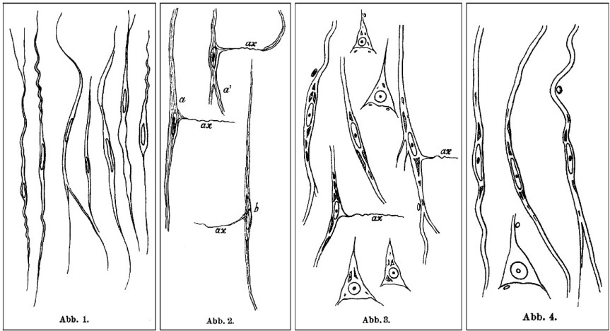
Every neuroscience or biology textbook will tell you that neurons communicate with one another in two ways: via chemical synapses (one neuron releases some substances that change the membrane potential of the receiving neuron) or via electrical synapses (neurons physically share some membrane proteins that allows the electrical impulse to go from one neuron to another). (Nota bene: for the taxonomic nitpickers, I decided that the other non-conventional forms of communication, like diffusion, fall in one or the other of the two categories above).

Now Qiu et al. (2015) have some evidence that there may be another way of neuronal chatting. Long ago (in 1924), Hans Berger observed that neurons have rhythmic and spontaneous electrical activity. The rhythmic activity of an entire population of neurons is called a wave and can be detected with rudimentary tools such as EEG. Qiu et al. reasoned that the speed of such a traveling wave is too slow to be explained by conventional neuronal communication (of which we know quite a lot).

So the researchers ran some computer simulations to test if diffuse electrical fields can explain the speed of a wave, instead of the conventional fast communications. In other words, can local, weak, slow endogenous fields sweep the brain by recruiting nearby neurons? The computer simulations said it is possible, with a very slow speed of 10 cm/s.

Then they recorded the electrical activity of mouse hippocampus isolated in a Petri dish. When the researchers blocked the electrical field there was a decrease in the speed of the wave, meaning the field is required for the wave speed observed.

To be fair, this is not a new idea; even in the early eighties these fields were suggested, because the wave persisted even when the other two ways of communicating have been blocked. And in the early noughts it was shown that a neural network is much more sensitive to electrical field manipulation than individual neurons. What makes this paper interesting is that the authors show that the endogenous electrical fields are not too weak to underlie the wave, as previously thought. The work has implications for the study of epilepsy, sleep, and memory.

Reference: Qiu C, Shivacharan RS, Zhang M, & Durand DM (2 December 2015). Can Neural Activity Propagate by Endogenous Electrical Field?, Journal of Neuroscience, 35(48): 15800-15811; doi: 10.1523/JNEUROSCI.1045-15.2015 Article | Full Text via Research Gate | ScienceAlert cover

By Neuronicus, 11 February 2016

CCL11 found in aged but not young blood inhibits adult neurogenesis

vil - Copy
Portion of Fig. 1 from Villeda et al. (2011, doi: 10.1038/nature10357) describing the parabiosis procedure. Basically, under complete anesthesia, the peritoneal membranes and the skins of the two mice were sutured together. The young mice were 3–4 months (yellow) and old mice were 18–20 months old (grey).

My last post was about parabiosis and its sparse revival as a technique in physiology experiments. Parabiosis is the surgical procedure that joins two living animals allowing them to share their circulatory systems. Here is an interesting paper that used the method to tackle blood’s contribution to neurogenesis.

Adult neurogenesis, that is the birth of new neurons in the adult brain, declines with age. This neurogenesis has been observed in some, but not all brain regions, called neurogenic niches.

Because these niches occur in blood-rich areas of the brain, Villeda et al. (2011) wondered if, in addition with the traditional factors required for neurogenesis like enrichment or running, blood factors may also have something to do with neurogenesis. The authors made a young and an old mouse to share their blood via parabiosis (see pic.).

Five weeks after the parabiosis procedure, the young mouse had decreased neurogenesis and the old mouse had increased neurogenesis compared to age-matched controls. To make sure their results are due to something in the blood, they injected plasma from an old mouse into a young mouse and that also resulted in reduced neurogenesis. Moreover, the reduced neurogenesis was correlated with impaired learning as shown by electrophysiological recordings from the hippocampus and from behavioral fear conditioning.

So what in the blood does it? The authors looked at 66 proteins found in the blood (I don’t know the blood make-up, so I can’t tell if 66 is a lot or not ) and noticed that 6 of these had increased levels in the blood of ageing mice whether linked by parabiosis or not. Out of these six, the authors focus on CCL11 (unclear to me why that one, my bet is that they tried the others too but didn’t have enough data). CCLL11 is a small signaling protein involved in allergies. So the authors injected it into young mice and Lo and Behold! there was decreased neurogenesis in their hippocampus. Maybe the vampires were onto something, whadda ya know? Just kidding… don’t go around sucking young people’s blood!

This paper covers a lot of work and, correspondingly, has no less than 23 authors and almost 20 Mb of supplemental documents! The story it tells is very interesting and as complete as it gets, covering many aspects of the problems investigated and many techniques to address those problems. Good read.

Reference: Villeda SA, Luo J, Mosher KI, Zou B, Britschgi M, Bieri G, Stan TM, Fainberg N, Ding Z, Eggel A, Lucin KM, Czirr E, Park JS, Couillard-Després S, Aigner L, Li G, Peskind ER, Kaye JA, Quinn JF, Galasko DR, Xie XS, Rando TA, Wyss-Coray T. (31 Aug 2011). The ageing systemic milieu negatively regulates neurogenesis and cognitive function. Nature. 477(7362):90-94. doi: 10.1038/nature10357. Article | FREE Fulltext PDF

By Neuronicus, 6 January 2016

The FIRSTS: Betz pyramidal neurons (1874)

Plate86betz
Betz cell in the dog cortex. Copyright: RA Bergman, AK Afifi, PM Heidger, & MP D’Alessandro. Pic taken from here.

Bigger that Purkinje cerebellar neurons, the Betz pyramidal neurons (aka the giant pyramidal neurons) can have up to 100 micrometers in diameter. They are located in the fifth layer of the grey matter in the primary motor cortex. And they were discovered by a Ukrainian who did not receive the just place he deserves in the history of neuroscience, as most books on the subject ignore him. So let’s give him some attention.

Vladimir Alekseyevich Betz (1834–1894) was a professor of anatomy and a histologist at the Kiev University. Just like with Pavlov, sometimes there is nothing spectacular or weird or bizarre in the life of a great thinker. Betz was a child of a relatively wealthy family, went to good schools, then to Medical School, where he showed interest in the anatomy department. He continued his postgraduate studies in the West (that is Germany and Austria) after which he returned home where he got a position as a professor at his Alma Mater where he stayed until he died of heart problems at the age of 60.

Vladimir_Betz
Vladimir Alekseyevich Betz (1834 – 1894), License: PD

During his PhD, which was on the blood flow in the liver, Betz discovered an interest in histology. He was unsatisfied with the quality of the existing staining methods, so he worked for years to improve the fixation and staining methods of the brain tissue. His new methods allowed the cutting and preserving very thin slices and then he described what he saw. But Betz’s genius was in linking his cortical cytoarchitechtonic findings with physiological function, dividing the cortex into the motor and sensory areas. He also made revolutionary observations of the anatomical organization and development and various pathologies.

Original reference (which I did not find): Betz W (1874). Anatomischer Nachweis zweier Gehirncentra. Centralblatt für die medizinischen Wissenschaften. 12:578-580, 595-599.

Reference: Kushchayev SV, Moskalenko VF, Wiener PC, Tsymbaliuk VI, Cherkasov VG, Dzyavulska IV, Kovalchuk OI, Sonntag VK, Spetzler RF, & Preul MC (Jan 2012, Epub 10 Nov 2011). The discovery of the pyramidal neurons: Vladimir Betz and a new era of neuroscience. Brain, 135(Pt 1):285-300. doi: 10.1093/brain/awr276.  ArticleFREE FULLTEXT PDF

By Neuronicus, 17 December 2015

Herpes viruses infect neurons

virus EBV
FIG. 3 from Jha et al. (2015). Wild-type EBV infection of primary human fetal neurons. Fluorescence microscopy was carried out at 2, 4, 6, and 8 days post infection to monitor for GFP expression (the fluorescent label). Microscopy images were captured at x20 magnification.

For some mysterious reason, whether Epstein-Barr virus (EBV) and Kaposi’s sarcoma-associated herpesvirus (KSHV) can infect neurons has not been established until now. Probably because some viruses from the same family do not infect neurons, so it was assumed that EPV and KSHV do not either.

Jha et al. (2015) cultured Sh-Sy5y neuroblastoma cells, teratocarcinoma Ntera2 neurons, and primary human fetal neurons in Petri dishes and then exposed them to these viruses. After infection, the authors did some fluorescence microscopy (they tagged the viruses with fluorescent dyes), real-time PCR (to confirm there was viral RNA in the cells), immunofluorescence assays (to see if the viral proteins are expressed) and Western blot analyses (to see if the specific viral antigens are made). All these showed that the viruses were happily multiplying in the cells.

Now here comes the significance of the study: EBV and KSHV are viruses associated with all sorts of nasty diseases like mononucleosis and cancers. EBV has also been associated with neurological disorders, like multiple sclerosis, Alzheimer’s, neuropathies, lymphomas etc. But the critical word here is “associated”. That is, they found these viruses in people suffering from those diseases. So the knowledge that these viruses infect neurons could point to a mechanism behind these associations. Unfortunately, EBV is present in 90-95% of the population of the world. Which means that you will find this virus in, let’s say, 9 out of 10 people suffering from Alzheimer’s, assuming normal distributions and random sampling. So the virus’s presence maybe completely unrelated to the disease. By the same rationale, you would find the virus in 9 out of 10 people found guilty of theft, for example. It would be then interesting to see find what is NOT associated with.

Caveat: I have not read the association studies, so my argument holds only if what they report is that people with disease X also have EBV. If they made, however, a comparisons and found out that people with disease X are significantly more likely to be infected with EBV than the ones without the disease, then the argument does not hold.

Reference: Jha HC, Mehta D, Lu J, El-Naccache D, Shukla SK, Kovacsics C, Kolson D, Robertson ES (1 Dec 2015). Gammaherpesvirus Infection of Human Neuronal Cells. MBio,  6(6). pii: e01844-15. doi: 10.1128/mBio.01844-15. Article | FREE FULLTEXT PDF | PsyPost cover

By Neuronicus, 7 December 2015

The FIRSTS: Axon description (1865)

Human medulla oblongata sectioned at the level of the olivary nuclei. Drawing by Deiters (1965). Original caption: Fig. 15. Durchschnitt der medulla oblongata des Menschen in der Höhe der Olive (OL). R.R Raphe, Hyi). Nervus hypoglossus. Vag. Nervus vagus, deren Kerne in V und H liegen, aber in der Zeichnung nicht feiner ausgeführt sind. Den Haupttlißil der Figur nimmt die Formatio reticularis ein mit ihren zerstreuten Ganglienzellen, und die Olive mit den zu ihr hinzutretenden Fasern des Stratum zonale; C.c crura cerebelli ad medullam oblongatam; P Pyramidenstrang.
Human medulla oblongata sectioned at the level of the olivary nuclei. Drawing by Deiters (1865). Original caption: “Fig. 15. Durchschnitt der medulla oblongata des Menschen in der Höhe der Olive (OL). R.R Raphe, Hyi). Nervus hypoglossus. Vag. Nervus vagus, deren Kerne in V und H liegen, aber in der Zeichnung nicht feiner ausgeführt sind. Den Haupttlißil der Figur nimmt die Formatio reticularis ein mit ihren zerstreuten Ganglienzellen, und die Olive mit den zu ihr hinzutretenden Fasern des Stratum zonale; C.c crura cerebelli ad medullam oblongatam; P Pyramidenstrang”.

In 1863, using the microscope, a german neuroanatomist from the University of Bonn by the name of Otto Friedrich Karl Deiters describes in exquisite detail the branch-like processes of the neuron (i. e. dendrites) and the long, single “axis cylinder” (i.e. axon). Deiters’ nucleus is named after him (the place where a good portion of the cranial nerve VIII ends).

The book with the findings is published in German, posthumously (in 1865), with preface and under the editorial guidance of Max Schultze, another famous German anatomist. I got the information from Debanne et al. (2011), which is nice review on axon physiology (my German is kindda rusty due to lack of use). But I got my hands on the original German book (see link below) and, like a kid that doesn’t know how to read yet, all I could do was marvel at the absolutely stunning drawings by OFK Deiters. Which are truly and unequivocally beautiful. See for yourself.

Neurons with axons and dendrites. Drawings buy Deiters (1865.)
Neurons with axons and dendrites. Drawings by Deiters (1865.)

Reference: Debanne D, Campanac E, Bialowas A, Carlier E, & Alcaraz G (April 2011). Axon physiology. Physiological Reviews, 91(2):555-602. doi: 10.1152/physrev.00048.2009. Article | FREE FULLTEXT PDF

Original citation: Deiters OFK (1865). Untersuchungen über Gehirn und Rückenmark des Menschen und der Säugethiere. Ed. Max Schultze, Braunschweig: Vieweg, 1865. doi: 10.5962/bhl.title.15270. Book | PDF

By Neuronicus, 24 October 2015

You were not my first choice either!

lego
Sexually receptive female mice prefer a Lego brick over a male if their prefrontal oxytocin neurons are silenced.

Over the past five years or so, dopamine stepped down from the role of the “love molecule” in favor of oxytocin, a hormone previously known mostly for its crucial role in pregnancy, labor, delivery, lactation, and breastfeeding. Since some interesting discoveries in monogamous vs. polygamous voles (a type of rodent) pointing to oxytocin as essential for bonding, many studies implicated the chemical in all sorts of behaviors, from autistic to trusting, from generosity to wound healing.

Nakajima, Görlich, & Heintz (2015) add to that body of knowledge by finding that only a small group of cells in the medial prefrontal cortex express oxytocin receptors: a subpopulation of somatostatin cortical interneurons. Moreover, these neurons are gender dimorphic, meaning they differ from male to female: the female ones have twice as many action potentials upon application of oxytocin as compared to male’s.

And here is the more interesting part:
– Females in the sexually receptive phase of their estrus whose oxytocin neurons were silenced preferred to interact with a Lego brick over a male mouse (which, as you might have guessed, in not what they typically choose).
– Females that were not in their sexually receptive phase when their oxytocin neurons were silenced still preferred to interact with a mouse (male or female) over the Lego brick.
– Silencing of other neurons had no effect on their choice.
– Silencing had no effect on the males.

Hm… there are such things out there as oxytocin inter-nasal sprays… How soon do you think until the homeopaths, naturopaths, and other charlatans market oxytocin as a potent aphrodisiac? And it will take some deaths until the slow machine of beaurocracy turns its wheels and tightens the regulations on the accessibility to the hormone. Until then… as the cartoons say, don’t try this at home! Go buy some flowers or something for your intended one… it would work better, trust me on this.

Reference: Nakajima M, Görlich A, & Heintz N (9 October 2014). Oxytocin modulates female sociosexual behavior through a specific class of prefrontal cortical interneurons. Cell. 159(2): 295–305. doi:10.1016/j.cell.2014.09.020. Article | FREE FULLTEXT PDF

By Neuronicus, 23 October 2015

Giving up? Your parvalbumin neurons may have something to do with it

Cartoon from http://i393.photobucket.com/albums/pp20/saisi24/dontgivedup.jpg, licensing unknown
Cartoon from Photobucket, licensing unknown.

One of the most ecologically-valid rodent models of depression is the learned helplessness paradigm. You get a rat or a mouse and you confine it in a cage with an electrified grid. Then you apply mild foot shocks at random intervals and of random duration for an hour (which is one session). The mouse initially tries to escape, but there is no escape; the whole floor is electrified. After a couple of sessions, the mouse doesn’t try to escape anymore; it gives up. Even when you put the mouse in a cage with an open door, so it can flee to no-pain freedom, it doesn’t attempt to do so. The interpretation is that the mouse has learned that it cannot control the environment, no matter what he does, he’s helpless, so why bother? Hence the name of the behavioral paradigm: learned helplessness.

All antidepressants on the market have been tested at one point or another against this paradigm; if the drug got the mouse to try to escape more, then the drug passed the test.

Just like in the higher vertebrate realm, there are a few animals who keep trying to escape longer than the others, before they too finally give up; we call these resilient.

Perova, Delevich, & Li (2015) looked at a type of neuron that may have something to do with the capacity of some of the mice to be resilient; the parvalbumin interneurons (PAI) from the medial prefrontal cortex (mPFC). These neurons produce GABA, the major inhibitory neurotransmitter in the brain, and modulates the activity of the nearby neurons. Thanks to the ability to genetically engineer mice to have a certain kind of cell fluoresce, the researchers were able to identify and subsequently record from and manipulate the function of the PAIs. These PAIs’ response to stimulation was weaker in helpless animals compared to resilient or controls. Also, inactivation of the PAI via a designer virus promotes helplessness.

Reference: Perova Z, Delevich K, & Li B (18 Feb 2015). Depression of Excitatory Synapses onto Parvalbumin Interneurons in the Medial Prefrontal Cortex in Susceptibility to Stress. The Journal of Neuroscience, 35(7):3201–3206. doi: 10.1523/JNEUROSCI.2670-14.2015. Article | FREE FULLTEXT PDF

By Neuronicus, 21 October 2015

Making new neurons from glia. Fully functional, too!

NeuroD1 transforms glial cells into neurons. Summary of the first portion of the Guo et al. (2014) paper.
Fig. 1. NeuroD1 transforms glial cells into neurons. Summary of the first portion of the Guo et al. (2014) paper.

Far more numerous than the neurons, the glial cells have many roles in the brain, one of which is protecting an injury site from being infected. In doing so, they fill up the injury space, but they also prohibit other neurons to grow there.

Guo et al. (2015) managed to turn these glial cells into neurons. Functioning neurons, that is, fully integrated within the rest of the brain network! They did it in a mouse model of stab injury and a mouse model of Alzeihmer’s in vivo. Because a mouse is not a man, they also metamorphosized human astrocytes into functioning glutamatergic neurons in a Petri dish, that is in vitro.

It is an elegant paper that crossed all the Ts and dotted all the Is. They went to a lot of double checking in different ways (see Fig. 1) to make sure their fantastic claim is for real (this kind of double, triple, quadruple checking is what gets a paper into the Big Name journals, like Cell). Needles to say, the findings show a tremendous therapeutic potential for people with central nervous system injuries, like paralyses, strokes, Alzheimer’s, Parkinson’s, Huntington, tumor resections, and many many more. Certainly worth a read!

Reference: Guo Z, Zhang L, Wu Z, Chen Y, Wang F, & Chen G (6 Feb 2014, Epub 19 Dec 2013). In vivo direct reprogramming of reactive glial cells into functional neurons after brain injury and in an Alzheimer’s disease model. Cell Stem Cell, 14(2):188-202. doi: 10.1016/j.stem.2013.12.001. Article | FREE FULLTEXT PDF | Cell cover

By Neuronicus, 18 October 2015

I can watch you learning

Human Stereotaxic System. Photo credit: The Mind Project

Recording directly form the healthy living human brain has always been a coveted goal of many neuroscientists, thus bypassing the limitations of non-invasive techniques or animal work. But, understandably, nobody would seek or grant approval for inserting an electrode in the healthy living human brain, on moral and ethical grounds. The next best thing is to insert an electrode into the not so healthy living human brain.

Ison, Quiroga, & Fried (2015) got lucky and gained access to 14 patients with intractable epilepsy that had electrodes implanted in their brain to find where the seizure focus is (for possible surgical resection later on). Using these electrodes, they recorded the activity of single neurons within the medial temporal lobe (MTL, a brain area paramount for learning) while the patients performed some simple association tasks. First, they presented images of places, people, and animals to the patients to see “which (if any) of the recorded neurons responded to a picture” (p. 220). When they got a neuron responding to something, they rushed out, did some data and image processing, and after an hour they started the experiment. Which was showing the patient the picture to which the neuron responded to (e.g. Stimulus 1 = patient’s daughter) overimposed on a background that the neuron did not respond to (e.g. Stimulus 2 = the Eiffel tower). After one single trial (although there was some variability), the patients learned the associations (i.e. Stimulus 3 = daughter in front of Eiffel tower) and this learning was mirrored by how the neuron responded. Namely, the neuron increased its activity by 200% to 400% (counted in spikes per second) when shown the previously un-responded to image alone (i.e. Stimulus 2).

Excerpt from Fig. 5 from Ison, Quiroga, & Fried (2015).
Excerpt from Fig. 5 from Ison, Quiroga, & Fried (2015). “Average normalized neural activity (black squares) and behavioral responses (green circles) to the non-preferred stimulus as a function of trial number. Data were aligned to the learning time (relative trial number 0)”, i.e. when they showed the composite image between Stimulus 1 and Stimulus 2. “Note that the neural activity follows the sudden increase in behavioral learning”.

The authors recorded from over 600 neurons from various MTL regions, out of which 51 responded to a Stimulus 1. From these, only half learned, that is, they increased their activity when Stimulus 2 was shown. For the picky specialist, the cells were both Type 1 and Type 2 neurons, located 6 in the hippocampus, 4 in the entorhinal cortex, 11 in the parahippocampal cortex, and 1 in the amygdala. And the authors controlled for familiarity, attentional demands, and other extraneous variables (with some very fancy and hard to follow stats, I might add).

The paper settles an old psychology dispute. Do we learn an association gradually or at once? In other words, do we learn gradually that A and B occur together, or do we learn that the first time we are shown A and B together and the next trials serve just to refine and consolidate the new knowledge? Ison, Quiroga, & Fried (2015) data show that learning happens at once, in an all-or-none fashion.

Reference: Ison, M. J., Quian Quiroga, R., & Fried, I. (1 July 2015). Rapid Encoding of New Memories by Individual Neurons in the Human Brain. Neuron, 87(1): 220-30. doi: 10.1016/j.neuron.2015.06.016. Article | FREE PDF

By Neuronicus, 4 October 2015

The FIRSTS: Adult neurogenesis (1962)

New neurons in the granular layer of the hippocampus. Fig. 30 from Altman & Das (1965).

I am starting a new category today: the Firsts. It will feature articles that showed something really interesting for the first time. Yes, all articles show something for the first time, that’s why they are published. But I have noticed either a lack of acknowledgment (“it is known that x”) or a disregard for the old papers (“doesn’t count if it’s before, say, 2001”), particularly among the new generation of scientists. So I will feature both the really big ones (e.g., first proof of adult neurogenesis) or the more obscure, but nonetheless, first in their field (e.g., first synthesis of morphine).

Today, first proof of adult neurogenesis. Altman (1962) wanted to see the kinetics of glial proliferation after brain injury. Glial cells are the other type of cells in the brain and they outnumber the neurons 10 to 1. Altman lesioned the rat lateral geniculate nucleus (a portion of the thalamus that deals primarily with vision) and then injected the rats with thymidine-H3, a dye that labels the newly formed cells. In addition to the expected glial proliferation, he also observed (by microscope and careful histology) that some neurons were also stained with the dye, which means that they were born after the injection. The new neurons were in many regions of the brain (so not only those associated with the lesioned area), including the cortical areas.

Altman followed up and three years later published the first comprehensive study of postnatal (not adult) neurogenesis in dendate gyrus of the hippocampus.

References:

  1. Altman, J. (30 March 1962). Are New Neurons Formed in the Brains of Adult Mammals?. Science, 135 (3509): 1127-1128. DOI: 10.1126/science.135.3509.1127. Article | PDF
  2. Altman, J, & Das, G. D. (June 1965). Autoradiographic and histological evidence of postnatal hippocampal neurogenesis in rats. The Journal of Comparative Neurology, 124 (3): 319 –335. DOI: 10.1002/cne.901240303. Article | PDF

by Neuronicus, 30 September 2015

I’m not like you, inside and out

Credit: Dawn of the Planet of the Apes
Screenshot from “Dawn of the Planet of the Apes” (Director: Tim Burton, 2001)

One mistake than many neuroscientists make (myself included) is the implicit assumption that the human brain is a rodent brain scaled-up, plus a few more bits. Here is a remainder that “a rat is not a monkey is not a human”, in the famous words of A. D. (Bud) Craig (2009).

Mohan et al. (2015) analyzed a portion of the brain (Brodmann area 21) obtained from 28 individuals that had to undergo neurosurgery and have it removed for various illnesses. Using some good microscopy, fancy statistics, and 3-D modeling, they reconstructed the shape of individual neurons from that region. The main finding is that 88% of human pyramidal neurons were distinctly different than their mouse or macaque counterparts. Also, they managed to record the electrical activity of these neurons in less than 10 minutes after resection. So it appears that this morphological distinctness of ours results in unique electrical properties of human neurons, which may account for the “distinct cognitive capabilities of humans”, as the authors put it.

Approximate location of Brodmann Area 21, corresponding to gyrus temporalis medium. Credit: Brain template to _DJ_; Area tracing to Neuronicus

Citation: Mohan, H., Verhoog, M. B., Doreswamy, K. K., Eyal, G., Aardse, R., Lodder, B. N., Goriounova, N. A., Asamoah, B., B. Brakspear, A. B. C., Groot, C., van der Sluis, S., Testa-Silva, G., Obermayer, J., Boudewijns, Z. S., Narayanan, R. T., Baayen, J. C., Segev, I., Mansvelder, H. D., de Kock, C. P. (28 August 2015; Epub ahead of print). Dendritic and Axonal Architecture of Individual Pyramidal Neurons across Layers of Adult Human Neocortex. Cerebral Cortex, 1-15. doi: 10.1093/cercor/bhv188. Article + FREE PDF

The song of a fly… the courtship of another

Drosophila melanogaster image illustrating sexual dimorphism and mating behavior. Credit: TheAlphaWolf (Wikimedia Commons)

Did you know that flies sing? True to the dictum that I just made up – ‘where is song, there is lust’ – it turns out not only that flies can sing, but they even have courtship songs! Granted, since they don’t have a larynx, the male flies sing by vibrating their wings in a certain way, which is unique to each fly species, and females listen with the feather-looking bit on top of their antennae, called arista. The behavior has generated enough research that a fairly hefty review about it has been published two years ago in Nature Reviews Neuroscience, pointing to a gene central to the male courtship circuitry and expressed only in the fly’s neurons, the fru gene (I bet it was called that way because when you make mutants you get fru/fru …).

Zhou et al. (2015) used a series of complicated experiments to successively activate or inhibit the neurons which express the fru gene, in order to identify the neural circuitry underlying hearing and processing the courtship songs. This circuitry is different in males and females, which makes sense since the serenading male expects different behaviors from his audience, depending on their sex; the listening males hurry to compete for the intended female and the females slow down and… listen carefully. Mind wondering: if I was the one serenading, wouldn’t I want to drive away the competitors, instead of drawing them in towards the object of my desire? Perhaps I want the competitors to also engage in courtship behavior so I can show off my wing vibrating prowess… Anyway, digression aside, in addition to figuring out which neuron does what, the authors managed to elicit courtship behavior in the listening males by optogenetically stimulating the 3rd and 4th order neurons in the newly identified circuit.

Besides being strangely interesting in itself, the research fills a gap in the understanding how courtship behavior is recognized, at least in fruit flies, which may be very useful information for other species as well, humans included.

Reference: Zhou, C., Franconville, R., Vaughan, A. G., Robinett, C. C., Jayaraman, V., & Baker, B. S. (21 September 2015). Central neural circuitry mediating courtship song perception in male Drosophila. Elife, 4:1-15. doi: 10.7554/eLife.08477. Article + FREE PDF

For the interested specialist, the MATLAB source code for analyzing calcium-imaging data can be found here.

By Neuronicus, 24 September 2015