
There are some neurons in the human brain that fire both when the person is doing some behavior and when watching that behavior performed by someone else. These cells are called mirror neurons and were first discovered in 1988 (see NOTE) by a group of researchers form the University of Parma, Italy, led by Giacomo Rizzolatti.
The discovery was done by accident. The researchers were investigating the activity of neurons in the rostral part of the inferior premotor cortex (riPM) of macaque monkeys with electrophysiological recordings. They placed a box in front of the monkey which had various objects in it. When the monkey pressed a switch, the content of the box was illuminated, then a door would open and the monkey reached for an object. Under each object was hidden a small piece of food. Several neurons were discharging when the animal was grasping the object. But the researchers noticed that some of these neurons ALSO fired when the monkey was motionless and watching the researcher grasping the objects!
The authors then did more motions to see when exactly the two neurons were firing, whether it’s related to the food or threatening gestures and so on. And then they recorded from some 182 more neurons while the monkey or the experimenter were performing hand actions with different objects. Importantly, they also did an electromyogram (EMG) and saw that when the neurons that were firing when the monkey was observing actions, the muscles did not move at all.
They found that some neurons responded to both when doing and seeing the actions, whereas some other neurons responded only when doing or only when seeing the actions. The neurons that are active when observing are called mirror neurons now. In 1996 they were identified also in humans with the help of positron emission tomography (PET).
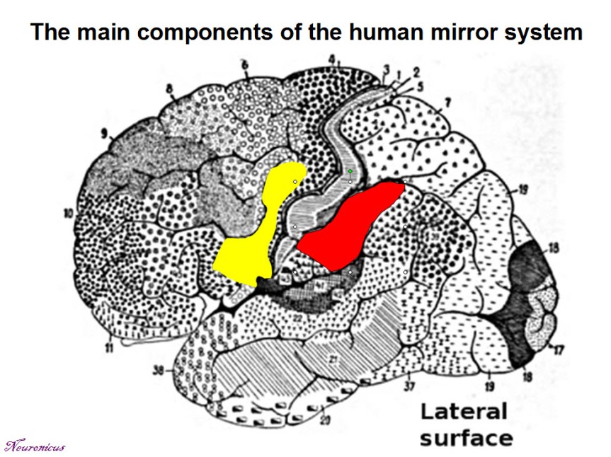
- In yellow, the frontal region; in red, the parietal region. Credits: Brain diagram by Korbinian Brodmann under PD license; Tracing by Neuronicus under PD license; Area identification and color coding after Rizzolatti & Fabbri-Destro (2010) © Springer-Verlag 2009.
It is tragicomical that the authors first submitted their findings to the most prestigious scientific journal, Nature, believing that their discovery is worth it, and rightfully so. But, Nature rejected their paper because of its “lack of general interest” (Rizzolatti & Fabbri-Destro, 2010)! Luckily for us, the editor of Experimental Brain Research, Otto Creutzfeld, did not share Nature‘s opinion.
Thousands of experiments followed the tremendous discovery of mirror neurons, even trying to manipulate their activity. Many researchers believe that the activity of the mirror neurons is fundamental for understanding the intentions of others, the development of theory of mind, empathy, the process of socialization, language development and even human self-awareness.
NOTE: Whenever possible, I try to report both the date of the discovery and the date of publication. Sometimes, the two dates can differ quite a bit. In this case, the discovery was done in 1988 and the publishing in 1992.
References:
- di Pellegrino G, Fadiga L, Fogassi L, Gallese V, & Rizzolatti G (October 1992). Understanding motor events: a neurophysiological study. Experimental Brain Research, 91(1):176-180. DOI: 10.1007/BF00230027. ARTICLE | Research Gate FULLTEXT PDF
- Rizzolatti G & Fabbri-Destro M (Epub 18 Sept 2009; January 2010). Mirror neurons: From discovery to autism. Experimental Brain Research, 200(3): 223-237. DOI: 10.1007/s00221-009-2002-3. ARTICLE | Research Gate FULLTEXT PDF
By Neuronicus, 15 July 2016


