Earliest memories

I found a rather old-ish paper that attempts to settle a curiosity regarding human memory: how far back can we remember?

MacDonald et al. (2000) got 96 participants to fill a 15-minute questionnaire about their demographics and their earliest memories. The New Zealand subjects were in their early twenties, a third of Maori descent, a third of European descent and the last third of Asian descent.

The Maori had the earliest memories, some of them as early as before they turned 1 year old, though the mean was 2 years and 8 months. Next came the Europeans with the mean of 3 years and a half, followed by the Asians with the mean of 4 and 9 months. Overall, most memories seem to occur between 3 and 4 years. There was no difference in gender except for the Asian group where the females reported much later memories, around 6 years.

The subjects were also required to indicate the source of the memory as being personal recollection, family story or photographs. About 86% reported it as personal recollection. The authors argue that even without the remaining 14% the results looks the same. I personally would have left those 14% out if they really don’t make a difference, it would have made the results much neater.

There are a few caveats that one must keep in mind with this kind of studies, the questionnaire studies. One of them is the inherent veracity problem: you rely on human honesty because there is no way to check the data for truth. The fact that the memory may be true or false would not matter for this study, but whether is a personal recollection or a family story would matter. So take the results at face value. Besides, human memory is extremely easy to manipulate, therefore some participants may actually believe that they ‘remember’ an event when in fact it was learned much later from relatives. I also have very early memories and while one of them I believe was told ad nauseam by family members at every family gathering so many times that I incorporated it as actual recollection, there are a couple that I couldn’t tell you for the life of me whether I remember them truly or they too have been subjected to family re-reminiscing.

Another issue might be the very small sample sizes with sub-groups. The authors divided their participants in many subgroups (whether they spoke English first, whether they were raised mainly by the mother etc.) that some subgroups ended up having 2 or 3 members, which is not enough to make a statistical judgement. Which also leads me to multiple comparisons adjustments, which should be more visible.

So not exactly the best paper ever written. Nevertheless, it’s an interesting paper in that even if it doesn’t really establish (in my opinion) when do most people have their earliest true memories, it does point to cultural differences in individuals’ earliest recollections. The authors speculate that that may be due to the emphasis put on detailed stories about personal experiences told by the mother in the early years in some cultures (here Maori) versus a lack of these stories in other cultures (here Asian).

105-copy

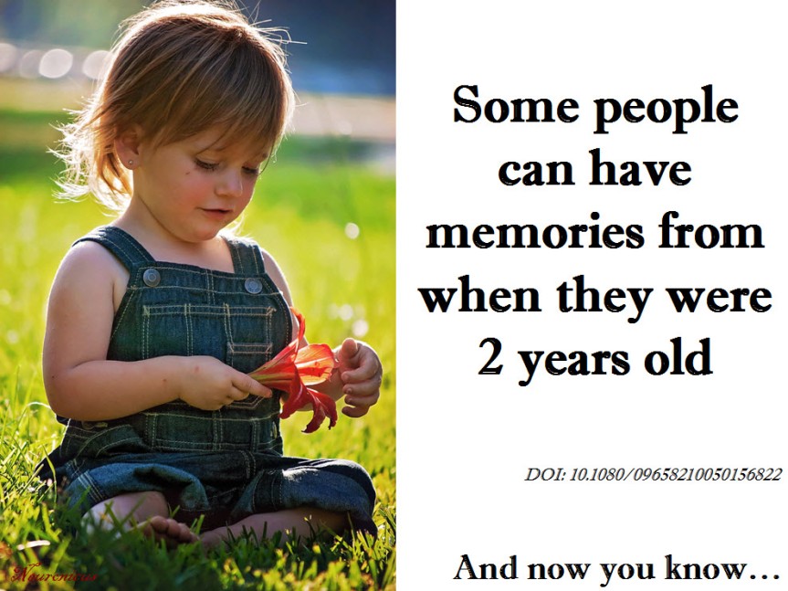
Reference: MacDonald S, Uesiliana K, & Hayne H. (Nov 2000). Cross-cultural and gender differences in childhood amnesia. Memory. 2000 Nov;8(6):365-76. PMID: 11145068, DOI: 10.1080/09658210050156822. ARTICLE | FULLTEXT PDF

By Neuronicus, 28 November 2016

Save

Amusia and stroke

Although a complete musical anti-talent myself, that doesn’t prohibit me from fully enjoying the works of the masters in the art. When my family is out of earshot, I even bellow – because it cannot be called music – from the top of my lungs alongside the most famous tenors ever recorded. A couple of days ago I loaded one of my most eclectic playlists. While remembering my younger days as an Iron Maiden concert goer (I never said I listen only to classical music :D) and screaming the “Fear of the Dark” chorus, I wondered what’s new on the front of music processing in the brain.

And I found an interesting recent paper about amusia. Amusia is, as those of you with ancient Greek proclivities might have surmised, a deficit in the perception of music, mainly the pitch but sometimes rhythm and other aspects of music. A small percentage of the population is born with it, but a whooping 35 to 69% of stroke survivors exhibit the disorder.

So Sihvonen et al. (2016) decided to take a closer look at this phenomenon with the help of 77 stroke patients. These patients had an MRI scan within the first 3 weeks following stroke and another one 6 months poststroke. They also completed a behavioral test for amusia within the first 3 weeks following stroke and again 3 months later. For reasons undisclosed, and thus raising my eyebrows, the behavioral assessment was not performed at 6 months poststroke, nor an MRI at the 3 months follow-up. It would be nice to have had behavioral assessment with brain images at the same time because a lot can happen in weeks, let alone months after a stroke.

Nevertheless, the authors used a novel way to look at the brain pictures, called voxel-based lesion-symptom mapping (VLSM). Well, is not really novel, it’s been around for 15 years or so. Basically, to ascertain the function of a brain region, researchers either get people with a specific brain lesion and then look for a behavioral deficit or get a symptom and then they look for a brain lesion. Both approaches have distinct advantages but also disadvantages (see Bates et al., 2003). To overcome the disadvantages of these methods, enter the scene VLSM, which is a mathematical/statistical gimmick that allows you to explore the relationship between brain and function without forming preconceived ideas, i.e. without forcing dichotomous categories. They also looked at voxel-based morphometry (VBM), which a fancy way of saying they looked to see if the grey and white matter differ over time in the brains of their subjects.

After much analyses, Sihvonen et al. (2016) conclude that the damage to the right hemisphere is more likely conducive to amusia, as opposed to aphasia which is due mainly to damage to the left hemisphere. More specifically,

“damage to the right temporal areas, insula, and putamen forms the crucial neural substrate for acquired amusia after stroke. Persistent amusia is associated with further [grey matter] atrophy in the right superior temporal gyrus (STG) and middle temporal gyrus (MTG), locating more anteriorly for rhythm amusia and more posteriorly for pitch amusia.”

The more we know, the better chances we have to improve treatments for people.

104-copy

unless you’re left-handed, then things are reversed.

References:

1. Sihvonen AJ, Ripollés P, Leo V, Rodríguez-Fornells A, Soinila S, & Särkämö T. (24 Aug 2016). Neural Basis of Acquired Amusia and Its Recovery after Stroke. Journal of Neuroscience, 36(34):8872-8881. PMID: 27559169, DOI: 10.1523/JNEUROSCI.0709-16.2016. ARTICLE  | FULLTEXT PDF

2.Bates E, Wilson SM, Saygin AP, Dick F, Sereno MI, Knight RT, & Dronkers NF (May 2003). Voxel-based lesion-symptom mapping. Nature Neuroscience, 6(5):448-50. PMID: 12704393, DOI: 10.1038/nn1050. ARTICLE

By Neuronicus, 9 November 2016

Save

Pic of the Day: Russell on stupid

russell-copy-2

Reference: Russell, B. (10 May 1933). “The Triumph of Stupidity”. In: H. Ruja (Ed.), Mortals and Others: Bertrand Russell’s American Essays, Volume 2, 1931–1935.

The history of the quote and variations of it by others can be found on the Quote Investigator.

By Neuronicus, 6 November 2016

Pic of the Day: Neil on teaching creationism

104neil-copy
Dr. deGrasse Tyson’s picture is from Wikimedia released under PD and the quote is from a “Letter to the Editor” of New York Times retrieved from the Hayden Planetarium website on Nov. 2, 2016.