Air pollution and mortality

The authors used a chemical transport model called GEOS-Chem to estimate the global exposure levels to airborne particulate matter derived from fossil fuel combustion. It’s a worldwide used model for estimating dispersion, for example the simulation of surface pollination. The model has been validated, meaning its predictions were compared with the reality and they matched. Levels of fossil fuels emission were inputted from publicly available data “from multiple sectors (power generation, industry, ships, aircraft, ground transportation, backup generators, kerosene, oil/gas extraction), detailed oxidant-aerosol chemistry, and reanalysis meteorology from the NASA Global Modeling and Assimilation Office” (page 5 of the manuscript). The mortality due to air pollution was computed by starting with the classical risk assessment analysis from epidemiological research and then adjusting for other variables like in-door particulate exposure (smoking) and so on. It sounds straightforward enough but the math and stats involved are very complicated. You have to take into consideration concentration, exposure time, and other factors that affect the outcome.

In addition, the authors calculated the mortality caused by fossil-fuel air pollution in children between 0 and 4 years of age, which was 876 in North America, 747 in South America, and 605 in Europe, annually. Pollution data is from 2012, mortality from 2015. The authors are hopeful that aggressive regulation designed to curb emissions in the last few years may have already lowered these numbers. Policy-makers, take note!

REFERENCE: Vohra K, Vodonos A, Schwartz J, Marais EA, Sulprizio MP, & Mickley LJ (Apr 2021, Epub 9 Feb 2021). Global mortality from outdoor fine particle pollution generated by fossil fuel combustion: Results from GEOS-Chem. Environmental Research, Volume 195, 110754. PMID: 33577774, DOI: 10.1016/j.envres.2021.110754, ARTICLE.

By Neuronicus, 11 March 2021

Penis Size

For whoever needs to be reassured…

L is for length and W is for… circumference. I could have corrected the pic, but it generated far more interesting comments as it is on social media.

REFERENCE: Veale D, Miles S, Bramley S, Muir G., & Hodsoll J (Jun 2015).  Am I normal? A systematic review and construction of nomograms for flaccid and erect penis length and circumference in up to 15 521 men. British Journal of Urology International, 115(6):978-986. PMID: 25487360, DOI: 10.1111/bju.13010. ARTICLE | FREE FULLTEXT PDF | Editorial | Science cover

By Neuronicus, 11 November, 2020

Chloroquine-induced psychosis

In the past few days, a new hot subject has gripped the attention of various media and concerned the medical doctors, as if they don’t have enough to deal with: chloroquine. That is because the President of the U.S.A., Donald Trump, endorsed chloroquine as treatment of COVID-19, a “game changer”, despite his very own director of the National Institute of Allergy and Infectious Diseases (NIAID), Dr. Anthony Fauci, very emphatically and vehemently denying that the promise of (hydroxy)chloroquine is beyond anecdotal (see the White House briefing transcript here).

161 - Copy

Many medical doctors spoke out urging caution against the drug, particularly against the combination the President endorses: hydroxychloroquine + azithromycin. As I understand it, this combo can be lethal as it can lead to fatal arrhythmia.

As for the (hydroxy)cloroquine’s possibility to help treat COVID-19, the jury is still out. Far out. Meaning that there have been a few interesting observations of the drugs working in a Petri dish (Liu et al. 2020, Wang et al., 2020), but as any pharma company knows, there is a long and perilous way from Petri dishes to pharmacies. To be precise, only 1 in 5000 drugs get from pre-clinical trials to approval and it takes about 12 years for this process to be completed (Kaljevic et al., 2004). The time is so long not because red tape, as some would deplore, but because it takes time to see what it does in humans (Phase 0), what doses are safe and don’t kill you (Phase 1), does it work at all for the intended disease (Phase 2), compare it with other drugs and evaluate the long-term side effects (Phase 3) and, finally, to see the risks and benefits of this drug (Phase 4). While we could probably get rid of Phase 0 and 4 when there is such a pandemic, there is no way I would submit my family to anything that hasn’t passed phases 1, 2, and 3. And those take years. With all the money that a nation-state has, it would still take 18 months to do it semi-properly.

Luckily for all of us, chloroquine is a very old and established anti-malarial medicine, and as such we can safely dispense of Phases 0, 1, and 4, which is fine. So we can start Phase 2 with (hydroxy)chloroquine. And that is exactly what WHO and several others are doing right now. But we don’t have enough data. We haven’t done it yet. So one can hope as much as they want, but that doesn’t make it faster.

Unfortunately – and here we go to the crux of the post -, following the President’s endorsement, many started to hoard chloroquine. Particularly the rich who can afford to “convince” an MD to write them a script for it. In countries where chloroquine is sold without prescription, like Nigeria, where it is used for arthritis, people rushed to clear the pharmacies and some didn’t just stockpiled it, but they took it without reason and without knowing the dosage. And they died. [EDIT, 23 March 2020. If you think that wouldn’t ever happen in the land of the brave, think again, as the first death to irresponsible taking chloroquine just happened in the USA]. In addition, the chloroquine hoarding in US by those who can afford it (is about $200 for 50 pills) lead to lack of supply for those who really need it, like lupus or rheumatology patients.

For those who blindly hoard or take chloroquine without prescription, I have a little morsel of knowledge to impart. Remember I am not an MD; I hold a PhD in neuroscience. So I’ll tell you what my field knows about chloroquine.

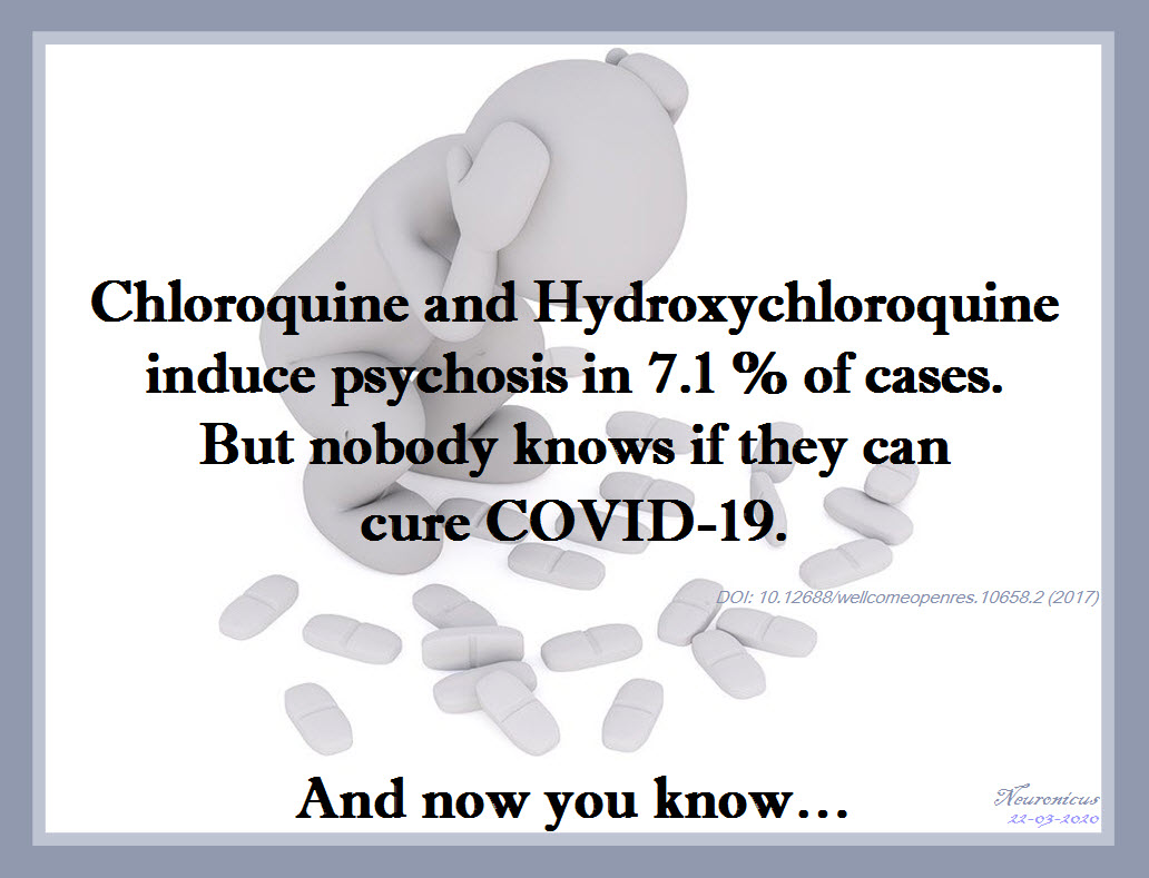
Both chloroquine and hydroxychloroquine can cause severe psychosis.

That’s right. More than 7.1 % of people who took chloroquine as prophylaxis or for treatment of malaria developed “mental and neurological manifestations” (Bitta et al.,  2017). “Hydroxychloroquine was associated with the highest prevalence of mental neurological manifestations” (p. 12). The phenomenon is well-reported, actually having its own syndrome name: “chloroquine-induced psychosis”. It was observed more than 50 years ago, in 1962 (Mustakallio et al., 1962). The mechanisms are unclear, with several hypotheses being put forward, like the drugs disrupting the NMDA transmission, calcium homeostasis, vacuole exocytosis or some other mysterious immune or transport-related mechanism. Because the symptoms are so acute, so persistent and so diverse than more than one brain neurotransmitter system must be affected.

Chloroquine-induced psychosis has sudden onset, within 1-2 days of ingestion. The syndrome presents with paranoid ideation, persecutory delusions, hallucinations, fear, confusion, delirium, altered mood, personality changes, irritability, insomnia, suicidal ideation, and violence (Biswas et al., 2014, Mascolo et al., 2018). All these at moderately low or therapeutically recommended doses (Good et al., 1982). One or two pills can be lethal in toddlers (Smith & Klein-Schwartz, 2005). The symptoms persist long after the drug ingestion has stopped (Maxwell et al., 2015).

Still want to take it “just in case”?

162 chloro - Copy

P.S. A clarification: the chemical difference between hydroxychloroquine and chloroquine is only one hydroxyl group (OH). Both are antimalarial and both have been tested in vitro for COVID-19. There are slight differences between them in terms of toxicity, safety and even mechanisms, but for the intents of this post I have treated them as one drug, since both produce psychosis.

REFERENCES:

1) Biswas PS, Sen D, & Majumdar R. (2014, Epub 28 Nov 2013). Psychosis following chloroquine ingestion: a 10-year comparative study from a malaria-hyperendemic district of India. General Hospital Psychiatry, 36(2): 181–186. doi: 10.1016/j.genhosppsych.2013.07.012, PMID: 24290896 ARTICLE

2) Bitta MA, Kariuki SM, Mwita C, Gwer S, Mwai L, & Newton CRJC (2 Jun 2017). Antimalarial drugs and the prevalence of mental and neurological manifestations: A systematic review and meta-analysis. Version 2. Wellcome Open Research, 2(13): 1-20. PMCID: PMC5473418, PMID: 28630942, doi: 10.12688/wellcomeopenres.10658.2 ARTICLE|FREE FULLTEXT PDF

4) Good MI & Shader RI. Lethality and behavioral side effects of chloroquine (1982). Journal of Clinical Psychopharmacology, 2(1): 40–47. doi: 10.1097/00004714-198202000-00005, PMID: 7040501. ARTICLE

3) Kraljevic S, Stambrook PJ, & Pavelic K (Sep 2004). Accelerating drug discovery. EMBO Reports, 5(9): 837–842. doi: 10.1038/sj.embor.7400236, PMID: 15470377, PMCID: PMC1299137. ARTICLE| FREE FULLTEXT PDF

4) Mascolo A, Berrino PM, Gareri P, Castagna A, Capuano A, Manzo C, & Berrino L. (Oct 2018, Epub 9 Jun 2018). Neuropsychiatric clinical manifestations in elderly patients treated with hydroxychloroquine: a review article. Inflammopharmacology, 26(5): 1141-1149. doi: 10.1007/s10787-018-0498-5, PMID: 29948492. ARTICLE

5) Maxwell NM, Nevin RL, Stahl S, Block J, Shugarts S, Wu AH, Dominy S, Solano-Blanco MA, Kappelman-Culver S, Lee-Messer C, Maldonado J, & Maxwell AJ (Jun 2015, Epub 9 Apr 2015). Prolonged neuropsychiatric effects following management of chloroquine intoxication with psychotropic polypharmacy. Clinical Case Reports, 3(6): 379-87. doi: 10.1002/ccr3.238, PMID: 26185633. ARTICLE | FREE FULLTEXT PDF

6) Mustakallio KK, Putkonen T, & Pihkanen TA (1962 Dec 29). Chloroquine psychosis? Lancet, 2(7270): 1387-1388. doi: 10.1016/s0140-6736(62)91067-x, PMID: 13936884. ARTICLE

7) Smith ER & Klein-Schwartz WJ (May 2005). Are 1-2 dangerous? Chloroquine and hydroxychloroquine exposure in toddlers. The Journal of Emergency Medicine, 28(4): 437-443. doi: 10.1016/j.jemermed.2004.12.011, PMID: 15837026. ARTICLE

Studies about chloroquine and hydoxychloroquine on SARS-Cov2 in vitro:

  • Gautret P, Lagier J-C, Parola P, Hoang VT, Meddeb L, Mailhe M, Doudier B, Courjon J, Giordanengo V, Esteves Vieira V, Tissot Dupont H,Colson SEP, Chabriere E, La Scola B, Rolain J-M, Brouqui P,  Raoult D. (20 March 2020). Hydroxychloroquine and azithromycin as a treatment of COVID-19: results of an open-label non-randomized clinical trial. International Journal of Antimicrobial Agents, PII:S0924-8579(20)30099-6, https://doi.org/10.1016/j.ijantimicag.2020.105949. ARTICLE | FREE FULLTEXT PDF

These studies are also not peer reviewed or at the very least not properly peer reviewed. I say that so as to take them with a grain of salt. Not to criticize in the slightest. Because I do commend the speed with which these were done and published given the pandemic. Bravo to all the authors involved (except maybe the last one f it proves to be fraudulent). And also a thumbs up to the journals which made the data freely available in record time. Unfortunately, from these papers to a treatment we still have a long way to go.

By Neuronicus, 22 March 2020

High fructose corn syrup IS bad for you

Because I cannot leave controversial things well enough alone – at least not when I know there shouldn’t be any controversy – my ears caught up with my tongue yesterday when the latter sputtered: “There is strong evidence for eliminating sugar from commonly used food products like bread, cereal, cans, drinks, and so on, particularly against that awful high fructose corn syrup”. “Yeah? You “researched” that up, haven’t you? Google is your bosom friend, ain’t it?” was the swift reply. Well, if you get rid of the ultra-emphatic air-quotes flanking the word ‘researched’ and replace ‘Google’ with ‘Pubmed’, then, yes, I did researched it and yes, Pubmed is my bosom friend.

Initially, I wanted to just give you all a list with peer-reviewed papers that found causal and/or correlational links between high fructose corn syrup (HFCS) and weight gain, obesity, type 2 diabetes, cardiovascular disease, fatty liver disease, metabolic and endocrine anomalies and so on. But there are way too many of them; there are over 500 papers on the subject in Pubmed only. And most of them did find that HFCS does nasty stuff to you, look for yourselves here. Then I thought to feature a paper showing that HFCS is differently metabolized than the fructose from fruits, because I keep hearing that lie perpetrated by the sugar and corn industries that “sugar is sugar” (no, it’s not! Demonstrably so!), but I doubt my yesterday’s interlocutor would care about liver’s enzymatic activity and other chemical processes with lots of acronyms. So, finally, I decided to feature a straight forward, no-nonsense paper, published recently, done at a top tier university, with human subjects, so I won’t hear any squabbles.

Price et al. (2018) studied 49 healthy subjects aged age 18–40 yr, of normal and stable body weight, and free from confounding medications or drugs, whose physical activity and energy-balanced meals were closely monitored. During the study, the subjects’ food and drink intake as well as their timing were rigorously controlled. The researchers varied only the beverages between groups, in such a way that one group received a drink sweetened with HFCS-55 (55% fructose, 45% glucose, as the one used in commercially available drinks) with every controlled meal, whereas the other group received an identical drink in size (adjusted for their energy requirements in such a way that it provided the same 25% of it), but sweetened with aspartame. The study lasted two weeks. No other beverage was allowed, including fruit juice. Urine samples were collected daily and blood samples 4 times per day.

There was a body weight increase of 810 grams (1.8 lb) in subjects consuming HFCS-sweetened beverages for 2 weeks when compared with aspartame controls. The researches also found differences in the levels of a whole host of acronyms (ppTG, ApoCIII, ApoE, OEA, DHEA, DHG, if you must know) involved in a variety of nasty things, like obesity, fatty liver disease, atherosclerosis, cardiovascular disease, stroke, diabetes, even Alzheimer’s.

This study is the third part of a larger NIH-funded study which investigates the metabolic effects of consuming sugar-sweetened beverages in about 200 participants over 5 years, registered at clinicaltrials.gov as NCT01103921. The first part (Stanhope et al., 2009) reported that consuming fructose-sweetened, not glucose-sweetened, beverages increases visceral adiposity and lipids and decreases insulin sensitivity in overweight/obese humans” (title), and the second part (Stanhope et al., 2015) found that “consuming beverages containing 10%, 17.5%, or 25% of energy requirements from HFCS produced dose-dependent increases in circulating lipid/lipoprotein risk factors for cardiovascular disease and uric acid within 2 weeks” (Abstract). They also found a dose-dependant increase in body weight, but in those subjects the results were not statistically significant (p = 0.09) after correcting for multiple comparisons. But I’ll bet that if/when the authors will publish all the data in one paper at the end of clinical trials they will have more statistical power and the trend in weight gain more obvious, as in the present paper.  Besides, it looks like there may be more than three parts to this study anyway.

The adverse effects of a high sugar diet, particularly in HFCS, are known to so many researchers in the field that they have been actually compiled in a name: the “American Lifestyle-Induced Obesity Syndrome model, which included consumption of a high-fructose corn syrup in amounts relevant to that consumed by some Americans” (Basaranoglu et al., 2013). It doesn’t refer only to increases in body weight, but also type 2 diabetes, cardiovascular disease, hypertriglyceridemia, fatty liver disease, atherosclerosis, gout, etc.

The truly sad part is that avoiding added sugars in diets in USA is impossible unless you do all – and I mean all – your cooking home, including canning, jamming, bread-making, condiment-making and so on, not just “Oh, I’ll cook some chicken or ham tonight” because in that case you end up using canned tomato sauce (which has added sugar), bread crumbs (which have added sugar), or ham (which has added sugar), salad dressing (which has sugar) and so on. Go on, check your kitchen and see how many ingredients have sugar in them, including any meat products short of raw meat. If you never read the backs of the bottles, cans, or packages, oh my, are you in for a big surprise if you live in USA…

There are lot more studies out there on the subject, as I said, of various levels of reading difficulty. This paper is not easy to read for someone outside the field, that’s for sure. But the main gist of it is in the abstract, for all to see.

150 hfcs - Copy

P.S. 1. Please don’t get me wrong: I am not against sugar in desserts, let it be clear. Nobody makes a more mean sweetalicious chocolate cake or carbolicious blueberry muffin than me, as I have been reassured many times. But I am against sugar in everything. You know I haven’t found in any store, including high-end and really high-end stores a single box of cereal of any kind without sugar? Just for fun, I’d like to be a daredevil and try it once. But there ain’t. Not in USA, anyway. I did find them in EU though. But I cannot keep flying over the Atlantic in the already crammed at premium luggage space unsweetened corn flakes from Europe which are probably made locally, incidentally and ironically, with good old American corn.

P.S. 2 I am not so naive, blind, or zealous to overlook the studies that did not find any deleterious effects of HFCS consumption. Actually, I was on the fence about HFCS until about 10 years ago when the majority of papers (now overwhelming majority) was showing that HFCS consumption not only increases weight gain, but it can also lead to more serious problems like the ones mentioned above. Or the few papers that say all added sugar is bad, but HFCS doesn’t stand out from the other sugars when it comes to disease or weight gain. But, like with most scientific things, the majority has it its way and I bow to it democratically until the new paradigm shift. Besides, the exposés of Kearns et al. (2016a, b, 2017) showing in detail and with serious documentation how the sugar industry paid prominent researchers for the past 50 years to hide the deleterious effects of added sugar (including cancer!) further cemented my opinion about added sugar in foods, particularly HFCS.

References:

  1. Price CA, Argueta DA, Medici V, Bremer AA, Lee V, Nunez MV, Chen GX, Keim NL, Havel PJ, Stanhope KL, & DiPatrizio NV (1 Aug 2018, Epub 10 Apr 2018). Plasma fatty acid ethanolamides are associated with postprandial triglycerides, ApoCIII, and ApoE in humans consuming a high-fructose corn syrup-sweetened beverage. American Journal of Physiology. Endocrinology and Metabolism, 315(2): E141-E149. PMID: 29634315, PMCID: PMC6335011 [Available on 2019-08-01], DOI: 10.1152/ajpendo.00406.2017. ARTICLE | FREE FULTEXT PDF
  1. Stanhope KL1, Medici V2, Bremer AA2, Lee V2, Lam HD2, Nunez MV2, Chen GX2, Keim NL2, Havel PJ (Jun 2015, Epub 22 Apr 2015). A dose-response study of consuming high-fructose corn syrup-sweetened beverages on lipid/lipoprotein risk factors for cardiovascular disease in young adults. The American Journal of Clinical Nutrition, 101(6):1144-54. PMID: 25904601, PMCID: PMC4441807, DOI: 10.3945/ajcn.114.100461. ARTICLE | FREE FULTEXT PDF
  1. Stanhope KL1, Schwarz JM, Keim NL, Griffen SC, Bremer AA, Graham JL, Hatcher B, Cox CL, Dyachenko A, Zhang W, McGahan JP, Seibert A, Krauss RM, Chiu S, Schaefer EJ, Ai M, Otokozawa S, Nakajima K, Nakano T, Beysen C, Hellerstein MK, Berglund L, Havel PJ (May 2009, Epub 20 Apr 2009). Consuming fructose-sweetened, not glucose-sweetened, beverages increases visceral adiposity and lipids and decreases insulin sensitivity in overweight/obese humans. The Journal of Clinical Investigation,119(5):1322-34. PMID: 19381015, PMCID: PMC2673878, DOI:10.1172/JCI37385. ARTICLE | FREE FULTEXT PDF

(Very) Selected Bibliography:

Bocarsly ME, Powell ES, Avena NM, Hoebel BG. (Nov 2010, Epub 26 Feb 2010). High-fructose corn syrup causes characteristics of obesity in rats: increased body weight, body fat and triglyceride levels. Pharmacology, Biochemistry, and Behavior, 97(1):101-6. PMID: 20219526, PMCID: PMC3522469, DOI: 10.1016/j.pbb.2010.02.012. ARTICLE | FREE FULLTEXT PDF

Kearns CE, Apollonio D, Glantz SA (21 Nov 2017). Sugar industry sponsorship of germ-free rodent studies linking sucrose to hyperlipidemia and cancer: An historical analysis of internal documents. PLoS Biology, 15(11):e2003460. PMID: 29161267, PMCID: PMC5697802, DOI: 10.1371/journal.pbio.2003460. ARTICLE | FREE FULTEXT PDF

Kearns CE, Schmidt LA, Glantz SA (1 Nov 2016). Sugar Industry and Coronary Heart Disease Research: A Historical Analysis of Internal Industry Documents. JAMA Internal Medicine, 176(11):1680-1685. PMID: 27617709, PMCID: PMC5099084, DOI: 10.1001/jamainternmed.2016.5394. ARTICLE | FREE FULTEXT PDF

Mandrioli D, Kearns CE, Bero LA (8 Sep 2016). Relationship between Research Outcomes and Risk of Bias, Study Sponsorship, and Author Financial Conflicts of Interest in Reviews of the Effects of Artificially Sweetened Beverages on Weight Outcomes: A Systematic Review of Reviews. PLoS One, 11(9):e0162198.PMID: 27606602, PMCID: PMC5015869, DOI: 10.1371/journal.pone.0162198. ARTICLE | FREE FULTEXT PDF

By Neuronicus, 22 March 2019

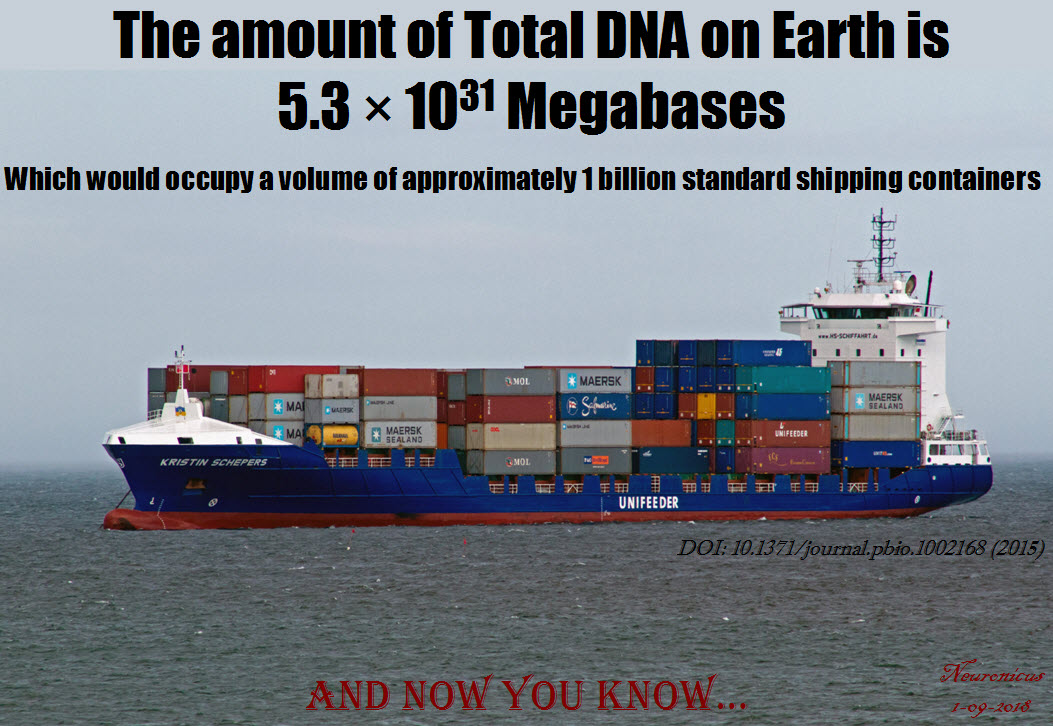
Pic of the day: Total amount of DNA on Earth

139 DNA amount, better font - Copy

Approximately… give or take…

REFERENCE: Landenmark HKE, Forgan DH, & Cockell CS (11 Jun 2915). An Estimate of the Total DNA in the Biosphere. PLoS Biology, 13(6): e1002168. PMCID: PMC4466264, PMID: 26066900, DOI: 10.1371/journal.pbio.1002168. ARTICLE | FREE FULLTEXT PDF

By Neuronicus, 1 September 2018

The FIRSTS: The cause(s) of dinosaur extinction

A few days ago, a follower of mine gave me an interesting read from The Atlantic regarding the dinosaur extinction. Like many of my generation, I was taught in school that dinosaurs died because an asteroid hit the Earth. That led to a nuclear winter (or a few years of ‘nuclear winters’) which killed the photosynthetic organisms, and then the herbivores didn’t have anything to eat so they died and then the carnivores didn’t have anything to eat and so they died. Or, as my 4-year-old puts it, “[in a solemn voice] after the asteroid hit, big dusty clouds blocked the sun; [in an ominous voice] each day was colder than the previous one and so, without sunlight to keep them alive [sad face, head cocked sideways], the poor dinosaurs could no longer survive [hands spread sideways, hung head] “. Yes, I am a proud parent. Now I have to do a sit-down with the child and explain that… What, exactly?

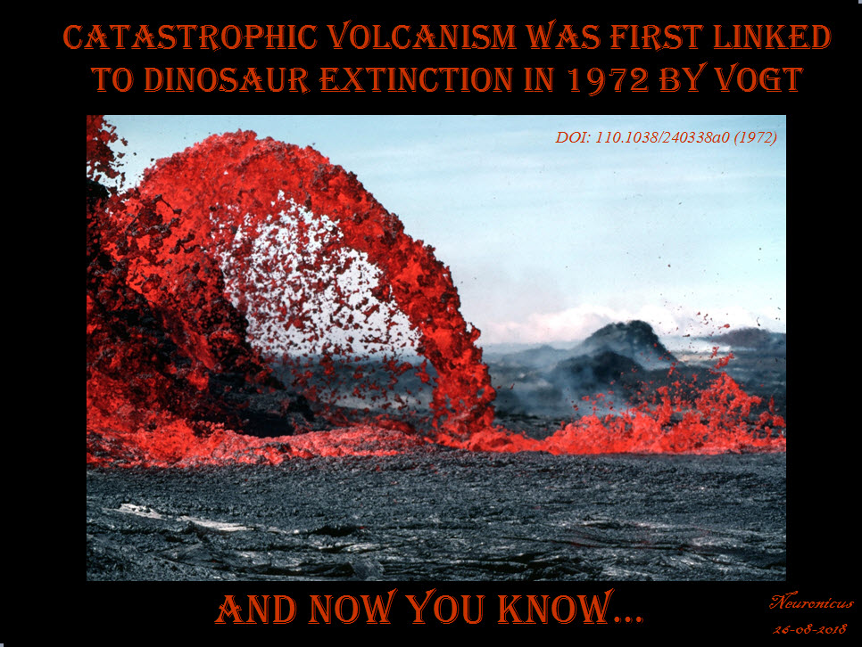
Well, The Atlantic article showcases the struggles of a scientist – paleontologist and geologist Gerta Keller – who doesn’t believe the mainstream asteroid hypothesis; rather she thinks there is enough evidence to point out that extreme volcano eruptions, like really extreme, thousands of times more powerful than anything we know in the recorded history, put out so much poison (soot, dust, hydrofluoric acid, sulfur, carbon dioxide, mercury, lead, and so on) in the atmosphere that, combined with the consequent dramatic climate change, killed the dinosaurs. The volcanoes were located in India and they erupted for hundreds of thousands of years, but most violent eruptions, Keller thinks, were in the last 40,000 years before the extinction. This hypothesis is called the Deccan volcanism from the region in India where these nasty volcanoes are located, first proposed by Vogt (1972) and Courtillot et al. (1986).
138- Vogt - Copy.jpg

So which is true? Or, rather, because this is science we’re talking about, which hypothesis is more supported by the facts: the volcanism or the impact?

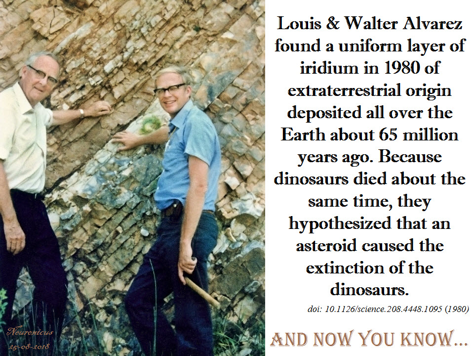
The impact hypothesis was put forward in 1980 when Walter Alvarez, a geologist, noticed a thin layer of clay in rocks that were about 65 million years old, which coincided with the time when the dinosaurs disappeared. This layer is on the KT boundary (sometimes called K-T, K-Pg, or KPB, looks like the biologists are not the only ones with acronym problems) and marks the boundary between the Cretaceous and Paleogenic geological periods (T is for Triassic, yeah, I know). Walter asked his father, the famous Nobel Prize physicist Louis Alvarez, to take a look at it and see what it is. Alvarez Sr. analyzed it and decided that the clay contains a lot of iridium, dozens of times more than expected. After gathering more samples from Europe and New Zealand, they published a paper (Alvarez et al., 1980) in which the scientists reasoned that because Earth’s iridium is deeply buried in its bowels and not in its crust, this iridium at the K-Pg boundary is of extraterrestrial origin, which could be brought here only by an asteroid/comet. This is also the paper in which it was put forth for the first time the conjecture that the asteroid impact killed the dinosaurs, based on the uncanny coincidence of timing.
138-alvarez - Copy

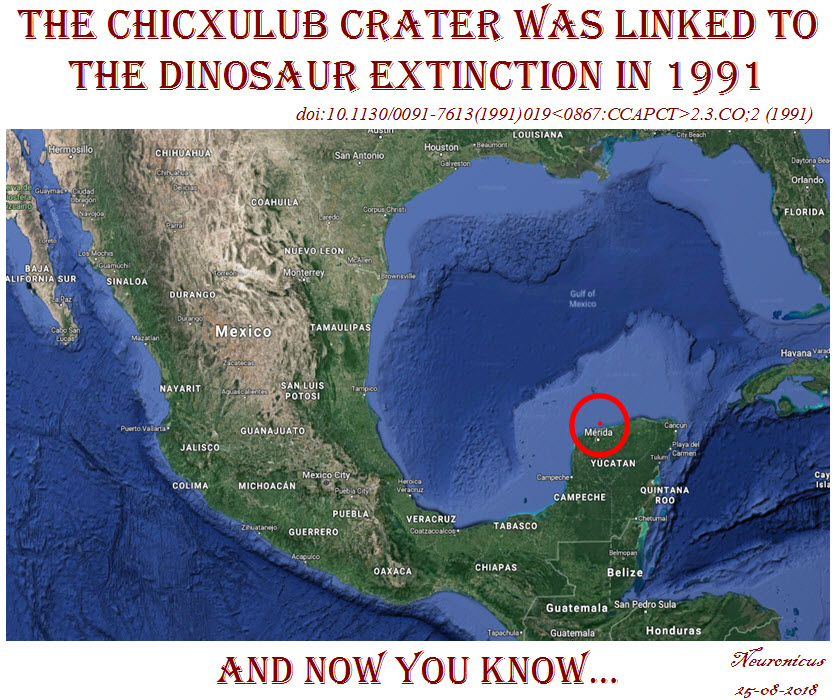
The discovery of the Chicxulub crater in Mexico followed a more sinuous path because the geophysicists who first discovered it in the ’70s were working for an oil company, looking for places to drill. Once the dinosaur-died-due-to-asteroid-impact hypothesis gained popularity outside academia, the geologists and the physicists put two-and-two together, acquired more data, and published a paper (Hildebrand et al., 1991) where the Chicxulub crater was for the first time linked with the dinosaur extinction. Although the crater was not radiologically dated yet, they had enough geophysical, stratigraphic, and petrologic evidence to believe it was as old as the iridium layer and the dinosaur die-out.
138-chicxulub - Copy

But, devil is in the details, as they say. Keller published a paper in 2007 saying the Chicxulub event predates the extinction by some 300,000 years (Keller et al., 2007). She looked at geological samples from Texas and found the glass granule layer (indicator of the Chicxhulub impact) way below the K-Pg boundary. So what’s up with the iridium then? Keller (2014) believes that is not of extraterrestrial origin and it might well have been spewed up by a particularly nasty eruption or the sediments got shifted. Schulte et al. (2010), on the other hand, found high levels of iridium in 85 samples from all over the world in the KPG layer. Keller says that some other 260 samples don’t have iridium anomalies. As a response, Esmeray-Senlet et al. (2017) used some fancy Mass Spectrometry to show that the iridium profiles could have come only from Chicxulub, at least in North America. They argue that the variability in iridium profiles around the world is due to regional geochemical processes. And so on, and so on, the controversy continues.

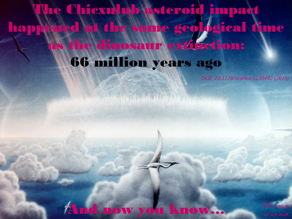
Actual radioisotope dating was done a bit later in 2013: date of K-Pg is 66.043 ± 0.043 MA (millions of years ago), date of the Chicxulub crater is 66.038 ±.025/0.049 MA. Which means that the researchers “established synchrony between the Cretaceous-Paleogene boundary and associated mass extinctions with the Chicxulub bolide impact to within 32,000 years” (Renne et al., 2013), which is a blink of an eye in geological times.
138-66 chixhulub - Copy

Now I want you to understand that often in science, though by far not always, matters are not so simple as she is wrong, he is right. In geology, what matters most is the sample. If the sample is corrupted, so will be your conclusions. Maybe Keller’s or Renne’s samples were affected by a myriad possible variables, some as simple as shifting the dirt from here to there by who knows what event. After all, it’s been 66 million years since. Also, methods used are just as important and dating something that happened so long ago is extremely difficult due to intrinsic physical methodological limitations. Keller (2014), for example, claims that Renne couldn’t have possibly gotten such an exact estimation because he used Argon isotopes when only U-Pb isotope dilution–thermal ionization mass spectrometry (ID-TIMS) zircon geochronology could be so accurate. But yet again, it looks like he did use both, so… I dunno. As the over-used always-trite but nevertheless extremely important saying goes: more data is needed.

Even if the dating puts Chicxulub at the KPB, the volcanologists say that the asteroid, by itself, couldn’t have produced a mass extinction because there are other impacts of its size and they did not have such dire effects, but were barely noticeable at the biota scale. Besides, most of the other mass extinctions on the planet have been already associated with extreme volcanism (Archibald et al., 2010). On the other hand, the circumstances of this particular asteroid could have made it deadly: it landed in the hydrocarbon-rich areas that occupied only 13% of the Earth’s surface at the time which resulted in a lot of “stratospheric soot and sulfate aerosols and causing extreme global cooling and drought” (Kaiho & Oshima, 2017). Food for thought: this means that the chances of us, humans, to be here today are 13%!…

I hope that you do notice that these are very recent papers, so the issue is hotly debated as we speak.

It is possible, nay probable, that the Deccan volcanism, which was going on long before and after the extinction, was exacerbated by the impact. This is exactly what Renne’s team postulated in 2015 after dating the lava plains in the Deccan Traps: the eruptions intensified about 50,000 years before the KT boundary, from “high-frequency, low-volume eruptions to low-frequency, high-volume eruptions”, which is about when the asteroid hit. Also, the Deccan eruptions continued for about half a million years after KPB, “which is comparable with the time lag between the KPB and the initial stage of ecological recovery in marine ecosystems” (Renne et al., 2016, p. 78).

Since we cannot get much more accurate dating than we already have, perhaps the fossils can tell us whether the dinosaurs died abruptly or slowly. Because if they got extinct in a few years instead of over 50,000 years, that would point to a cataclysmic event. Yes, but which one, big asteroid or violent volcano? Aaaand, we’re back to square one.

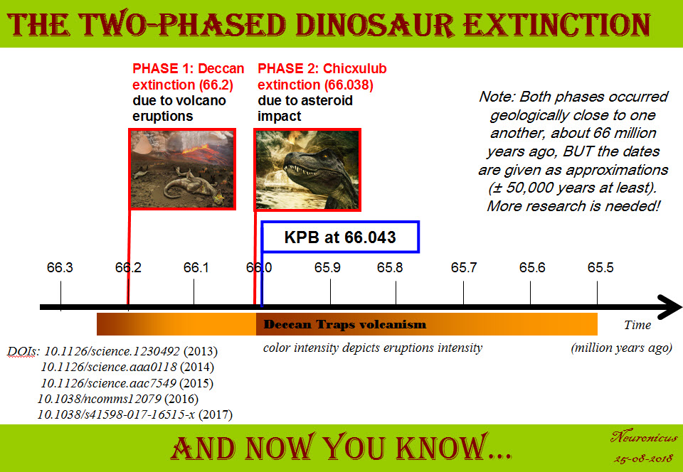
Actually, the last papers on the matter points to two extinctions: the Deccan extinction and the Chicxulub extinction. Petersen et al., (2016) went all the way to Antarctica to find pristine samples. They noticed a sharp increase in global temperatures by about 7.8 ºC at the onset of Deccan volcanism. This climate change would surely lead to some extinctions, and this is exactly what they found: out of 24 species of marine animals investigated, 10 died-out at the onset of Deccan volcanism and the remaining 14 died-out when Chicxulub hit.

In conclusion, because this post is already verrrry long and is becoming a proper college review, to me, a not-a-geologist/paleontologist/physicist-but-still-a-scientist, things happened thusly: first Deccan traps erupted and that lead to a dramatic global warming coupled with spewing poison in the atmosphere. Which resulted in a massive die-out (about 200,000 years before the bolide impact, says a corroborating paper, Tobin, 2017). The surviving species (maybe half or more of the biota?) continued the best they could for the next few hundred thousand years in the hostile environment. Then the Chicxulub meteorite hit and the resulting megatsunami, the cloud of super-heated dust and soot, colossal wildfires and earthquakes, acid rain and climate cooling, not to mention the intensification of the Deccan traps eruptions, finished off the surviving species. It took Earth 300,000 to 500,000 years to recover its ecosystem. “This sequence of events may have combined into a ‘one-two punch’ that produced one of the largest mass extinctions in Earth history” (Petersen et al., 2016, p. 6).
138-timeline dinosaur - Copy

By Neuronicus, 25 August 2018

P. S. You, high school and college students who will use this for some class assignment or other, give credit thusly: Neuronicus (Aug. 26, 2018). The FIRSTS: The cause(s) of dinosaur extinction. Retrieved from https://scientiaportal.wordpress.com/2018/08/26/the-firsts-the-causes-of-dinosaur-extinction/ on [date]. AND READ THE ORIGINAL PAPERS. Ask me for .pdfs if you don’t have access, although with sci-hub and all… not that I endorse any illegal and fraudulent use of the above mentioned server for the purpose of self-education and enlightenment in the quest for knowledge that all academics and scientists praise everywhere around the Globe!

EDIT March 29, 2019. Astounding one-of-a-kind discovery is being brought to print soon. It’s about a site in North Dakota that, reportedly, has preserved the day of the Chicxhulub impact in amazing detail, with tons of fossils of all kinds (flora, mammals, dinosaurs, fish) which seems to put the entire extinction of dinosaurs in one day, thus favoring the asteroid impact hypothesis. The data is not out yet. Can’t wait til it is! Actually, I’ll have to wait some more after it’s out for the experts to examine it and then I’ll find out. Until then, check the story of the discovery here and here.

REFERENCES:

1. Alvarez LW, Alvarez W, Asaro F, & Michel HV (6 Jun 1980). Extraterrestrial cause for the cretaceous-tertiary extinction. PMID: 17783054. DOI: 10.1126/science.208.4448.1095 Science, 208(4448):1095-1108. ABSTRACT | FULLTEXT PDF

2. Archibald JD, Clemens WA, Padian K, Rowe T, Macleod N, Barrett PM, Gale A, Holroyd P, Sues HD, Arens NC, Horner JR, Wilson GP, Goodwin MB, Brochu CA, Lofgren DL, Hurlbert SH, Hartman JH, Eberth DA, Wignall PB, Currie PJ, Weil A, Prasad GV, Dingus L, Courtillot V, Milner A, Milner A, Bajpai S, Ward DJ, Sahni A. (21 May 2010) Cretaceous extinctions: multiple causes. Science,328(5981):973; author reply 975-6. PMID: 20489004, DOI: 10.1126/science.328.5981.973-aScience. FULL REPLY

3. Courtillot V, Besse J, Vandamme D, Montigny R, Jaeger J-J, & Cappetta H (1986). Deccan flood basalts at the Cretaceous/Tertiary boundary? Earth and Planetary Science Letters, 80(3-4), 361–374. doi: 10.1016/0012-821x(86)90118-4. ABSTRACT

4. Esmeray-Senlet, S., Miller, K. G., Sherrell, R. M., Senlet, T., Vellekoop, J., & Brinkhuis, H. (2017). Iridium profiles and delivery across the Cretaceous/Paleogene boundary. Earth and Planetary Science Letters, 457, 117–126. doi:10.1016/j.epsl.2016.10.010. ABSTRACT

5. Hildebrand AR, Penfield GT, Kring DA, Pilkington M, Camargo AZ, Jacobsen SB, & Boynton WV (1 Sept. 1991). Chicxulub Crater: A possible Cretaceous/Tertiary boundary impact crater on the Yucatán Peninsula, Mexico. Geology, 19 (9): 867-871. DOI: https://doi.org/10.1130/0091-7613(1991)019<0867:CCAPCT>2.3.CO;2. ABSTRACT

6. Kaiho K & Oshima N (9 Nov 2017). Site of asteroid impact changed the history of life on Earth: the low probability of mass extinction. Scientific Reports,7(1):14855. PMID: 29123110, PMCID: PMC5680197, DOI:10.1038/s41598-017-14199-x. . ARTICLE | FREE FULLTEXT PDF

7. Keller G, Adatte T, Berner Z, Harting M, Baum G, Prauss M, Tantawy A, Stueben D (30 Mar 2007). Chicxulub impact predates K–T boundary: New evidence from Brazos, Texas, Earth and Planetary Science Letters, 255(3–4): 339-356. DOI: 10.1016/j.epsl.2006.12.026. ABSTRACT

8. Keller, G. (2014). Deccan volcanism, the Chicxulub impact, and the end-Cretaceous mass extinction: Coincidence? Cause and effect? Geological Society of America Special Papers, 505:57–89. doi:10.1130/2014.2505(03) ABSTRACT

9. Petersen SV, Dutton A, & Lohmann KC. (5 Jul 2016). End-Cretaceous extinction in Antarctica linked to both Deccan volcanism and meteorite impact via climate change. Nature Communications, 7:12079. doi: 10.1038/ncomms12079. PMID: 27377632, PMCID: PMC4935969, DOI: 10.1038/ncomms12079. ARTICLE | FREE FULLTEXT PDF 

10. Renne PR, Deino AL, Hilgen FJ, Kuiper KF, Mark DF, Mitchell WS 3rd, Morgan LE, Mundil R, & Smit J (8 Feb 2013). Time scales of critical events around the Cretaceous-Paleogene boundary. Science, 8;339(6120):684-687. doi: 10.1126/science.1230492. PMID: 23393261, DOI: 10.1126/science.1230492 ABSTRACT 

11. Renne PR, Sprain CJ, Richards MA, Self S, Vanderkluysen L, Pande K. (2 Oct 2015). State shift in Deccan volcanism at the Cretaceous-Paleogene boundary, possibly induced by impact. Science, 350(6256):76-8. PMID: 26430116. DOI: 10.1126/science.aac7549 ABSTRACT

12. Schoene B, Samperton KM, Eddy MP, Keller G, Adatte T, Bowring SA, Khadri SFR, & Gertsch B (2014). U-Pb geochronology of the Deccan Traps and relation to the end-Cretaceous mass extinction. Science, 347(6218), 182–184. doi:10.1126/science.aaa0118. ARTICLE

13. Schulte P, Alegret L, Arenillas I, Arz JA, Barton PJ, Bown PR, Bralower TJ, Christeson GL, Claeys P, Cockell CS, Collins GS, Deutsch A, Goldin TJ, Goto K, Grajales-Nishimura JM, Grieve RA, Gulick SP, Johnson KR, Kiessling W, Koeberl C, Kring DA, MacLeod KG, Matsui T, Melosh J, Montanari A, Morgan JV, Neal CR, Nichols DJ, Norris RD, Pierazzo E,Ravizza G, Rebolledo-Vieyra M, Reimold WU, Robin E, Salge T, Speijer RP, Sweet AR, Urrutia-Fucugauchi J, Vajda V, Whalen MT, Willumsen PS.(5 Mar 2010). The Chicxulub asteroid impact and mass extinction at the Cretaceous-Paleogene boundary. Science, 327(5970):1214-8. PMID: 20203042, DOI: 10.1126/science.1177265. ABSTRACT

14. Tobin TS (24 Nov 2017). Recognition of a likely two phased extinction at the K-Pg boundary in Antarctica. Scientific Reports, 7(1):16317. PMID: 29176556, PMCID: PMC5701184, DOI: 10.1038/s41598-017-16515-x. ARTICLE | FREE FULLTEXT PDF 

15. Vogt, PR (8 Dec 1972). Evidence for Global Synchronism in Mantle Plume Convection and Possible Significance for Geology. Nature, 240(5380), 338–342. doi:10.1038/240338a0 ABSTRACT

No Link Between Mass Shootings & Mental Illness

By Neuronicus, 25 February 2018

On Valentine’s Day another horrifying school mass shooting happened in USA, leaving 17 people dead. Just like after the other mass shootings, a lot of people – from media to bystanders, from gun lovers to gun critics, from parents to grandparents, from police to politicians – talk about the link between mental illness and mass shootings. As one with advanced degrees in both psychology and neuroscience, I am tired to explain over and over again that there is no significant link between the two! Mass shootings happen because an angry person has had – or made to think they had – enough sorrow, stress, rejection and/or disappointment and HAS ACCESS TO A MASS KILLING WEAPON. Yeah, I needed the caps. Sometimes scientists too need to shout to be heard.

So here is the abstract of a book chapter called straightforwardly “Mass Shootings and Mental Illness”. The entire text is available at the links in the reference below.

From Knoll & Annas (2015):

“Common Misperceptions

  • Mass shootings by people with serious mental illness represent the most significant relationship between gun violence and mental illness.
  • People with serious mental illness should be considered dangerous.
  • Gun laws focusing on people with mental illness or with a psychiatric diagnosis can effectively prevent mass shootings.
  • Gun laws focusing on people with mental illness or a psychiatric diagnosis are reasonable, even if they add to the stigma already associated with mental illness.

Evidence-Based Facts

  • Mass shootings by people with serious mental illness represent less than 1% of all yearly gun-related homicides. In contrast, deaths by suicide using firearms account for the majority of yearly gun-related deaths.
  • The overall contribution of people with serious mental illness to violent crimes is only about 3%. When these crimes are examined in detail, an even smaller percentage of them are found to involve firearms.
  • Laws intended to reduce gun violence that focus on a population representing less than 3% of all gun violence will be extremely low yield, ineffective, and wasteful of scarce resources. Perpetrators of mass shootings are unlikely to have a history of involuntary psychiatric hospitalization. Thus, databases intended to restrict access to guns and established by guns laws that broadly target people with mental illness will not capture this group of individuals.
  • Gun restriction laws focusing on people with mental illness perpetuate the myth that mental illness leads to violence, as well as the misperception that gun violence and mental illness are strongly linked. Stigma represents a major barrier to access and treatment of mental illness, which in turn increases the public health burden”.

REFERENCE: Knoll, James L. & Annas, George D. (2015). Mass Shootings and Mental Illness. In book: Gun Violence and Mental Illness, Edition: 1st, Chapter: 4, Publisher: American Psychiatric Publishing, Editors: Liza H. Gold, Robert I. Simon. ISBN-10: 1585624985, ISBN-13: 978-1585624980. FULLTEXT PDF via ResearchGate | FULLTEXT PDF via Psychiatry Online

The book chapter is not a peer-reviewed document, even if both authors are Professors of Psychiatry. To quiet putative voices raising concerns about that, here is a peer-reviewed paper with open access that says basically the same thing:

Swanson et al. (2015) looked at large scale (thousands to tens of thousands of individuals) data to see if there is any relationship between violence, gun violence, and mental illness. They concluded that “epidemiologic studies show that the large majority of people with serious mental illnesses are never violent. However, mental illness is strongly associated with increased risk of suicide, which accounts for over half of US firearms–related fatalities”. The last sentence is reminiscent of the finding that stricter gun control laws lower suicide rate.

REFERENCE: Swanson JW, McGinty EE, Fazel S, Mays VM (May 2015). Mental illness and reduction of gun violence and suicide: bringing epidemiologic research to policy. Annals of Epidemiology, 25(5): 366–376. doi: 10.1016/j.annepidem.2014.03.004, PMCID: PMC4211925. FULLTEXT | FULLTEXT PDF.

Further peer-reviewed bibliography (links to fulltext pdfs):

  1. Guns, anger, and mental disorders: Results from the National Comorbidity Survey Replication (NCS-R): “a large number of individuals in the United States have anger traits and also possess firearms at home (10.4%) or carry guns outside the home (1.6%).”
  2. News Media Framing of Serious Mental Illness and Gun Violence in the United States, 1997-2012: “most news coverage occurred in the wake of mass shootings, and “dangerous people” with serious mental illness were more likely than “dangerous weapons” to be mentioned as a cause of gun violence.”
  3. The Link Between Mental Illness and Firearm Violence: Implications for Social Policy and Clinical Practice: “Firearm violence is a significant and preventable public health crisis. Mental illness is a weak risk factor for violence despite popular misconceptions reflected in the media and policy”.
  4. Using Research Evidence to Reframe the Policy Debate Around Mental Illness and Guns: Process and Recommendations: “restricting firearm access on the basis of certain dangerous behaviors is supported by the evidence; restricting access on the basis of mental illness diagnoses is not”.
  5. Mental Illness, Mass Shootings, and the Politics of American Firearms: “notions of mental illness that emerge in relation to mass shootings frequently reflect larger cultural stereotypes and anxieties about matters such as race/ethnicity, social class, and politics. These issues become obscured when mass shootings come to stand in for all gun crime, and when “mentally ill” ceases to be a medical designation and becomes a sign of violent threat”.

131 gun - Copy

By Neuronicus, 25 February 2018

The oldest known anatomically modern humans in Europe

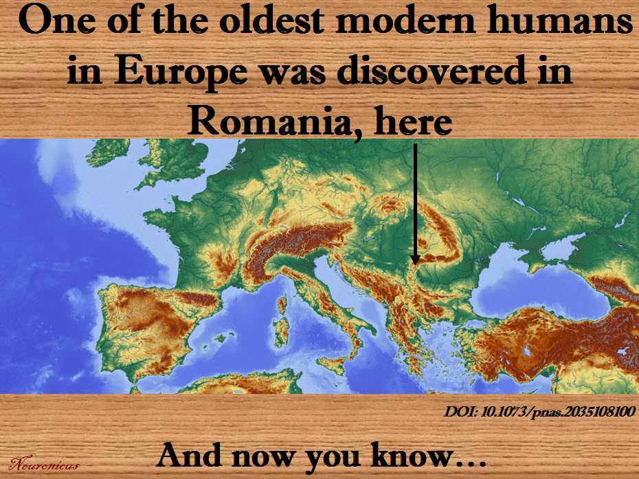
A couple of days ago, on December 1st, was the National Day of Romania, a small country in the South-East of Europe. In its honor, I dug out a paper that shows that some of the earliest known modern humans in Europe were also… dug out there.

Trinkaus et al. (2003) investigated the mandible of an individual found in 2002 by a Romanian speological expedition in Peștera cu Oase (the Cave with Bones), one of the caves in the SouthWest of the country, not far from where Danube meets the Carpathians.

First the authors did a lot of very fine measurement of various aspects of the jaw, including the five teeth, and then compared them with those found in other early humans and Neanderthals. The morphological features place the Oase 1 individual as an early modern human with some Neanderthal features. The accelerator mass spectrometry radiocarbon (14C) direct dating makes him the oldest early modern human discovered to that date in Europe; he’s 34,000–36,000 year old. I’m assuming is a he for no particular reason; the paper doesn’t specify anywhere whether they know the jaw owner’s gender and age. A later paper (Fu et al., 2015) says Oase 1 is even older: 37,000–42,000-year-old.

After this paper it seemed to be a race to see what country can boast to have the oldest human remains on its territory. Italy and UK successfully reassessed their own previous findings thusly: UK has a human maxilla that was incorrectly dated in 1989 but new dating makes it 44,200–39,000 year old, carefully titling their paper “The earliest evidence for anatomically modern humans in northwestern Europe” (Higham et al., 2011) while Italy’s remains that they thought for decades to be Neanderthal turned out to be 45,000-43,000 years old humans, making “the Cavallo human remains […] the oldest known European anatomically modern humans” (Benmazzi et al., 2011).

I wonder what prompted the sudden rush in reassessing the old untouched-for-decades fossils… Probably good old fashioned national pride. Fair enough. Surely it cannot have anything to do with the disdain publicly expressed by some Western Europe towards Eastern Europe, can it? Surely scientists are more open minded than some petty xenophobes, right?

Well, the above thought wouldn’t have even crossed my mind, nor would I have noticed that the Romanians’ discovery has been published in PNAS and the others in Nature, had it not been for the Fu et al. (2015) paper, also published in Nature. This paper does a genetic analysis of the Oase 1 individual and through some statistical inferences that I will not pretend to fully understand they arrive to two conclusions. First, Oase 1 had a “Neanderthal ancestor as recently as four to six generations back”. OK. Proof of interbreeding, nothing new here. But the second conclusion I will quote in full: “However, the Oase individual does not share more alleles with later Europeans than with East Asians, suggesting that the Oase population did not contribute substantially to later humans in Europe.”

Now you don’t need to know much about statistics or about basic logic either to know that from 1 (one) instance alone you cannot generalize to a whole population. That particular individual from the Oase population hasn’t contributed to later humans in Europe, NOT the entire population. Of course it is possible that that is the case, but you cannot scientifically draw that conclusion from one instance alone! This is in the abstract, so everybody can see this, but I got access to the whole paper, which I have read in the hopes against hope that maybe I’m missing something. Nope. The authors did not investigate any additional DNA and they reiterate that the Oase population did not contribute to modern-day Europeans. So it’s not a type-O. From the many questions that are crowding to get out like ‘How did it get past reviewers?’, ‘Why was it published in Nature (interesting paper, but not that interesting, we knew about interbreeding so what makes it so new and exciting)?’, the one that begs to be asked the most is: ‘Why would they say this, when stating the same thing about the Oase 1 individual instead about the Oase population wouldn’t have diminished their paper in any way?’ .

I must admit that I am getting a little paranoid in my older age. But with all the hate that seems to come out and about these days EVERYWHERE towards everything that is “not like me” and “I don’t want it to be like me”, one cannot but wonder… Who knows, maybe it is really just as simple as an overlooked mistake or some harmless national pride so all is good and life goes on, especially since the authors of all four papers discussed above are from various countries and institutions all across the Globe. Should that be the case, I offer my general apologies for suspecting darker motives behind these papers, but I’m not holding my breath.

106-copy

References:

1) Trinkaus E, Moldovan O, Milota S, Bîlgăr A, Sarcina L, Athreya S, Bailey SE, Rodrigo R, Mircea G, Higham T, Ramsey CB, & van der Plicht J. (30 Sep 2003, Epub 22 Sep 2003). An early modern human from the Peştera cu Oase, Romania. Proceedings of the National Academy of Sciences U S A,  100(20):11231-11236. PMID: 14504393, PMCID: PMC208740, DOI: 10.1073/pnas.2035108100. ARTICLE  | FREE FULLTEXT PDF

 2) Higham T, Compton T, Stringer C, Jacobi R, Shapiro B, Trinkaus E, Chandler B, Gröning F, Collins C, Hillson S, O’Higgins P, FitzGerald C, & Fagan M. (2 Nov 2011). The earliest evidence for anatomically modern humans in northwestern Europe. Nature. 479(7374):521-4. PMID: 22048314, DOI: 10.1038/nature10484. ARTICLE | FULLTEXT PDF via ResearchGate

3) Benazzi S, Douka K, Fornai C, Bauer CC, Kullmer O, Svoboda J, Pap I, Mallegni F, Bayle P, Coquerelle M, Condemi S, Ronchitelli A, Harvati K, & Weber GW. (2 Nov 2011). Early dispersal of modern humans in Europe and implications for Neanderthal behaviour. Nature, 479(7374):525-8. PMID: 22048311, DOI: 10.1038/nature10617. ARTICLE | FULLTEXT PDF via ResearchGate

4) Fu Q, Hajdinjak M, Moldovan OT, Constantin S, Mallick S, Skoglund P, Patterson N, Rohland N, Lazaridis I, Nickel B, Viola B, Prüfer K, Meyer M, Kelso J, Reich D, & Pääbo S. (13 Aug 2015, Epub 22 Jun 2015). An early modern human from Romania with a recent Neanderthal ancestor. Nature. 524(7564):216-9. PMID: 26098372, PMCID: PMC4537386, DOI:10.1038/nature14558. ARTICLE | FREE FULLTEXT PDF

By Neuronicus, 3 December 2016

Save

Save

Save

Save

Save

A third way of neuronal communication

electrical fields - CopyEvery neuroscience or biology textbook will tell you that neurons communicate with one another in two ways: via chemical synapses (one neuron releases some substances that change the membrane potential of the receiving neuron) or via electrical synapses (neurons physically share some membrane proteins that allows the electrical impulse to go from one neuron to another). (Nota bene: for the taxonomic nitpickers, I decided that the other non-conventional forms of communication, like diffusion, fall in one or the other of the two categories above).

Now Qiu et al. (2015) have some evidence that there may be another way of neuronal chatting. Long ago (in 1924), Hans Berger observed that neurons have rhythmic and spontaneous electrical activity. The rhythmic activity of an entire population of neurons is called a wave and can be detected with rudimentary tools such as EEG. Qiu et al. reasoned that the speed of such a traveling wave is too slow to be explained by conventional neuronal communication (of which we know quite a lot).

So the researchers ran some computer simulations to test if diffuse electrical fields can explain the speed of a wave, instead of the conventional fast communications. In other words, can local, weak, slow endogenous fields sweep the brain by recruiting nearby neurons? The computer simulations said it is possible, with a very slow speed of 10 cm/s.

Then they recorded the electrical activity of mouse hippocampus isolated in a Petri dish. When the researchers blocked the electrical field there was a decrease in the speed of the wave, meaning the field is required for the wave speed observed.

To be fair, this is not a new idea; even in the early eighties these fields were suggested, because the wave persisted even when the other two ways of communicating have been blocked. And in the early noughts it was shown that a neural network is much more sensitive to electrical field manipulation than individual neurons. What makes this paper interesting is that the authors show that the endogenous electrical fields are not too weak to underlie the wave, as previously thought. The work has implications for the study of epilepsy, sleep, and memory.

Reference: Qiu C, Shivacharan RS, Zhang M, & Durand DM (2 December 2015). Can Neural Activity Propagate by Endogenous Electrical Field?, Journal of Neuroscience, 35(48): 15800-15811; doi: 10.1523/JNEUROSCI.1045-15.2015 Article | Full Text via Research Gate | ScienceAlert cover

By Neuronicus, 11 February 2016

Mechanisms of stress resilience

71 stress - CopyLast year a new peer-reviewed journal called Neurobiology of Stress made its debut. The journal is published by Elsevier, who, in an uncharacteristic move, has provided Open Access for its first three issues. So hurry up and download the papers.

The very first issue is centered around the idea of resilience. That is, exposed to the same stressors, some people are more likely to develop stress-induced diseases, whereas others seem to be immune to the serious effects of stress.

Much research has been carried out to uncover the effects of chronic stress or of an exposure to a single severe stressor, which vary from cardiovascular disorders, obesity, irritable bowel syndrome, immune system dysfunctions to posttraumatic stress disorder, generalized anxiety, specific phobias, or depression. By comparison, there is little, but significant data on resilience: the ability to NOT develop those nasty stress-induced disorders. Without doubt, one reason for this scarcity is the difficulty in finding such rare subjects in our extremely stressful society. Therefore most of the papers in this issue focus on animal models.

Nevertheless, there is enough data on resilience to lead to no less that twenty reviews on the subject. It was difficult to choose one as most are very interesting, tackling various aspects of resilience, from sex differences to prenatal exposure to stress, from epigenetic to neurochemical modifications, from social inequalities to neurogenesis and so on.

So I chose for today a more general review of Pfau & Russo (2015), entitled “Peripheral and central mechanisms of stress resilience”. After it introduces the reader to four animal models of resilience, the paper looks at the neruoendocrine responses to stress and identifies some possible chemical mediators of resilience (like certain hormones), then at the immune responses to stress (bad, bad cytokines), and finally at the brain responses to stress (surprisingly, not focusing on amygdala, hypothalamus or hippocampus, but on the dopamine system originating from ventral tegmental area).

I catalogue the review as a medium difficulty read because it requires a certain amount of knowledge of the stress field beforehand. But do check out the other ones in the issue, too!

Reference: Pfau ML & Russo SJ (1 Jan 2015). Peripheral and central mechanisms of stress resilience. Neurobiology of Stress, 1:66-79. PMID: 25506605, PMCID: PMC4260357, DOI: 10.1016/j.ynstr.2014.09.004. Article | FREE FULLTEXT PDF

By Neuronicus, 24 January 2016

Your blood is better than my blood

pictures-004
Siamese tomatoes. Taken from here.

Parabiosis is a surgical procedure that lets two animals to share the same blood; it’s a case of reverse conjoined twins restricted to the circulatory system.

The procedure is over 150 years old and is a useful technique in physiology, though rarely used, probably due to the perceived cruelty towards the animals, although today is performed under anesthesia and aseptic condition. It delivered good data; for example, it was a parabiosis experiment with rodents that showed is not the sugar in the blood that causes cavities but the sugar in the mouth. Similarly, parabiosis has been proven useful in cancer, diabetes, and ageing research.

Scudellari (2015) wrote a News piece for Nature describing some advancements in the ageing field using the parabiosis technique. Namely, by joining the circulatory systems of a young and an old mouse, researchers have observed that the old mouse is faster, smarter, with rejuvenated muscles and glossier fur. Now the race is to find out what in the blood does it. Researchers caution that the young blood is not effectively reversing ageing, but may have factors circulating in it that promote tissue repair. Already a muscle-rejuvenating protein has been identified.

I am not going through the original papers themselves as I usually do (they are provided as links in the Reference paper). Instead, I am featuring the news piece by Scudellari because in addition of looking at parabiosis and ageing result, it also provides a nice historical account of the use of parabiosis. Enjoy!

Reference: Scudellari, M. (22 Jan 2015). Ageing research: Blood to blood. Nature, 517: 426-429. Article | FREE Fulltext PDF

By Neuronicus, 4 January 2015

Yeast can make morphine

poppy - Copy

Opiates like morphine and heroin can be made at home by anybody with a home beer-brewing kit and the right strain of yeast. In 2015, two published papers and a Ph.D. dissertation described the relatively easy way to convince yeast to make morphine from sugar (the links are provided in the Reference paper). That is the bad news.

The good news is that scientists have been policing themselves (well, most of them, anyway) long before regulations are put in place to deal with technological advancements by, for example, limiting access to the laboratory, keeping things under lock and key, publishing incomplete data, and generally being very careful with what they’re doing.

Complementing this behavior, an article published by Oye et al. (2015) outlines other measures that can be put in place so that this new piece of knowledge doesn’t increase the accessibility to opiates, thereby increasing the number of addicts, which is estimated to more than 16 million people worldwide. For example, researchers can make the morphine-producing yeast dependent on unusual nutrients or engineer the existing strain to produce less-marketable varieties of opiates or prohibit the access to made-to-order DNA sequences for this type of yeast and so on.

You may very well ask “Why did the scientists made this kind of yeast anyway?”. Because some medicines are either very expensive or laborious to produce by the pharmaceutical companies, the researchers have sought a method to make these drugs more easily and cheaply by engineering bacteria, fungi, or plants to produce them for us. Insulin is a good example of an expensive and hard-to-get-by drug that we managed to engineer yeast strains to produce it for us. And opiates are still the best analgesics out there.

Reference: Oye KA, Lawson JC, & Bubela T (21 May 2015). Drugs: Regulate ‘home-brew’ opiates. Nature, 521(7552):281-3. doi: 10.1038/521281a. Article | FREE Fulltext PDF

By Neuronicus, 2 January 2016

Beer spoiling bacteria, oh no! But we know now how you’re made, suckers!

64 - Copy

Over 250 years ago today, on 31 December 1759, Arthur Guinness started brewing one of the most loved adult drinks today, the Guinness beer.

As with all food and drink products, beer can be also suffer spoiling due to various bacteria. The genomes of two of these culprits – Megasphaera cerevisiae PAT 1T and Lactobacillus brevis BSO 464 – have been sequenced in 2015 by two different groups.

Funny thing though: the papers that announce the completion of the genome sequencing (see bellow References) do not talk abut the significance of their discovery. The usual template for a biology paper (or as a matter of fact any science paper) is:

Introduction: x is important because y,
Methods and Results: here is what we did to understand x,
Conclusion: now we can better tackle y.

Not these papers, which basically say, in less than a page: “This bacterium spoils beer; here is its genome. You’re welcome!”

Well played, geneticists, well played… And we are, indeed, grateful. Oh, yes, we are…

References:

1. Kutumbaka KK, Pasmowitz J, Mategko J, Reyes D, Friedrich A, Han S, Martens-Habbena W, Neal-McKinney J, Janagama HK, & Nadala C, Samadpour M (10 Sep 2015). Draft Genome Sequence of the Beer Spoilage Bacterium Megasphaera cerevisiae Strain PAT 1T. Genome Announcements, 3(5). pii: e01045-15. doi: 10.1128/genomeA.01045-15. Article | FREE Fulltext PDF | FREE GENOME

2. Bergsveinson J, Pittet V, Ewen E, Baecker N, & Ziola B (3 Dec 2015). Genome Sequence of Rapid Beer-Spoiling Isolate Lactobacillus brevis BSO 464. Genome Announcements, 3(6). pii: e01411-15. doi: 10.1128/genomeA.01411-15. Article | FREE Fulltext PDF | FREE GENOME

By Neuronicus, 31 December 2015

Vagus nerve stimulation improves recovery after stroke

61NIh NIMH vagus-nerve-stimulation-public
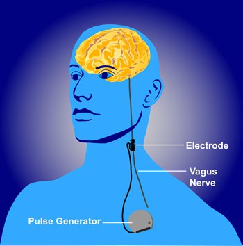
Vagus nerve stimulation by a wireless device implanted subcutaneously. License: PD. Credit: NIH/NIMH

A stroke is a serious medical condition that is characterized by the death of brain cells following bleeding in the brain or lack of blood supply to those cells. Three quarters of the survivors have weakness in the arm which can be permanent. Physical therapy helps, but not much.

Dawson et al. (2015) report a novel way to increase arm mobility after stroke. Previous findings in animal studies showed promising results by stimulating the vagus nerve (VNS). This nerve is the tenth out of the twelve cranial nerves and has many functions, primarily heart and digestive control. The authors implanted a small wireless device in the neck of nine stroke survivors and delivered very short (half a second) mild (0.8 mA) electrical pulses during rehabilitation therapy. Ten matched controls received rehab therapy only.

The authors measured motor recovery by several tests, one of which is Fugl–Meyer assessment-UE. At this test, the rehab only group improved by 3 points and the VNS + rehab group by about 9 points and this difference was statistically significant (I believe this scale’s upper limit is 66, but I’m not 100% sure).

Although the authors offer a possible mechanism through which VNS might produce cortical plasticity (through release of acetylcholine and norepinephrine driven by activation of nucleus tractus solitarius) the truth is that we don’t really know how it works. Nevertheless, it seems that VNS paired with rehab is better than rehab alone, and that in itself certainly warrants further studies, perhaps the next step being a fully double-blind experiment.

Reference: Dawson J, Pierce D, Dixit A, Kimberley TJ, Robertson M, Tarver B, Hilmi O, McLean J, Forbes K, Kilgard MP, Rennaker RL, Cramer SC, Walters M, & Engineer N. (Epub 8 Dec 2015). Safety, Feasibility, and Efficacy of Vagus Nerve Stimulation Paired With Upper-Limb Rehabilitation After Ischemic Stroke. Stroke. 2016; 47:00-00. DOI: 10.1161/STROKEAHA.115.010477. Article | FREE FULLTEXT PDF

By Neuronicus, 11 December 2015

Herpes viruses infect neurons

virus EBV
FIG. 3 from Jha et al. (2015). Wild-type EBV infection of primary human fetal neurons. Fluorescence microscopy was carried out at 2, 4, 6, and 8 days post infection to monitor for GFP expression (the fluorescent label). Microscopy images were captured at x20 magnification.

For some mysterious reason, whether Epstein-Barr virus (EBV) and Kaposi’s sarcoma-associated herpesvirus (KSHV) can infect neurons has not been established until now. Probably because some viruses from the same family do not infect neurons, so it was assumed that EPV and KSHV do not either.

Jha et al. (2015) cultured Sh-Sy5y neuroblastoma cells, teratocarcinoma Ntera2 neurons, and primary human fetal neurons in Petri dishes and then exposed them to these viruses. After infection, the authors did some fluorescence microscopy (they tagged the viruses with fluorescent dyes), real-time PCR (to confirm there was viral RNA in the cells), immunofluorescence assays (to see if the viral proteins are expressed) and Western blot analyses (to see if the specific viral antigens are made). All these showed that the viruses were happily multiplying in the cells.

Now here comes the significance of the study: EBV and KSHV are viruses associated with all sorts of nasty diseases like mononucleosis and cancers. EBV has also been associated with neurological disorders, like multiple sclerosis, Alzheimer’s, neuropathies, lymphomas etc. But the critical word here is “associated”. That is, they found these viruses in people suffering from those diseases. So the knowledge that these viruses infect neurons could point to a mechanism behind these associations. Unfortunately, EBV is present in 90-95% of the population of the world. Which means that you will find this virus in, let’s say, 9 out of 10 people suffering from Alzheimer’s, assuming normal distributions and random sampling. So the virus’s presence maybe completely unrelated to the disease. By the same rationale, you would find the virus in 9 out of 10 people found guilty of theft, for example. It would be then interesting to see find what is NOT associated with.

Caveat: I have not read the association studies, so my argument holds only if what they report is that people with disease X also have EBV. If they made, however, a comparisons and found out that people with disease X are significantly more likely to be infected with EBV than the ones without the disease, then the argument does not hold.

Reference: Jha HC, Mehta D, Lu J, El-Naccache D, Shukla SK, Kovacsics C, Kolson D, Robertson ES (1 Dec 2015). Gammaherpesvirus Infection of Human Neuronal Cells. MBio,  6(6). pii: e01844-15. doi: 10.1128/mBio.01844-15. Article | FREE FULLTEXT PDF | PsyPost cover

By Neuronicus, 7 December 2015

I am blind, but my other personality can see

58depression-388872_960_720

This is a truly bizarre report.

A woman named BT suffered an accident when she was 20 years old and she became blind. Thirteen year later she was referred to Bruno Waldvogel (one of the two authors of the paper) for psychotherapy by a psychiatry clinic who diagnosed her with dissociative identity disorder, formerly known as multiple personality disorder.

The cortical blindness diagnosis has been established after extensive ophtalmologic tests in which she appeared blind but not because of damage to the eyes. So, by inference, it had to be damage to the brain. Remarkably (we shall see later why), she had no oculomotor reflexes in response to glare. Moreover, visual evoked potentials (VEP is an EEG in the occipital region) showed no activity in the primary visual area of the brain (V1).

During the four years of psychotherapy, BT showed more than 10 distinct personalities. One of them, a teenage male, started to see words on a magazine and pretty soon could see everything. With the help of hypnotherapeutic techniques, more and more personalities started to see.

“Sighted and blind states could alternate within seconds” (Strasburger & Waldvogel, 2015).

The VEP showed no or very little activity when the blind personality was “on” and showed normal activity when the sighted personality was “on”. Which is extremely curious, because similar studies in people with psychogenic blindness or anesthetized showed intact VEPs.

There are a couple of conclusions from this: 1) BT was misdiagnosed, as is unlikely to be any brain damage because some personalities could see, and 2) Multiple personalities – or dissociate identities, as they are now called – are real in the sense that they can be separated at the biological level.

BEAR_10_04
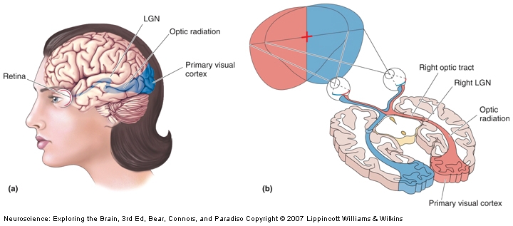
The visual pathway that mediates conscious visual perception. a) A side view of the human brain with the retinogeniculocortical pathway shown inside (blue). b) A horizontal section through the brain exposing the same pathway.

Fascinating! The next question is, obviously, what’s the mechanism behind this? The authors say that it’s very likely the LGN (the lateral geniculate nucleus of the thalamus) which is the only relay between retina and V1 (see pic). It can be. Surely is possible. Unfortunately, so are other putative mechanisms, as 10% of the neurons in the retina also go to the superior colliculus, and some others go directly to the hypothalamus, completely bypassing the thalamus. Also, because it is impossible to have a precise timing on the switching between personalities, even if you MRI the woman it would be difficult to establish if the switching to blindness mode is the result of a bottom-up or a top-down modulation (i.e. the visual information never reaches V1, it reaches V1 and is suppressed there, or some signal form other brain areas inhibits V1 completely, so is unresponsive when the visual information arrives).

Despite the limitations, I would certainly try to get the woman into an fMRI. C’mon, people, this is an extraordinary subject and if she gave permission for the case study report, surely she would not object to the scanning.

Reference: Strasburger H & Waldvogel B (Epub 15 Oct 2015). Sight and blindness in the same person: Gating in the visual system. PsyCh Journal. doi: 10.1002/pchj.109.  Article | FULLTEXT PDF | Washington Post cover

By Neuronicus, 29 November 2015

Lucy’s 9 vertebrae are actually 8

Lucy
Lucy. Left: picture of the real skeleton. Middle and Right: reconstructions. Courtesy of Wikipedia

As Google reminded us, today is the 41st anniversary of the finding of Lucy, the first discovered member of the species Australopithecus afarensis. Lucy lived in Ethiopia about 3.2 million years ago and the most extraordinary fact about her is that her fossil represents the first evidence of bipedalism in a hominin (we are also hominins).

Lucy is one “missing link” (not ‘missing’ anymore, obviously) between the common ancestor of humans and chimpanzees and humans because she has ape-like features (jaw, forehead, long arms, small cranium) and human-like features (knee, ankle, lumbar curve, pelvic bones) and walked upright.

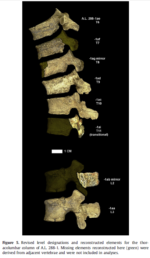
Meyer et al. (2015) wanted to do a comprehensive reconstruction of Lucy for display at the American Museum of Natural History in New York. During this work they noticed that one vertebrae of the total of nine found is kindda small compared to the other ones. So they set to measure vertebrae form all sorts of other species, alive and extinct, and after some factor analysis they concluded that out of Lucy’s nine found vertebrae, the little one is not actually hers, but belongs to a different species from the genus Theropithecus (a baboon ancestor).

This finding is functionally uninformative and their “work does not refute previous work on Lucy or its importance for human evolution, but rather highlights the importance of studying original fossils, as well as the efficacy of the scientific method.” In other words, give the poor anthropologists not reconstructions but the original fossils to work with (most people worked with Lucy’s reconstructions which missed some details, thus allowing this pesky vertebra to remain miss-cataloged for 40 years).

lucy1
The new alignment from doi: 10.1016/j.jhevol.2015.05.007

This is the first paper of pure anthropology that I have read in full and let me tell you that I found a lot of curious things, unrelated to Lucy. Like, for example, from an anthropologist’s point of view, an adult is someone with the third molar completely erupted. We should then look into the people’s mouths before giving them the keys to the wine cellar, because some 21-year olds are definitely not adults. Also, instead of a medical doctor, get an anthropologist to teach anatomy, because oh boy do these people know their skeletons! Here is an excerpt from the Methods section: “The overall size of the A.L. 288-1am partial vertebra was calculated as the geometric mean of six linear dimensions: lamina superoinferior height and dorsoventral thickness, pars interarticularis width, interarticular facet height, and superior and inferior articular interfacet maximum transverse widths. The pars interarticularis geometric mean includes three variables from the pars interarticularis: lamina superoinferior height and dorsoventral thickness, and pars interarticularis width” (p. 175).

All in all, nice!

Reference: Meyer MR, Williams SA, Smith MP, Sawyer GJ (August 2015, Epub 6 Jun 2015). Lucy’s back: Reassessment of fossils associated with the A.L. 288-1 vertebral column. Journal of Human Evolution, 85:174-80. doi: 10.1016/j.jhevol.2015.05.007. Article | FREE FULLTEXT PDF

By Neuronicus, 24 November 2015


Pesticides reduce pollination

2410-closeup-of-a-bee-with-pollen-flying-by-a-flower-pv
Close-up of a bee with pollen flying by a flower. Credit: Jon Sullivan. License: PD

Bees have difficult times these days, what with that mysterious colony collapse disorder on top of various viral, bacterial and parasitical diseases. Of course, the widespread use of pesticides did not help the thriving of the hive, as many pesticides have various deleterious effects on the bees, from poor foraging or less reproduction to even death.

The relatively new (’90s) class of insecticide – the neonicotinoids – has been met with great hope because has low toxic effects on birds and mammals, as opposed to the organophosphates, for example. Why that should be the case, is a mystery for me, because the neonicotinoids bind to the nicotinic receptors present in both peripheral and central nervous system in an irreversible manner, which does not put the neonicotinoids in a favorable light.

Now Stanley et al. (2015) have found that exposure to the neonicotinoid thiamethoxam reduces the pollination provided by the bumblebees to apples. They checked it using 24 bumblebee colonies and the exposure was at low levels over 13 days, trying to mimic realistic in-field exposure. The apples visited by the bumblebees exposed to insecticide had 36% reduction in apple seeds.

Almost 90% of the flowering plants need pollination to reproduce, so any threat to pollination can cause serious problems. Over the paste few years, virtually all USA corn had been treated with neonicotinoids; EU banned the thiamethoxam use in 2013. And, to make matters worse, neonicotinoids are but only one class of the many toxins affecting the bees.

Related post: Golf & Grapes OR Grandkids (but not both!)

Reference: Stanley DA, Garratt MP, Wickens JB, Wickens VJ, Potts SG, & Raine NE. (Epub 18 Nov 2015). Neonicotinoid pesticide exposure impairs crop pollination services provided by bumblebees. Nature, doi: 10.1038/nature16167. Article

By Neuronicus, 21 November 2015

Will you trust a pigeon pathologist? That’s right, he’s a bird. Stop being such an avesophobe!

pigeon

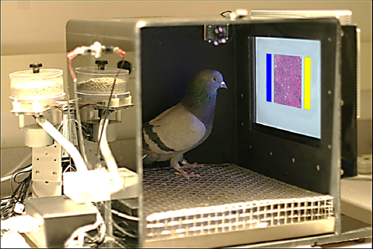
From Levenson et al. (2015), doi: 10.1371/journal.pone.0141357. License: CC BY 4.0

Pigeons have amazing visual skills. They can remember facial expressions, recall almost 2000 images, recognizes all the letters of the alphabet (well, even I can do that), and even tell apart a Monet form a Picasso! (ok, birdie, you got me on that one).

Given their visual prowess, Levenson et al. (2015) figured that pigeons might be able to distinguish medically-relevant images (a bit of a big step in reasoning there, but let’s go with it). They got a few dozen pigeons, starved them a bit so the birds show motivation to work for food, and started training them on recognizing malignant versus non-malignant breast tumors histology pictures. These are the same exact pictures your radiologist looks at after a mammogram and your pathologist after a breast biopsy; they were not retouched in any way for the pigeon’s benefit (except to make it more difficult, see below). Every time the pigeon pecked on the correct image, it got a morsel of food (see picture). Training continued for a few weeks on over 100 images.

For biopsies, the birds had an overwhelming performance, reaching 99% accuracy, regardless of the magnification of the picture, and for mammograms, up to 80% accuracy, just like their human counterparts. Modifying the pictures’ attributes, like rotation, compression or color lowered somewhat their accuracy, but they were still able to score only marginally less than humans and considerably better than any computer software. More importantly, the pigeons were able to generalize, after training, to correctly classify previously unseen pictures.

Let’s be clear: I’m not talking about some fancy breed here, but your common beady-eyed, suspicious-sidling, feral-looking rock pigeon. Yes, the one and only pest that receives stones and bread in equal measures, the former usually accompanied by vicious swearings uttered by those that encountered their slushy “gifts” under the shoes, on the windshield or in the coffee and the latter offered by more kindly disposed and yet utterly naive individuals in the misguided hopes of befriending nature. Columba livia by its scientific name, at the same time an exasperating pest and an excellent pathologist! Who knew?!

The authors even suggest using pigeons instead of training and paying clinicians. Hmmm… But who do I sue if my mother’s breast cancer gets missed by the bird, in one of those 1% chances? Because somehow making a pigeon face the guillotine does not seem like justice to me. Or is this yet another plot to get the clinicians off the hook for misdiagnoses? Leave the medical profession alone, birdies – is morally sensitive as it is -, and search employment in the police or Google; they always need better performance in the ever-challenging task of face-recognition in surveillance videos.

P.S. The reason why you didn’t recognized the word “avesophobe” in the title is because I just invented it, to distinguish the hate for birds from a more serious affliction, ornithophobia, the fear of birds.

Reference: Levenson RM, Krupinski EA, Navarro VM, & Wasserman EA (18 Nov 2015). Pigeons (Columba livia) as Trainable Observers of Pathology and Radiology Breast Cancer Images. PLoS One, 10(11):e0141357. doi: 10.1371/journal.pone.0141357.  Article | FREE FULLTEXT PDF

By Neuronicus, 19 November 2015