One reason why I don’t post more often is that I have such a hard time deciding what to cover (Hint: send me stuff YOU find awesome). Most of the cool and new stuff is already covered by big platforms with full-time employees and I try to stay away of the media-grabbers. Mostly. Some papers I find so cool that it doesn’t matter that professional science journalists have already covered them and I too jump on the wagon with my meager contribution. Anyway, here is a glimpse on how my train of thought goes on inspiration-less days.
Inner monologue: Check the usual journals’ current issues. Nothing catches my eye. Maybe I’ll feature a historical. Open Wikipedia front page and see what happened today throughout history. Aha, apparently Babinski died in 1932. He’s the one who described the Babinski’s sign. Normally, when the sole of the foot is stroked, the big toe flexes inwards, towards the sole. If it extends upwards, then that’s a sure sign of neurological damage, the Babinski’s sign. But healthy infants can have that sign too not because they have neurological damage, but because their corticospinal neurons are not fully myelinated. Myelin, who discovered that? Probably Schwann. Quick search on PubMed. Too many. Restrict to ‘history”. I hate the search function on PubMed, it brings either to many or no hits, no matter the parameters. Ah, look, Virchow. Interesting. Aha. Find the original reference. Aha. Springer charges 40 bucks for a paper published in 1854?! The hell with that! I’m not even going to check if I have institutional access. Get the pdf from other sources. It’s in German. Bummer. Go to Highwire. Find recent history of myelin. Mielinization? Myelination? Myelinification? All have hits… Get “Fundamental Neuroscience” off of the shelf and check… aha, myelination. Ok. Look at the pretty diagram with the saltatory conduction! Enough! Go back to Virchow. Does it have pictures, maybe I can navigate the legend? Nope. Check if any German speaking friends are online. Nope, they’re probably asleep, which is what I should be doing. Drat. Refine Highwire search. Evrika! “Hystory of Myelin” by Boullerne, 2016. Got the author manuscript. Hurray. Read. Write.
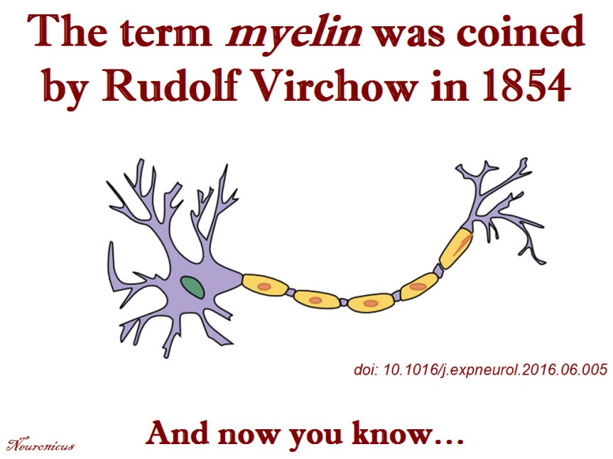
Myelinated fibers, a.k.a. white matter has been observed and described by various anatomists, as early as the 16th century, Boullerne (2016) informs us. But the name of myelin was given only in 1854 by Rudolph Virchow, a physician with a rich academic and public life. Although Virchow introduced the term to distinguish between bone marrow and the medullary substance, paradoxically, he managed to muddy waters even more because he did not restrict the usage of the term mylein to … well, myelin. He used it also to refer to substances in blood cells and egg’s yolk and spleen and, frankly, from the quotes provided in the paper, I cannot make heads or tails of what Virchow thought myelin was. The word myelin comes form the Greek myelos or muelos, which means marrow.
Boullerne (2016) obviously did a lot of research, as the 53-page account is full of quotes from original references. Being such a scholar on the history of myelin I have no choice but to believe her when she says: “In 1868, the neurologist Jean-Martin Charcot (1825-1893) used myelin (myéline) in what can be considered its first correct attribution.”
So even if Virchow coined the term, he was using it incorrectly! Nevertheless, in 1858 he correctly identified the main role of myelin: electrical insulation of the axon. Genial insight for the time.
I love historical reviews of sciency stuff. This one is a ‘must-have’ for any biologist or neuroscientist. Chemists and physicists, too, don’t shy away; the paper has something for you too, like myelin’s biochemistry or its birefringence properties.
Reference: Boullerne, AI (Sep 2016, Epub 8 Jun 2016). The history of myelin. Experimental Neurology, 283(Pt B): 431-45. doi: 10.1016/j.expneurol.2016.06.005. ARTICLE
Original Reference: Virchow R. (Dec 1854). Ueber das ausgebreitete Vorkommen einer dem Nervenmark analogen Substanz in den thierischen Geweben. Archiv für pathologische Anatomie und Physiologie und für klinische Medicin, 6(4): 562–572. doi:10.1007/BF02116709. ARTICLE
P.S. I don’t think is right that Springer can retain the copyright for the Virchow paper and charge $39.95 for it. I don’t think they have the copyright for it anyway, despite their claims, because the paper is 162 years old. I am aware of no German or American copyright law that extends for so long. So, if you need it for academic purposes, write to me and thou shalt have it.
By Neuronicus, 29 October 2016
Save