The FIRSTS: mRNA from one cell can travel to another cell and be translated there (2006)

I’m interrupting the series on cognitive biases (unskilled-and-unaware, superiority illusion, and depressive realism) to tell you that I admit it, I’m old. -Ish. Well, ok, I’m not that old. But this following paper made me feel that old. Because it invalidates some stuff I thought I knew about molecular cell biology. Mind totally blown.

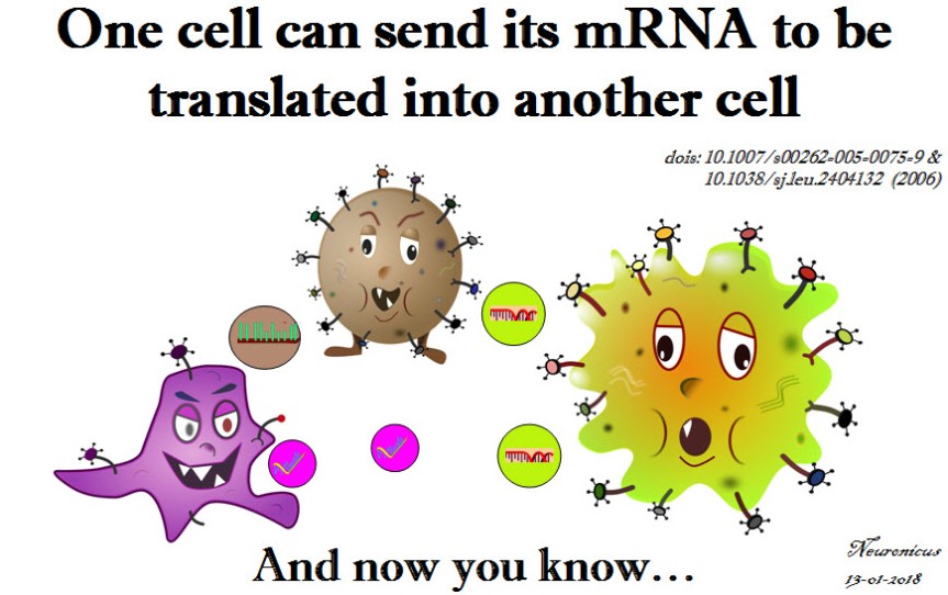
It all started with a paper freshly published two days ago and that I’ll cover tomorrow. It’s about what the title says: mRNA can travel between cells packaged nicely in vesicles and once in a target cell can be made into protein there. I’ll explain – briefly! – why this is such a mind-blowing thing.

Central_dogma - Copy
Fig. 1. Illustration of the central dogma of biology: information transfer between DNA, RNA, and protein. Courtesy of Wikipedia, PD

We’ll start with the central dogma of molecular biology (specialists, please bear with me): the DNA is transcribed into RNA and the RNA is translated into protein (see Fig. 1). It is an oversimplification of the complexity of information flow in a biological system, but it’ll do for our purposes.

DNA needs to be transcribed into RNA because RNA is a much more flexible molecule and thus can do many things. So RNA is the traveling mule between DNA and the place where its information becomes protein, i.e. ribosome. Hence the name mRNA. Just kidding; m stands for messenger RNA (not that I will ever be able to call that ever again: muleRNA is stuck in my brain now).

There are many kinds of RNA: some don’t even get out of the nucleus, some are chopped and re-glued (alternative splicing), some decide which bits of DNA (genes) are to be expressed, some are busy housekeepers and so on. Once an RNA has finished its business it is degraded in many inventive ways. It cannot leave the cell because it cannot cross the cell membrane. And that was that. Or so I’ve been taught.

Exceptions from the above were viruses whose ways of going from cell to cell are very clever. A virus is a stretch of nucleic acids (DNA and/or RNA) and some proteins encapsulated in a blob (capsid). Not a cell!

In the ’90s several groups were looking at some blobs (yes, most stuff in biology can be defined by the all-encompassing and enlightening term of ‘blob’) that cells spew out every now and then. These were termed extracellular vesicles (EV) for obvious reasons. Turned out that many kinds of cells were doing it and on a much more regular basis than previously thought. The contents of these EVs varied quite a bit, based on the type of cells studied. Proteins, mostly, and maybe some cytoplasmic debris. In the ’80s it was thought that this was one way for a cell to get rid of trash. But in 1982, Stegmayr & Ronquist showed that prostate cells release some EVs that result in sperm cell motility increase (Raposo & Stoorvogel, 2013) so, clearly, the EVs were more than trash. Soon it became evident that EVs were another way of cell-to-cell communication. (Note to self: the first time intercellular communication by EVs was demonstrated was in 1982, Stegmayr & Ronquist. Maybe I’ll dig out the paper to cover it sometime).

So. In 2005, Baj-Krzyworzeka et al. (2006) looked at some human cancer cells to see what they spew out and for what purpose. They saw that the cancer cells were transferring some of the tumor proteins packaged in EVs to monocytes. For devious purposes, probably. And then they made to what it looks to me like a serious leap in reasoning: since the EVs contain tumor proteins, why wouldn’t they also contain the mRNA for those proteins? My first answer to that would have been: “because it would be rapidly degraded”. And I would have been wrong. To my credit, if the experiment wouldn’t take up too many resources I still would have done it, especially if I would have some random primers lying around the lab. Luckily for the world, I was not in charge with this particular experiment and Baj-Krzyworzeka et al. (2005) proceeded with a real-time PCR (polymerase chain reaction) which showed them that the EVs released by the tumor cells also contained mRNA.

Now the 1 million dollar, stare-in-your-face question was: is this mRNA functional? Meaning, once delivered to the host cell, would it be translated into protein?

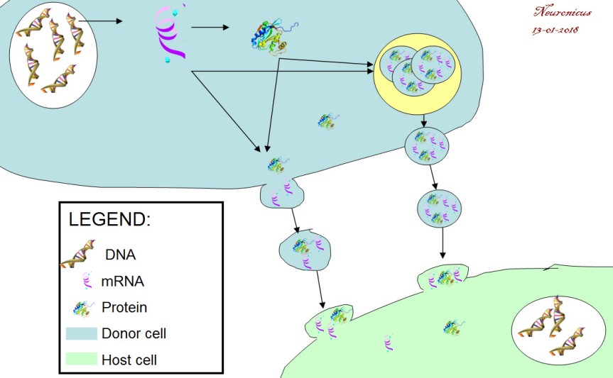
Six months later the group answered it. Ratajcza et al. (2006) used embryonic stem cells as the donor cells and hematopoietic progenitor cells as host cells. First, they found out that if you let the donors spit EVs at the hosts, the hosts are faring much better (better survival, upregulated good genes, phosphorylated MAPK to induce proliferation etc.). Next, they looked at the contents of EVs and found out that they contained proteins and mRNA that promote those good things (Wnt-3 protein, mRNA for transcription factors etc.). Next, to make sure that the host cells don’t show this enrichment all of a sudden out of the goodness of their little pluripotent hearts but is instead due to the mRNA from the donor cells, the authors looked at the expression of one of the transcription factors (Oct-4) in the hosts. They used as host a cell line (SKL) that does not express the pluripotent marker Oct-4. So if the hosts express this protein, it must have come only from outside. Lo and behold, they did. This means that the mRNA carried by the EVs is functional (Fig. 2).

128-1 - Copy
Fig. 2. Cell-to-cell mRNA transfer via extracellular vesicles (EVs). DNA is translated into RNA. A portion of RNA is transcribed into protein and another portion remains untranscribed. Both resultant protein and mRNA can get packaged into a vesicle: either a repackage into a microvesicle (a budding off of the cell membrane that shuttles cargo to and forth, about the size of 100-300nm) or packaged in a newly formed exosome (<100 nm) inside a multivesicular endosome (the yellow circle). The cell releases these vesicles in the intercellular space. The vesicles dock onto the host cell’s membrane and empty their cargo.

What bugs me is that these papers came out in a period where I was doing some heavy reading. How did I miss this?! Probably because they were published in cancer journals, not my field. But this is big enough you’d think others would mention it. (If you’re a recurrent reader of my blog, by now you should be familiarized with my stream-of-consciousness writing and my admittedly sometimes annoying in-parenthesis-meta-cognitions :D). So how did I miss this? How many more great discoveries have I missed? Am I the only one to discover such fundamental gaps in my knowledge? And thus the imposter syndrome takes root.

Just kidding, I don’t have the imposter syndrome. If anything, I got a superiority illusion complex. And I am absolutely sure that many, many scientists read things they consider fundamental to their way of thinking about the world all the time and wonder what other truly great discoveries are out there already that they missed.

Frankly, I should probably be grateful to this blog – and my friend GT who made me do it – because without nosing outside my field in search of material for it I would have probably remained ignorant of this awesome discovery. So, even if this is a decade old discovery for you, for me is one day old and I am a bit giddy about it.

This is a big deal because of the theoretical implications: a cell’s transcriptome (all the mRNA expressed in a cell) varies not only due to its needs, activity, and experiences, but also due to its neighbors’! A cell is, more or less, its transcriptome. Soooo… if we can change that at will, does that means we can change the type or function of the cell too? There are so many questions that such a discovery raises! And possibilities.

This is also a big deal because it opens up not a new therapy, or a new therapy direction, or a new drug class, but a new DELIVERY METHOD, the Holy Grail of Pharmacopeia. You just put your drug in one of these vesicles and let nature take its course. Of course, there are all sorts of roadblocks to overcome, like specificity, toxicity, etc. Looks like some are already conquered as there are several clinical trials out there that take advantage of this mechanism and I bet there will be more.

Stop by tomorrow for a freshly published paper on this mechanism in neurons.

127 - Copy

REFERENCES:

1) Baj-Krzyworzeka M, Szatanek R, Weglarczyk K, Baran J, Urbanowicz B, Brański P, Ratajczak MZ, & Zembala M. (Jul. 2006, Epub 9 Nov 2005). Tumour-derived microvesicles carry several surface determinants and mRNA of tumour cells and transfer some of these determinants to monocytes. Cancer Immunology, Immunotherapy, 55(7):808-818. PMID: 16283305, DOI: 10.1007/s00262-005-0075-9. ARTICLE

2) Ratajczak J, Miekus K, Kucia M, Zhang J, Reca R, Dvorak P, & Ratajczak MZ (May 2006). Embryonic stem cell-derived microvesicles reprogram hematopoietic progenitors: evidence for horizontal transfer of mRNA and protein delivery. Leukemia, 20(5):847-856. PMID: 16453000, DOI: 10.1038/sj.leu.2404132. ARTICLE | FREE FULLTEXT PDF 

Bibliography:

Raposo G & Stoorvogel W. (18 Feb. 2013). Extracellular vesicles: exosomes, microvesicles, and friends. The Journal of Cell Biology, 200(4):373-383. PMID: 23420871, PMCID: PMC3575529, DOI: 10.1083/jcb.201211138. ARTICLE | FREE FULLTEXT PDF

By Neuronicus, 13 January 2018

One parent’s gene better than the other’s

Not all people with the same bad genetic makeup that predisposes them to a particular disease go and develop that disease or, at any rate, not with the same severity and prognosis. The question is why? After all, they have the same genes…

Here comes a study that answers that very important question. Eloy et al. (2016) looked at the most common pediatric eye cancer (1 in 15,000) called retinoblastoma (Rb). In the hereditary form of this cancer, the disease occurs if the child carries mutant (i.e. bad) copies of the RB1 tumour suppressor gene located on chromosome 13 (13q14). These copies, called alleles, are inherited by the child from the mother or from the father. But some children with this genetic disadvantage do not develop Rb. They should, so why not?

The authors studied 57 families with Rb history. They took blood and tumour samples from the participants and then did a bunch of genetic tests: DNA, RNA, and methylation analyses.

They found out that when the RB1 gene is inherited from the mother, the child has only 9.7% chances of developing Rb, but when the gene is inherited from the father the child has only 67.5% chances of developing Rb.

The mechanism for this different outcomes may reside in the differential methylation of the gene. Methylation is a chemical process that suppresses the expression of a gene, meaning that less protein is produced from that gene. The maternal gene had less methylation, meaning that more protein was produced, which was able to offer some protection against the cancer. Seems counter-intuitive, you’d think less bad protein is a good thing, but there is a long and complicated explanation for that, which, in a very simplified form, posits that other events influence the function of the resultant protein.

Again, epigenetics seem to offer explanations for pesky genetic inheritance questions. Epigenetic processes, like DNA methylation, are modalities through which traits can be inherited that are not coded in the DNA itself.

RB - Copy

Reference: Eloy P, Dehainault C, Sefta M, Aerts I, Doz F, Cassoux N, Lumbroso le Rouic L, Stoppa-Lyonnet D, Radvanyi F, Millot GA, Gauthier-Villars M, & Houdayer C (29 Feb 2016). A Parent-of-Origin Effect Impacts the Phenotype in Low Penetrance Retinoblastoma Families Segregating the c.1981C>T/p.Arg661Trp Mutation of RB1. PLoS Genetics, 12(2):e1005888. eCollection 2016. PMID: 26925970, PMCID: PMC4771840, DOI: 10.1371/journal.pgen.1005888. ARTICLE | FREE FULLTEXT PDF

By Neuronicus, 24 July 2016

The FIRSTS: The rise and fall of Pokemon (2001-2005?)

90pok - Copy

Few people know that Pokemon refers not only to a game, but also to a gene. An oncogene, to be precise, with a rather strange story.

An oncogene is a gene that promotes cancer (from oncology). Conventionally, a gene name is written in lowercase italicized letters (pokemon), whereas the protein the gene makes is not italicized (POKEMON, Pokemon, or pokemon, depending on the species). Maeda et al. (2005) first established in a Petri dish that the Pokemon is required for the growth of malignant tumors. Then, through a series of classic molecular biology experiments, the scientists found out how exactly Pokemon acts to accomplish this (by suppressing the expression of anti-cancer genes). Next, they engineered mice with pokemon overexpressed and saw that the mice with a lot of Pokemon “developed aggressive tumours” (p. 282). Then the authors checked how is this gene behaving in human cancers and found out that “Pokemon is expressed at very high levels in a subset of human lymphomas” (p. 284).

And here is how the gene got its name, according to Pier Paolo Pandolfi, the leader of the research group. Bear with me because it’s complicated. [*Takes deep breath*]: PO in POK stands for POZ domain (poxvirus and zinc finger) and K in POK stands for Krüppel (zinc finger transcription factor) whereas EMON stands for erythroid myeloid ontogenic factor. POK-EMON. Simple, eh? Phew…

Truth be told, Pandolfi first named the gene pokemon at a conference in 2001 (Simonite, 2005). Then the name has been used by researchers at various scientific meetings and poster presentations.

But when the Maeda et al. paper was published in Nature in 2005 which discovered the mechanism through which the gene promotes cancer, a lot of people, scientists and journalists alike, in an attempt at humour, flooded the internet with eye-catching titles along the lines of “Pokemon causes cancer”, “Pokemon kills you” and the like. I mean, even the researchers themselves in the abstract of the paper state: “Pokemon is aberrantly overexpressed in human cancers”. In response, The Pokémon Company threatened to sue for trademark copyright infringement because they didn’t want the game to be associated with cancer, like the gene is, even if the researches said the name is an acronym (maybe they meant backronym?). In the end, the researchers changed the name of the pokemon gene to the far less enticing zbtb7.

As the question mark in the title of the post suggests, the pokeman gene may not be entirely dead yet because there are stubborn scientists that still use the name pokemon and not zbtb7. I hope they have the cash to take on Nintendo if they decide to sue after all.

Too bad the zbtb7 (a.k.a. pokemon) gene was not a beneficial gene… Because another group of researchers named their new-found gene in 2008 pikachurin and so far, Nintendo din not make any waves… That is, probably, because Pikachurin is a protein in the eye retina that is required for proper vision by speeding the electric signals. Zip zip zip Pikachurin goes…

References:

  1. Maeda T, Hobbs RM, Merghoub T, Guernah I, Zelent A, Cordon-Cardo C, Teruya-Feldstein J, & Pandolfi PP (20 Jan 2005). Role of the proto-oncogene Pokemon in cellular transformation and ARF repression. Nature, 433(7023):278-85. PMID: 15662416, DOI: 10.1038/nature03203. ARTICLE | FULLTEXT PDF at Univ. Barcelona
  2. Simonite T (15 Dec 2005). Pokémon blocks gene name. Nature, 438(7070):897. PMID: 16355177, DOI: 10.1038/438897a. ARTICLE 

By Neuronicus, 18 July 2016

The FIRSTS: the discovery of the telomerase (1985)

WIKI-Working_principle_of_telomerase, licensed under the Creative Commons Attribution-Share Alike 3.0 Unported license.
Telomerase at work. Credit: Fatma Uzbas. License: CC BY-S.A. 3.0

A telomere is a genetic sequence (TTAGGG for vertebrates) that is repeated at the end of the chromosomes many thousands of times and serves as a protective cap that keep the chromosome stable and protected from degradation. Every time a cell divides, the telomere length shortens. This shortening had been linked to aging or, in other words, the shorter the telomere, the shorter the lifespan. But in some cells, like the germ cells, stem cells, or malignant cells, there is an enzyme that adds the telomere sequence back on the chromosome after the cell has divided.

The telomerase has been discovered in 1984 by Carol W. Greider and Elizabeth Blackburn in a protozoan (i.e. a unicellular eukaryotic organism) commonly found in puddles and ponds called Tetrahymena. I wanted to give a synopsis of their experiments, but who better to explain the work then the authors themselves? Here is a video of Dr. Blackburn herself explaining step by step in 20 minutes the rationale and the findings of the experiments for which she and Carol W. Greider received the Nobel Prize in Physiology or Medicine in 2009. If 20 minutes of genetics just whet your appetite, perhaps you will want to watch the extended 3 hours lecture (Part 1, Part 2, Part 3).

Reference: Greider, C.W. & Blackburn, E.H. (December 1985). Identification of a specific telomere terminal transferase activity in Tetrahymena extracts. Cell. Vol. 43, Issue 2, Part 1, pg. 405-413. DOI: 10.1016/0092-8674(85)90170-9. Article | FREE FULLTEXT PDF

By Neuronicus, 25 October 2015