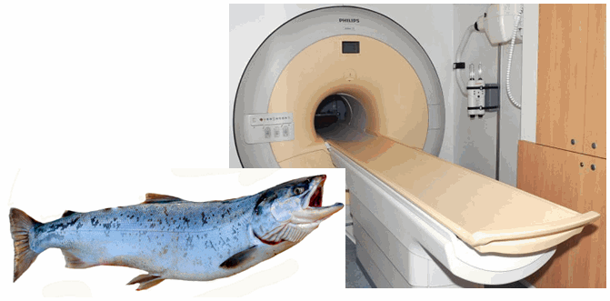
Dead salmon engaged in human perspective-taking, uncorrected fMRI study reports

salmon

Subject. One mature Atlantic Salmon (Salmo salar) participated in the fMRI study. The salmon was approximately 18 inches long, weighed 3.8 lbs, and was not alive at the time of scanning.

Task. The task administered to the salmon involved completing an open-ended mentalizing task. The salmon was shown a series of photographs depicting human individuals in social situations with a specified emotional valence. The salmon was asked to determine what emotion the individual in the photo must have been experiencing.”

Before explaining why you read what you just read and if it’s true (it is!), let me tell you that for many people, me included, the imaging studies seem very straightforward compared to, say, immunohistochemistry protocols. I mean, what do you have to do? You stick a human in a big scanner (fMRI, PET, or what-have-you), you start the image acquisition software and then some magic happens and you get pretty pictures of the human brain on your computer associated with some arbitrary numbers. Then you tell the humans to do something and you acquire more images which come with a different set of numbers. Finally, you compare the two sets of numbers and voila!: the neural correlates of whatever. Easy-peasy.

Well, it turns out it’s not so easy-peasy. Those numbers correspond to voxels, something like a pixel only 3D; a voxel is a small cube of brain (with the side of, say, 2 or 3 mm) comprising of hundreds of thousands to millions of brain cells. After this division, depending on your voxel size, you end up with a whooping 40,000 to 130,000 voxels or thereabouts for one brain. So a lot of numbers to compare.

When you do so many comparisons, by chance alone, you will find some that are significant. This is nature’s perverse way to show relationships where there are none and to screw-up a PhD. Those relationships are called false positives and the more comparisons you do, the more likely it is to find something statistically significant. So, in the ’90s, when the problem became very pervasive with the staggering amount of data generated by an fMRI scan, researchers came up with mathematical ways to dodge the problem, called multiple comparisons corrections (like application of the Gaussian Random Field Theory). Unfortunately, even 20 years later one could still find imaging studies with uncorrected results.

To show how important it is to perform that statistical correction, Bennet et al. (2010) did an fMRI study on perspective taking on one subject: a salmon. The subject was dead at the time of scanning. Now you can re-read the above excerpt from the Methods section.

Scroll down a bit to the Results section: “Out of a search volume of 8064 voxels a total of 16 voxels were significant”, p(uncorrected) < 0.001, showing that the salmon was engaging in active perspective-taking.

After the multiple comparisons correction, no voxel lit up, meaning that the salmon was not really imagining what the humans are feeling. Bummer…

The study has been extensively covered by media and I jumped on that wagon too – even if a bit late – because I never tire of this study as it’s absolutely funny and timeless. The authors even received the 2012 IgNobel prize for Neuroscience, as justly deserved. I refrained from fish puns because there are aplenty in the links I provided for you after the Reference. Feel free to come up with your own. Enjoy!

Reference: Bennett, CM, Baird AA, Miller MB & Wolford GL (2010). Neural correlates of interspecies perspective taking in the post-mortem Atlantic Salmon: An argument for multiple comparisons correction. Journal of Serendipitous Unexpected Results, 1, 1–5, presented as poster at the 2009 Human Brain Mapping conference. PDF | Nature cover | Neuroskeptic cover | Scientific American full story

By Neuronicus, 23 November 2015

How grateful would you feel after watching a Holocaust documentary? (Before you comment, READ the post first)

form Fox et al. (2015)

How would you feel if one of your favourite scientists published a paper that is, to put it in mild terms, not to their very best? Disappointed? Or perhaps secretly gleeful that even “the big ones” are not always producing pearl after pearl?

This is what happened to me after reading the latest paper of the Damasio group. Fox et al. (2015) decided to look for the neural correlates of gratitude. That is, stick people in fMRI, make them feel grateful, and see what lights up. All well and good, except they decided to go with a second-hand approach, meaning that instead of making people feel grateful (I don’t know how, maybe giving them something?), they made the participants watch stories in which gratitude may have been felt by other people (still not too too bad, maybe watching somebody helping the elderly). But, the researchers made an in-house documentary about the Holocaust and then had several actual Holocaust survivors tell their story (taken from the SC Shoah Foundation Institutes Visual History Archive), focusing on the part where their lives were saved or they were helped by others by giving them survival necessities. Then, the subjects were asked to immerse themselves in the story and tell how grateful they felt if they were the gift recipients.

I don’t know about you, but I don’t think that after watching a documentary about the Holocaust (done with powerfully evocative images and professional actor voice-overs, mind you!) and seeing people tell the horrors they’ve been through and then receiving some food or shelter from a Good Samaritan, gratitude would not have been my first feeling. Anger perhaps? That such an abominable thing as the Holocaust was perpetrated by my fellow humans? Sorrow? Sadness? Sick to my stomach? Compassion for the survivors? Maybe I am blatantly off-Gauss here, but I don’t think Damasio et co. measured what they thought they were measuring.

Anyway, for what is worth, the task produced significant activity in the medial prefrontal cortex (which is involved in so many behaviors that is not even worth listing them), along with the usual suspects in a task as ambiguous as this, like various portions of the anterior cingulate and orbitofrontal cortices.

Reference: Fox GR, Kaplan J, Damasio H, & Damasio A (30 September 2015). Neural correlates of gratitude. Frontiers in Psycholology, 6:1491. doi: 10.3389/fpsyg.2015.01491. Article | FREE FULLTEXT PDF

By Neuronicus, 27 October 2015

Save

The good and the bad of abstinence

The nucleus accumbens, a key region involved in reward processing and addiction. Credit: Zou et al. (2015)

Consumption of addictive drugs changes your brain and these changes underlie the consequent dependence. It is very difficult to quit, and certainly it is not a matter of lack of will power that the majority of drug users have such a great difficulty in trying to quit. But what happens to the brains of the lucky few who managed to kick the addiction out the window? Are their brains reverting to their pre-addiction states? As the paper below shows, sadly, no. But there is hope.

Zhou et al. ( 2015) used resting state fMRI (which means scanning your brain without requiring you to perform any task) to investigate the brains of 30 healthy controls and 30 heroin-addicts who were abstinent from the drug for more than 3 years. Specifically, they looked to see if the connectivity of certain brain regions involved in addiction is different after such a long abstinence time.

The bad news is that the abstinents still had some abnormal connectivity (for specialists: “stronger functional connectivity between the nucleus accumbens and the right ventromedial prefrontal cortex and relatively weaker connectivity between the nucleus accumbens and the left putamen, left precuneus, and supplementary motor area” p. 1697).

The good news: the longer the abstinence time, the greater the strength of the connection between nucleus accumbens and putamen (first structure involved in reward processing, the second in habit-learning), suggesting a partial neural recovery.

The study has some limitations: technical (did not control for heartbeat and respiration), methodological (most of the heroin abstinents were smokers, another addiction) and theoretical (one brain area does not support only one function, so its connectivity shouldn’t be over interpreted). With the caveat that the connectivity differences observed do not in any way point to a cause and effect relationship – that is we don’t know if these differences existed before the first heroin intake and caused it or they appeared after as a result of drug consumption, I think the paper is still is worth reading.

Reference: Zou F, Wu X, Zhai T, Lei Y, Shao Y, Jin X, Tan S, Wu B, Wang L, Yang Z (November 2015, Epub 17 Aug 2015). Abnormal resting-state functional connectivity of the nucleus accumbens in multi-year abstinent heroin addicts. Journal of Neuroscience Research, 93(11):1693-702. doi: 10.1002/jnr.23608. Article | FREE PDF

By Neuronicus, 9 October 2015

Dopamine role still not settled

vta pfc
No idea why the prefrontal cortex neuron is Australian, but here you go. Cartoon made by me with free (to the best of my knowledge) clipart elements. Feel free to use to your heart’s content.

There have been literally thousands of pages published about the dopamine function(s). Dopamine, which made its stage debut as the “pleasure molecule”, is a chemical produced by some neurons in your brain that is vital to its functioning. It has been involved in virtually all types of behavior and most diseases, from pain to pleasure, from mating to addiction, from working-memory to decision-making, from autism to Parkinson’s, from depression to schizophrenia.

Here is another account about what dopamine really does in the brain. Schwartenbeck et al. (2015) trained 26 young adults to play a game in which they had to decide whether to accept an initial offer of small change or to wait for a more substantial offer. If they waited too long, they would lose everything. After that, the subjects played the game in the fMRI. The authors argue that their clever game allows segregation between previously known roles of dopamine, like salience or reward prediction.

As expected with most fMRI studies, a brain salad lit up (that is, your task activated many other structures in addition to your region of interest), which the authors address only very briefly. Instead, they focus on the timing of activation of their near and dear midbrain dopamine neurons, which they cannot detect directly in the scanner because their cluster is too small, so they infer their location by proxy. Anyway, after some glorious mental (and mathematical) gymnastics Schwartenbeck et al. (2015) conclude that

1) “humans perform hierarchical probabilistic Bayesian inference” (p. 3434) (i.e. “I don’t have a clue what’s going on here, so I’ll go with my gut instinct on this one”) and

2) dopamine discharges reflect the confidence in those inferences (i.e. “how sure am I that doing this is going to bring me goodies?”)

With the obvious caveat that the MRI doesn’t have the resolution to isolate the midbrain dopamine clusters and that these clusters refer to two very distinct population of dopamine neurons (ventral tegmental area and substantia nigra) with different physiological, topographical, and anatomical properties, and distinct connections, the study adds to the body of knowledge of “for the love of Berridge and Schultz, what the hell are you DOIN’, dopamine neuron?”.

Reference: Schwartenbeck, P., FitzGerald, T. H., Mathys, C., Dolan, R., & Friston K. (October 2015, Epub 23 July 2014). The Dopaminergic Midbrain Encodes the Expected Certainty about Desired Outcomes. Cerebral Cortex, 25:3434–3445, doi:10.1093/cercor/bhu159. Article + FREE PDF

By Neuronicus, 8 October 2015

Pee now! NOW, I said! In a huge magnet. While we watch.

Photo credit: Free clipart from www.cliparthut.com
Photo credit: Free clipart from http://www.cliparthut.com

Like many studies that fill in unknown gaps in the body of knowledge, the paper below may not attract attention, except from the people in their narrow field. So let’s give it a little attention.

Michels et al. (2015) sought to map out the brain network underlying the control of urination using an fMRI scanner. They got 22 healthy adult males and they gave them furosemide, which is a diuretic, and then asked them to drink as much water as they want until they need to urinate. During this, “a condom catheter was attached to the penis of each subject” (p. 3370), to monitor the urine flow while in scanner. Then the testing would not start, oh no. The subjects were then submitted to an ultrasound to make sure the bladder was full. Then they were asked again how much they really needed to go pee. Then they go in the scanner in a supine position, where they are told to wait, then to imagine the starting of urination (but don’t pee just yet!), and finally, finally allowed to urinate. But then, cruelly, told to stop only 3 second into the act. And then the scanner cycle would repeat. Their champion peers (I cannot avoid the pun, I’m sorry) managed to pee 15 times in the scanner. I wonder how many subjects peed sans cue… (authors don’t mention that).

Fig. 1 from Michels et al. (2015) depicting the fMRI scan paradigm, which consisted of 2 randomly alternating blocks.
Fig. 1 from Michels et al. (2015) depicting the fMRI scan paradigm, which consisted of 2 randomly alternating blocks. SDV = strong desire to void.

Amazingly, under these conditions, there were seven men who could not urinate in the scanner. Authors call these non-voiders, or, as we commonly know them, the shy bladders or the bashful kidneys. Not surprisingly, non-voiders had lower activity in the pontine micturition center (PMC), a brain area, which, as its name implies, is responsible for urination. Also not surprisingly – for me at least, the authors find this interesting -, the non-voiders showed increased activity in the anterior midcingulate cortex (aMCC), which is an area involved in control. I guess you need some steely control to not pee after all that. The aMCC inhibits the urination-facilitation brain regions, such as the PMC.

Anyway, the main finding of the study is a detailed map of micturition supraspinal mechanisms, which consists of a slew of structures, each with its own function. I had never known how complicated peeing and not-peeing are until I read this paper. Jokes and cringes aside, this study is a welcome addition to understanding how we control our bodily functions and where to start looking when this control fails, shedding new light on the interplay between reflex and control.

Reference: Michels, L., Blok, B. F., Gregorini, F., Kurz, M., Schurch, B., Kessler, T. M., Kollias, S., & Mehnert, U. (October 2015, Epub Jun 26 2014). Supraspinal Control of Urine Storage and Micturition in Men-An fMRI Study. Cerebral Cortex, 25(10): 3369-80. doi: 10.1093/cercor/bhu140. Article | FREE FULLTEXT PDF

By Neuronicus, 1 October 2015

Zap the brain to get more lenient judges

Neuronicus3
Reference: Buckholtz et al. (2015). Credit: Neuronicus

Humans manage to live successfully in large societies mainly because we are able to cooperate. Cooperation rests on commonly agreed rules and, equally important, the punishment bestowed upon their violators. Researchers call this norm enforcement, while the rest of us call it simply justice, whether it is delivered in its formal way (through the courts of law) or in a more personal manner (shout at the litterer, claxon the person who cut in your lane etc.). It is a complicate process to investigate, but scientists managed to break it into simpler operations: moral permissibility (what is the rule), causal responsibility (did John break the rule), moral responsibility (did John intend to break the rule, also called blameworthiness or culpability), harm assessment (how much harm resulted from John breaking the rule) and sanction (give the appropriate punishment to John). Different brain parts deal with different aspects of norm enforcement.

The approximate area where the stimulation took place. Note that the picture depicts the left hemisphere, whereas the low punishment judgement occured when the stimulation was mostly on the right hemisphere.
The approximate area where the stimulation took place. Note that the picture depicts the left hemisphere, whereas the low punishment judgement occurred when the stimulation was mostly on the right hemisphere.

Using functional magnetic resonance imaging (fMRI), Buckholtz et al. found out that the dorsolateral prefrontal cortex (DLPFC) gets activated when 60 young subjects decided what punishment fits a crime. Then, they used repetitive transcranial magnetic stimulation (rTMS), which is a non-invasive way to disrupt the activity of the neurons, to see what happens if you inhibit the DLPFC. The subjects made the same judgments when it came to assigning blame or assessing the harm done, but delivered lower punishments.

Reference: Buckholtz, J. W., Martin, J. W., Treadway, M. T., Jan, K., Zald, D.H., Jones, O., & Marois, R. (23 September 2015). From Blame to Punishment: Disrupting Prefrontal Cortex Activity Reveals Norm Enforcement Mechanisms. Neuron, 87: 1–12, http://dx.doi.org/10.1016/j.neuron.2015.08.023. Article + FREE PDF

by Neuronicus, 22 September 2015