Old chimpanzees get Alzheimer’s pathology

Alzheimer’s Disease (AD) is the most common type of dementia with a progression that can span decades. Its prevalence is increasing steadily, particularly in the western countries and Australia. So some researchers speculated that this particular disease might be specific to humans. For various reasons, either genetic, social, or environmental.

A fresh e-pub brings new evidence that Alzheimer’s might plague other primates as well. Edler et al. (2017) studied the brains of 20 old chimpanzees (Pan troglodytes) for a whole slew of Alzheimer’s pathology markers. More specifically, they looked for these markers in brain regions commonly affected by AD, like the prefrontal cortex, the midtemporal gyrus, and the hippocampus.

Alzheimer’s markers, like Tau and Aβ lesions, were present in the chimpanzees in an age-dependent manner. In other words, the older the chimp, the more severe the pathology.

Interestingly, all 20 animals displayed some form of Alzheimer’s pathology. This finding points to another speculation in the field which is: dementia is just part of normal aging. Meaning we would all get it, eventually, if we would live long enough; some people age younger and some age older, as it were. This hypothesis, however, is not favored by most researchers not the least because is currently unfalsifiable. The longest living humans do not show signs of dementia so how long is long enough, exactly? But, as the authors suggest, “Aβ deposition may be part of the normal aging process in chimpanzees” (p. 24).

Unfortunately, “the chimpanzees in this study did not participate in formal behavioral or cognitive testing” (p. 6). So we cannot say if the animals had AD. They had the pathological markers, yes, but we don’t know if they exhibited the disease as is not uncommon to find these markers in humans who did not display any behavioral or cognitive symptoms (Driscoll et al., 2006). In other words, one might have tau deposits but no dementia symptoms. Hence the title of my post: “Old chimpanzees get Alzheimer’s pathology” and not “Old chimpanzees get Alzheimer’s Disease”

Good paper, good methods and stats. And very useful because “chimpanzees share 100% sequence homology and all six tau isoforms with humans” (p. 4), meaning we have now a closer to us model of the disease so we can study it more, even if primate research has taken significant blows these days due to some highly vocal but thoroughly misguided groups. Anyway, the more we know about AD the closer we are of getting rid of it, hopefully. And, soon enough, the aforementioned misguided groups shall have to face old age too with all its indignities and my guess is that in a couple of decades or so there will be fresh money poured into aging diseases research, primates be damned.

121-chimps get Alz - Copy

REFERENCE: Edler MK, Sherwood CC, Meindl RS, Hopkins WD, Ely JJ, Erwin JM, Mufson EJ, Hof PR, & Raghanti MA. (EPUB July 31, 2017). Aged chimpanzees exhibit pathologic hallmarks of Alzheimer’s disease. Neurobiology of Aging, PII: S0197-4580(17)30239-7, DOI: http://dx.doi.org/10.1016/j.neurobiolaging.2017.07.006. ABSTRACT  | Kent State University press release

By Neuronicus, 23 August 2017

Save

Save

CCL11 found in aged but not young blood inhibits adult neurogenesis

vil - Copy
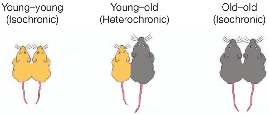
Portion of Fig. 1 from Villeda et al. (2011, doi: 10.1038/nature10357) describing the parabiosis procedure. Basically, under complete anesthesia, the peritoneal membranes and the skins of the two mice were sutured together. The young mice were 3–4 months (yellow) and old mice were 18–20 months old (grey).

My last post was about parabiosis and its sparse revival as a technique in physiology experiments. Parabiosis is the surgical procedure that joins two living animals allowing them to share their circulatory systems. Here is an interesting paper that used the method to tackle blood’s contribution to neurogenesis.

Adult neurogenesis, that is the birth of new neurons in the adult brain, declines with age. This neurogenesis has been observed in some, but not all brain regions, called neurogenic niches.

Because these niches occur in blood-rich areas of the brain, Villeda et al. (2011) wondered if, in addition with the traditional factors required for neurogenesis like enrichment or running, blood factors may also have something to do with neurogenesis. The authors made a young and an old mouse to share their blood via parabiosis (see pic.).

Five weeks after the parabiosis procedure, the young mouse had decreased neurogenesis and the old mouse had increased neurogenesis compared to age-matched controls. To make sure their results are due to something in the blood, they injected plasma from an old mouse into a young mouse and that also resulted in reduced neurogenesis. Moreover, the reduced neurogenesis was correlated with impaired learning as shown by electrophysiological recordings from the hippocampus and from behavioral fear conditioning.

So what in the blood does it? The authors looked at 66 proteins found in the blood (I don’t know the blood make-up, so I can’t tell if 66 is a lot or not ) and noticed that 6 of these had increased levels in the blood of ageing mice whether linked by parabiosis or not. Out of these six, the authors focus on CCL11 (unclear to me why that one, my bet is that they tried the others too but didn’t have enough data). CCLL11 is a small signaling protein involved in allergies. So the authors injected it into young mice and Lo and Behold! there was decreased neurogenesis in their hippocampus. Maybe the vampires were onto something, whadda ya know? Just kidding… don’t go around sucking young people’s blood!

This paper covers a lot of work and, correspondingly, has no less than 23 authors and almost 20 Mb of supplemental documents! The story it tells is very interesting and as complete as it gets, covering many aspects of the problems investigated and many techniques to address those problems. Good read.

Reference: Villeda SA, Luo J, Mosher KI, Zou B, Britschgi M, Bieri G, Stan TM, Fainberg N, Ding Z, Eggel A, Lucin KM, Czirr E, Park JS, Couillard-Després S, Aigner L, Li G, Peskind ER, Kaye JA, Quinn JF, Galasko DR, Xie XS, Rando TA, Wyss-Coray T. (31 Aug 2011). The ageing systemic milieu negatively regulates neurogenesis and cognitive function. Nature. 477(7362):90-94. doi: 10.1038/nature10357. Article | FREE Fulltext PDF

By Neuronicus, 6 January 2016

Herpes viruses infect neurons

virus EBV
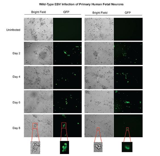
FIG. 3 from Jha et al. (2015). Wild-type EBV infection of primary human fetal neurons. Fluorescence microscopy was carried out at 2, 4, 6, and 8 days post infection to monitor for GFP expression (the fluorescent label). Microscopy images were captured at x20 magnification.

For some mysterious reason, whether Epstein-Barr virus (EBV) and Kaposi’s sarcoma-associated herpesvirus (KSHV) can infect neurons has not been established until now. Probably because some viruses from the same family do not infect neurons, so it was assumed that EPV and KSHV do not either.

Jha et al. (2015) cultured Sh-Sy5y neuroblastoma cells, teratocarcinoma Ntera2 neurons, and primary human fetal neurons in Petri dishes and then exposed them to these viruses. After infection, the authors did some fluorescence microscopy (they tagged the viruses with fluorescent dyes), real-time PCR (to confirm there was viral RNA in the cells), immunofluorescence assays (to see if the viral proteins are expressed) and Western blot analyses (to see if the specific viral antigens are made). All these showed that the viruses were happily multiplying in the cells.

Now here comes the significance of the study: EBV and KSHV are viruses associated with all sorts of nasty diseases like mononucleosis and cancers. EBV has also been associated with neurological disorders, like multiple sclerosis, Alzheimer’s, neuropathies, lymphomas etc. But the critical word here is “associated”. That is, they found these viruses in people suffering from those diseases. So the knowledge that these viruses infect neurons could point to a mechanism behind these associations. Unfortunately, EBV is present in 90-95% of the population of the world. Which means that you will find this virus in, let’s say, 9 out of 10 people suffering from Alzheimer’s, assuming normal distributions and random sampling. So the virus’s presence maybe completely unrelated to the disease. By the same rationale, you would find the virus in 9 out of 10 people found guilty of theft, for example. It would be then interesting to see find what is NOT associated with.

Caveat: I have not read the association studies, so my argument holds only if what they report is that people with disease X also have EBV. If they made, however, a comparisons and found out that people with disease X are significantly more likely to be infected with EBV than the ones without the disease, then the argument does not hold.

Reference: Jha HC, Mehta D, Lu J, El-Naccache D, Shukla SK, Kovacsics C, Kolson D, Robertson ES (1 Dec 2015). Gammaherpesvirus Infection of Human Neuronal Cells. MBio,  6(6). pii: e01844-15. doi: 10.1128/mBio.01844-15. Article | FREE FULLTEXT PDF | PsyPost cover

By Neuronicus, 7 December 2015

Obscure protein restores memory decline

In the not-too-distant-future, your grandma may give you a run for your money on your video games. Photo credit: http://www.funtoosh.com/pictures/
In the not-too-distant-future, your grandma may give you a run for your money on your video games. Photo credit: Funtoosh

Aging comes with all sorts of maladies, but one of the most frustrating is the feeling that you are not as sharp as you used to be. Cognitive decline has been previously linked, at least in part, to a dysregulation in the neuronal calcium homeostasis in the hippocampus, which is a brain region essential for learning and memory. One player that keeps in check the proper balance of calcium use is the protein FKBP1b, and, not surprisingly, its amounts are reduced in aging rats and Alzheimer’s suffering patients.

FKBP1b overexpression in hippocampal neurons reversed spatial memory deficits in aged rats. Fig. 3 (partial) from Gant, J. C., Chen, K. C., Kadish, I., Blalock, E. M., Thibault, O., Porter, N. M., Landfield, P. W. (29 July 2015). Reversal of Aging-Related Neuronal Ca2+ Dysregulation and Cognitive Impairment by Delivery of a Transgene Encoding FK506-Binding Protein 12.6/1b to the Hippocampus. The Journal of Neuroscience, 35(30):10878 –10887. doi: 10.1523/JNEUROSCI.1248-15.2015.
FKBP1b overexpression in hippocampal neurons reversed spatial memory deficits in aged rats. Fig. 3 (partial) from Gant et al. (2015): doi: 10.1523/JNEUROSCI.1248-15.2015.

Gant et al. (2015) sought to increase the expression of the FKBP1b protein in the hippocampus, in the hopes that its increase would result in better calcium homeostasis and, as a result, better memory performance in aging rats. They built a virus that carried the gene for making the FKBP1b protein and they injected this directly in the hippocampus. After they waited 5-6 weeks for the gene to be expressed, they tested the rats in the Morris water maze, a test for spatial memory. The old rats that received the injection performed as well as the young rats, and far better than the old rats who didn’t get the injection. Then the researchers made sure that the injection is the one responsible for the results, by checking the levels of the FKBP1b protein in the hippocampus (increased, as per specs), by recording from those neurons (they were awesome), and by imaging the calcium to make sure the balance was restored (ditto).

Reference: Gant, J. C., Chen, K. C., Kadish, I., Blalock, E. M., Thibault, O., Porter, N. M., Landfield, P. W. (29 July 2015). Reversal of Aging-Related Neuronal Ca2+ Dysregulation and Cognitive Impairment by Delivery of a Transgene Encoding FK506-Binding Protein 12.6/1b to the Hippocampus. The Journal of Neuroscience, 35(30):10878 –10887. doi: 10.1523/JNEUROSCI.1248-15.2015. Article + FREE PDF + Journal of Neuroscience cover

The song of a fly… the courtship of another

Drosophila melanogaster image illustrating sexual dimorphism and mating behavior. Credit: TheAlphaWolf (Wikimedia Commons)

Did you know that flies sing? True to the dictum that I just made up – ‘where is song, there is lust’ – it turns out not only that flies can sing, but they even have courtship songs! Granted, since they don’t have a larynx, the male flies sing by vibrating their wings in a certain way, which is unique to each fly species, and females listen with the feather-looking bit on top of their antennae, called arista. The behavior has generated enough research that a fairly hefty review about it has been published two years ago in Nature Reviews Neuroscience, pointing to a gene central to the male courtship circuitry and expressed only in the fly’s neurons, the fru gene (I bet it was called that way because when you make mutants you get fru/fru …).

Zhou et al. (2015) used a series of complicated experiments to successively activate or inhibit the neurons which express the fru gene, in order to identify the neural circuitry underlying hearing and processing the courtship songs. This circuitry is different in males and females, which makes sense since the serenading male expects different behaviors from his audience, depending on their sex; the listening males hurry to compete for the intended female and the females slow down and… listen carefully. Mind wondering: if I was the one serenading, wouldn’t I want to drive away the competitors, instead of drawing them in towards the object of my desire? Perhaps I want the competitors to also engage in courtship behavior so I can show off my wing vibrating prowess… Anyway, digression aside, in addition to figuring out which neuron does what, the authors managed to elicit courtship behavior in the listening males by optogenetically stimulating the 3rd and 4th order neurons in the newly identified circuit.

Besides being strangely interesting in itself, the research fills a gap in the understanding how courtship behavior is recognized, at least in fruit flies, which may be very useful information for other species as well, humans included.

Reference: Zhou, C., Franconville, R., Vaughan, A. G., Robinett, C. C., Jayaraman, V., & Baker, B. S. (21 September 2015). Central neural circuitry mediating courtship song perception in male Drosophila. Elife, 4:1-15. doi: 10.7554/eLife.08477. Article + FREE PDF

For the interested specialist, the MATLAB source code for analyzing calcium-imaging data can be found here.

By Neuronicus, 24 September 2015