The FIRSTS: Streptomycin was isolated and tested for its antibiotic properties by graduate students in 1943

A principal investigator tells a graduate student to discover new antibiotics using well known methods. Student discovers new antibiotic. Then, another graduate student performs the antibiotic tests to see if it works. Who takes the credit? The PI got the credit and the Nobel and the money. The discoverer sued and got some credit and some money. The tester got nothing. The tester was a woman. According to the woman’s daughter, “her mother’s colleagues told her it wasn’t important that her name be on the patent because she would one day get married and have a family”.

After the streptomycin controversy, the universities put forth regulations to make (somewhat) clearer who gets what in the process of discovery. Thanks to this and thanks to the women’s liberation movement and, more recently, the women’s fight for equality in workplaces, if this would happen today in a serious University, probably all three would share the Nobel. Probably.

Bibliography:

1)  Angelova, Lidiya. “Elizabeth Bugie – the invisible woman in the discovery of streptomycin”. Scientista | Women in STEM. Retrieved 2023-10-19.

2)  Eveleigh, Douglas E.; Bennett, Joan W. (2018-05-01), Whitaker; Barton (eds.), “Women Microbiologists at Rutgers in the Early Golden Age of Antibiotics”, Women in Microbiology, American Society of Microbiology, pp. 317–329, doi:10.1128/9781555819545.ch34, ISBN 978-1-55581-953-8

3)  Kingston W (July 2004). “Streptomycin, Schatz v. Waksman, and the balance of credit for discovery”. Journal of the History of Medicine and Allied Sciences. 59 (3): 441–462. doi: 10.1093/jhmas/jrh091. PMID 15270337. S2CID 27465970.

4) Schatz, Albert (1993). “The true story of the development of streptomycin”. Actinomycetes. 4 (2): 27–39. Couldn’t find the full text.

By Neuronicus, 19 October 2023

The Kallmann syndrome should be Maestre syndrome (or MSJJ)

Few of the Generation X people are unfamiliar with the super-hit/movie cult Twin Peaks. And from those, even fewer find the dwarf dancing in the Red Room not scary as hell. And yet even fewer know who the man singing “Sycamore Trees” in that room is. For memory refreshment, here is the clip.

The man with the unusual voice, to match all the rest of the… um… “unusualness”, is none other than Jimmy Scott, a phenomenal jazz singer. If you listen to him, particularly in the hit “I’m Afraid The Masquerade Is Over“, you’ll notice that, if you close your eyes, you might be confused on whether the voice belongs to an adult man or a very gifted prepubertal boy.

And that is because Jimmy Scott suffered his entire life from a rare and obscure disease, called the “Kallmann syndrome”. This is a genetic disorder that prevents a person to start or to fully complete puberty. And that is because they have low circulating sex hormones: testosterone in males and estrogen and progesterone in females. And that is because their hypothalamus does not produce enough or at the proper times gonadotropin-releasing hormone (GnRH). And that is because sometime during the first trimester of pregnancy, there was an abnormality in the development of the olfactory fibers. You might ask what does smell development has with sex hormones? Well, I’m glad you asked. The cells that will end up releasing GnRH during puberty need to migrate from where they are born (nasal epithelium) to hypothalamus. If something impedes said migration, like, say, a mass, then the cells cannot reach their destination and you get a person who cannot start or finish puberty, plus a few other symptoms (Teixeira et al., 2010).

As some of you might have already surmised, something else besides lack of puberty must happen to these people regarding their sense of smell. After all, their olfactory fibers got tangled during embryonic development, forming, probably, a benign tumor, a neuroma. Not surprisingly, the person ends up with severely diminished or absent sense of smell, information that can be clinically used for diagnosis (Yu et al., 2022).

The first one to notice that people with failure to start or fully complete puberty are also anosmic was Aureliano Maestre de San Juan, a Spanish scientist. Unfortunately, the syndrome he documented in 1856 was named not named after him, but after the German scientist Franz Joseph Kallmann who described it almost a century later, in 1944 (Martin et al., 2011). Kallmann not only he did not discover the syndrome himself, but he was a staunch supporter of “racial hygiene”, advocating for finding and sterilizing relatives of people with schizophrenia so to eradicate the disease from future generations. Ironically, he fled Germany in 1939 because he was of Jewish heritage into a country which enthusiastically embraced eugenics and performed their own sterilizations programs, the USA (Benbassat, 2016).

So, I hereby propose, in an obscure unvisited corner of the Internet, to rename the disease the Maestre syndrome or MSJJ, after the guy who actually noticed it the first time and published about it. Besides, it’s his birthday today, having been born on October 17, 1828.

REFERENCES (in order of appearance):

Yu B, Chen K, Mao J, Hou B, You H, Wang X, Nie M,Huang Q, Zhang R, Zhu Y, Sun B, Feng F, Zhou W, & Wu X (2022, Sep 22).The diagnostic value of the olfactory evaluation for congenital hypogonadotropic hypogonadism. Frontiers in Endocrinology (Lausanne). 2022; 13: 909623. doi: 10.3389/fendo.2022.909623, PMCID: PMC9523726, PMID: 36187095. ARTICLE | FREE FULLTEXT PDF

Teixeira L, Guimiot F, Dodé C, Fallet-Bianco C, Millar RP, Delezoide A-L, & Hardelin J-P (2010, Oct 1, Published online 2010 Sep 13). Defective migration of neuroendocrine GnRH cells in human arrhinencephalic conditions. Journal of Clinical Investigation, 120(10): 3668–3672. doi: 10.1172/JCI43699, PMCID: PMC2947242, PMID: 20940512. ARTICLE | FREE FULLTEXT PDF

Benbassat, CA (2016, Published online 2016 Apr 19). Kallmann Syndrome: Eugenics and the Man behind the Eponym. Rambam Maimonides Medical Journal, 7(2): e0015. doi: 10.5041/RMMJ.10242, PMCID: PMC4839542, PMID: 27101217. ARTICLE | FREE FULLTEXT ARTICLE

Martin C, Balasubramanian R, Dwyer AA, Au MG, Sidis Y, Kaiser UB, Seminara SB, Pitteloud N,  Zhou Q-Y, & Crowley, Jr WF (2011, Published online 2010 Oct 29). The Role of the Prokineticin 2 Pathway in Human Reproduction: Evidence from the Study of Human and Murine Gene Mutations. Endocrine Reviews, 32(2): 225–246. doi: 10.1210/er.2010-0007, PMCID: PMC3365793, PMID: 21037178. ARTICLE | FREE FULLTEXT PDF

Original references which I couldn’t find, as they appear in Martin et al. (2011):

Maestre de San Juan A. (1856). Teratologia: Falta total de los nervios olfactorios con anosmia en un individuo en quien existia una atrofia congénita de los testículos y miembro viril. Siglo Medico, 3:211–221 [Google Scholar]

Kallmann F, Schoenfeld W & Barrera S (1944). The genetic aspects of primary eunuchoidism. Am J Ment Defic, 48:203–236 [Google Scholar]

By Neuronicus, 17 October 2022

The FIRSTS: Guanine comes from guano (1846)

This is about naming things in science. You have been warned!

The DNA is made of four nucleobases: adenine (A), thymine (T), cytosine (C) and guanine (G). The “letters” of the code. Each of them has been named from where they were originally obtained by the scientists who first identified and/or isolated them.

Adenine was named thus because it was extracted from the pancreas of an ox, which is aden in Greek (the pancreas, not the ox), by the Nobel laureate Albrecht Kossel in 1885.

Thymine comes from thymic acid, which was extracted from the thymus gland of calves by the same Albrecht Kossel and Albert Neumann in 1893.

A year later, the duo named cytosine, another base obtained from the same thymus tissue. Cyto- pertains to cells in Greek.

Fifty years before that, Julion Bodo Unger, a German chemist, extracted the guanine from the guano of sea birds. Why was he looking at bird poop, curious minds inquire? Because he was studying it for its uses as fertilizer. The year of discovery was 1844 and the year of the naming was 1846.

And now you know…

153 guano - Copy

REFERENCE: Unger, JB (1846). Bemerkungen zu obiger Notiz (Comments on the above notice), Annalen der Chemie und Pharmacie, 58: 18-20. From page 20: … desshalb möchte ich den Namen Guanin vorschlagen, welcher an seine Herkunft erinnert.” ( “… therefore I would like to suggest the name guanine, which is reminiscent of its origin”.) (Wikipedia translation). Google Books | Google Book PDF

By Neuronicus, 3 June 2019

High fructose corn syrup IS bad for you

Because I cannot leave controversial things well enough alone – at least not when I know there shouldn’t be any controversy – my ears caught up with my tongue yesterday when the latter sputtered: “There is strong evidence for eliminating sugar from commonly used food products like bread, cereal, cans, drinks, and so on, particularly against that awful high fructose corn syrup”. “Yeah? You “researched” that up, haven’t you? Google is your bosom friend, ain’t it?” was the swift reply. Well, if you get rid of the ultra-emphatic air-quotes flanking the word ‘researched’ and replace ‘Google’ with ‘Pubmed’, then, yes, I did researched it and yes, Pubmed is my bosom friend.

Initially, I wanted to just give you all a list with peer-reviewed papers that found causal and/or correlational links between high fructose corn syrup (HFCS) and weight gain, obesity, type 2 diabetes, cardiovascular disease, fatty liver disease, metabolic and endocrine anomalies and so on. But there are way too many of them; there are over 500 papers on the subject in Pubmed only. And most of them did find that HFCS does nasty stuff to you, look for yourselves here. Then I thought to feature a paper showing that HFCS is differently metabolized than the fructose from fruits, because I keep hearing that lie perpetrated by the sugar and corn industries that “sugar is sugar” (no, it’s not! Demonstrably so!), but I doubt my yesterday’s interlocutor would care about liver’s enzymatic activity and other chemical processes with lots of acronyms. So, finally, I decided to feature a straight forward, no-nonsense paper, published recently, done at a top tier university, with human subjects, so I won’t hear any squabbles.

Price et al. (2018) studied 49 healthy subjects aged age 18–40 yr, of normal and stable body weight, and free from confounding medications or drugs, whose physical activity and energy-balanced meals were closely monitored. During the study, the subjects’ food and drink intake as well as their timing were rigorously controlled. The researchers varied only the beverages between groups, in such a way that one group received a drink sweetened with HFCS-55 (55% fructose, 45% glucose, as the one used in commercially available drinks) with every controlled meal, whereas the other group received an identical drink in size (adjusted for their energy requirements in such a way that it provided the same 25% of it), but sweetened with aspartame. The study lasted two weeks. No other beverage was allowed, including fruit juice. Urine samples were collected daily and blood samples 4 times per day.

There was a body weight increase of 810 grams (1.8 lb) in subjects consuming HFCS-sweetened beverages for 2 weeks when compared with aspartame controls. The researches also found differences in the levels of a whole host of acronyms (ppTG, ApoCIII, ApoE, OEA, DHEA, DHG, if you must know) involved in a variety of nasty things, like obesity, fatty liver disease, atherosclerosis, cardiovascular disease, stroke, diabetes, even Alzheimer’s.

This study is the third part of a larger NIH-funded study which investigates the metabolic effects of consuming sugar-sweetened beverages in about 200 participants over 5 years, registered at clinicaltrials.gov as NCT01103921. The first part (Stanhope et al., 2009) reported that consuming fructose-sweetened, not glucose-sweetened, beverages increases visceral adiposity and lipids and decreases insulin sensitivity in overweight/obese humans” (title), and the second part (Stanhope et al., 2015) found that “consuming beverages containing 10%, 17.5%, or 25% of energy requirements from HFCS produced dose-dependent increases in circulating lipid/lipoprotein risk factors for cardiovascular disease and uric acid within 2 weeks” (Abstract). They also found a dose-dependant increase in body weight, but in those subjects the results were not statistically significant (p = 0.09) after correcting for multiple comparisons. But I’ll bet that if/when the authors will publish all the data in one paper at the end of clinical trials they will have more statistical power and the trend in weight gain more obvious, as in the present paper.  Besides, it looks like there may be more than three parts to this study anyway.

The adverse effects of a high sugar diet, particularly in HFCS, are known to so many researchers in the field that they have been actually compiled in a name: the “American Lifestyle-Induced Obesity Syndrome model, which included consumption of a high-fructose corn syrup in amounts relevant to that consumed by some Americans” (Basaranoglu et al., 2013). It doesn’t refer only to increases in body weight, but also type 2 diabetes, cardiovascular disease, hypertriglyceridemia, fatty liver disease, atherosclerosis, gout, etc.

The truly sad part is that avoiding added sugars in diets in USA is impossible unless you do all – and I mean all – your cooking home, including canning, jamming, bread-making, condiment-making and so on, not just “Oh, I’ll cook some chicken or ham tonight” because in that case you end up using canned tomato sauce (which has added sugar), bread crumbs (which have added sugar), or ham (which has added sugar), salad dressing (which has sugar) and so on. Go on, check your kitchen and see how many ingredients have sugar in them, including any meat products short of raw meat. If you never read the backs of the bottles, cans, or packages, oh my, are you in for a big surprise if you live in USA…

There are lot more studies out there on the subject, as I said, of various levels of reading difficulty. This paper is not easy to read for someone outside the field, that’s for sure. But the main gist of it is in the abstract, for all to see.

150 hfcs - Copy

P.S. 1. Please don’t get me wrong: I am not against sugar in desserts, let it be clear. Nobody makes a more mean sweetalicious chocolate cake or carbolicious blueberry muffin than me, as I have been reassured many times. But I am against sugar in everything. You know I haven’t found in any store, including high-end and really high-end stores a single box of cereal of any kind without sugar? Just for fun, I’d like to be a daredevil and try it once. But there ain’t. Not in USA, anyway. I did find them in EU though. But I cannot keep flying over the Atlantic in the already crammed at premium luggage space unsweetened corn flakes from Europe which are probably made locally, incidentally and ironically, with good old American corn.

P.S. 2 I am not so naive, blind, or zealous to overlook the studies that did not find any deleterious effects of HFCS consumption. Actually, I was on the fence about HFCS until about 10 years ago when the majority of papers (now overwhelming majority) was showing that HFCS consumption not only increases weight gain, but it can also lead to more serious problems like the ones mentioned above. Or the few papers that say all added sugar is bad, but HFCS doesn’t stand out from the other sugars when it comes to disease or weight gain. But, like with most scientific things, the majority has it its way and I bow to it democratically until the new paradigm shift. Besides, the exposés of Kearns et al. (2016a, b, 2017) showing in detail and with serious documentation how the sugar industry paid prominent researchers for the past 50 years to hide the deleterious effects of added sugar (including cancer!) further cemented my opinion about added sugar in foods, particularly HFCS.

References:

  1. Price CA, Argueta DA, Medici V, Bremer AA, Lee V, Nunez MV, Chen GX, Keim NL, Havel PJ, Stanhope KL, & DiPatrizio NV (1 Aug 2018, Epub 10 Apr 2018). Plasma fatty acid ethanolamides are associated with postprandial triglycerides, ApoCIII, and ApoE in humans consuming a high-fructose corn syrup-sweetened beverage. American Journal of Physiology. Endocrinology and Metabolism, 315(2): E141-E149. PMID: 29634315, PMCID: PMC6335011 [Available on 2019-08-01], DOI: 10.1152/ajpendo.00406.2017. ARTICLE | FREE FULTEXT PDF
  1. Stanhope KL1, Medici V2, Bremer AA2, Lee V2, Lam HD2, Nunez MV2, Chen GX2, Keim NL2, Havel PJ (Jun 2015, Epub 22 Apr 2015). A dose-response study of consuming high-fructose corn syrup-sweetened beverages on lipid/lipoprotein risk factors for cardiovascular disease in young adults. The American Journal of Clinical Nutrition, 101(6):1144-54. PMID: 25904601, PMCID: PMC4441807, DOI: 10.3945/ajcn.114.100461. ARTICLE | FREE FULTEXT PDF
  1. Stanhope KL1, Schwarz JM, Keim NL, Griffen SC, Bremer AA, Graham JL, Hatcher B, Cox CL, Dyachenko A, Zhang W, McGahan JP, Seibert A, Krauss RM, Chiu S, Schaefer EJ, Ai M, Otokozawa S, Nakajima K, Nakano T, Beysen C, Hellerstein MK, Berglund L, Havel PJ (May 2009, Epub 20 Apr 2009). Consuming fructose-sweetened, not glucose-sweetened, beverages increases visceral adiposity and lipids and decreases insulin sensitivity in overweight/obese humans. The Journal of Clinical Investigation,119(5):1322-34. PMID: 19381015, PMCID: PMC2673878, DOI:10.1172/JCI37385. ARTICLE | FREE FULTEXT PDF

(Very) Selected Bibliography:

Bocarsly ME, Powell ES, Avena NM, Hoebel BG. (Nov 2010, Epub 26 Feb 2010). High-fructose corn syrup causes characteristics of obesity in rats: increased body weight, body fat and triglyceride levels. Pharmacology, Biochemistry, and Behavior, 97(1):101-6. PMID: 20219526, PMCID: PMC3522469, DOI: 10.1016/j.pbb.2010.02.012. ARTICLE | FREE FULLTEXT PDF

Kearns CE, Apollonio D, Glantz SA (21 Nov 2017). Sugar industry sponsorship of germ-free rodent studies linking sucrose to hyperlipidemia and cancer: An historical analysis of internal documents. PLoS Biology, 15(11):e2003460. PMID: 29161267, PMCID: PMC5697802, DOI: 10.1371/journal.pbio.2003460. ARTICLE | FREE FULTEXT PDF

Kearns CE, Schmidt LA, Glantz SA (1 Nov 2016). Sugar Industry and Coronary Heart Disease Research: A Historical Analysis of Internal Industry Documents. JAMA Internal Medicine, 176(11):1680-1685. PMID: 27617709, PMCID: PMC5099084, DOI: 10.1001/jamainternmed.2016.5394. ARTICLE | FREE FULTEXT PDF

Mandrioli D, Kearns CE, Bero LA (8 Sep 2016). Relationship between Research Outcomes and Risk of Bias, Study Sponsorship, and Author Financial Conflicts of Interest in Reviews of the Effects of Artificially Sweetened Beverages on Weight Outcomes: A Systematic Review of Reviews. PLoS One, 11(9):e0162198.PMID: 27606602, PMCID: PMC5015869, DOI: 10.1371/journal.pone.0162198. ARTICLE | FREE FULTEXT PDF

By Neuronicus, 22 March 2019

Pic of the day: Total amount of DNA on Earth

139 DNA amount, better font - Copy

Approximately… give or take…

REFERENCE: Landenmark HKE, Forgan DH, & Cockell CS (11 Jun 2915). An Estimate of the Total DNA in the Biosphere. PLoS Biology, 13(6): e1002168. PMCID: PMC4466264, PMID: 26066900, DOI: 10.1371/journal.pbio.1002168. ARTICLE | FREE FULLTEXT PDF

By Neuronicus, 1 September 2018

The FIRSTS: The cause(s) of dinosaur extinction

A few days ago, a follower of mine gave me an interesting read from The Atlantic regarding the dinosaur extinction. Like many of my generation, I was taught in school that dinosaurs died because an asteroid hit the Earth. That led to a nuclear winter (or a few years of ‘nuclear winters’) which killed the photosynthetic organisms, and then the herbivores didn’t have anything to eat so they died and then the carnivores didn’t have anything to eat and so they died. Or, as my 4-year-old puts it, “[in a solemn voice] after the asteroid hit, big dusty clouds blocked the sun; [in an ominous voice] each day was colder than the previous one and so, without sunlight to keep them alive [sad face, head cocked sideways], the poor dinosaurs could no longer survive [hands spread sideways, hung head] “. Yes, I am a proud parent. Now I have to do a sit-down with the child and explain that… What, exactly?

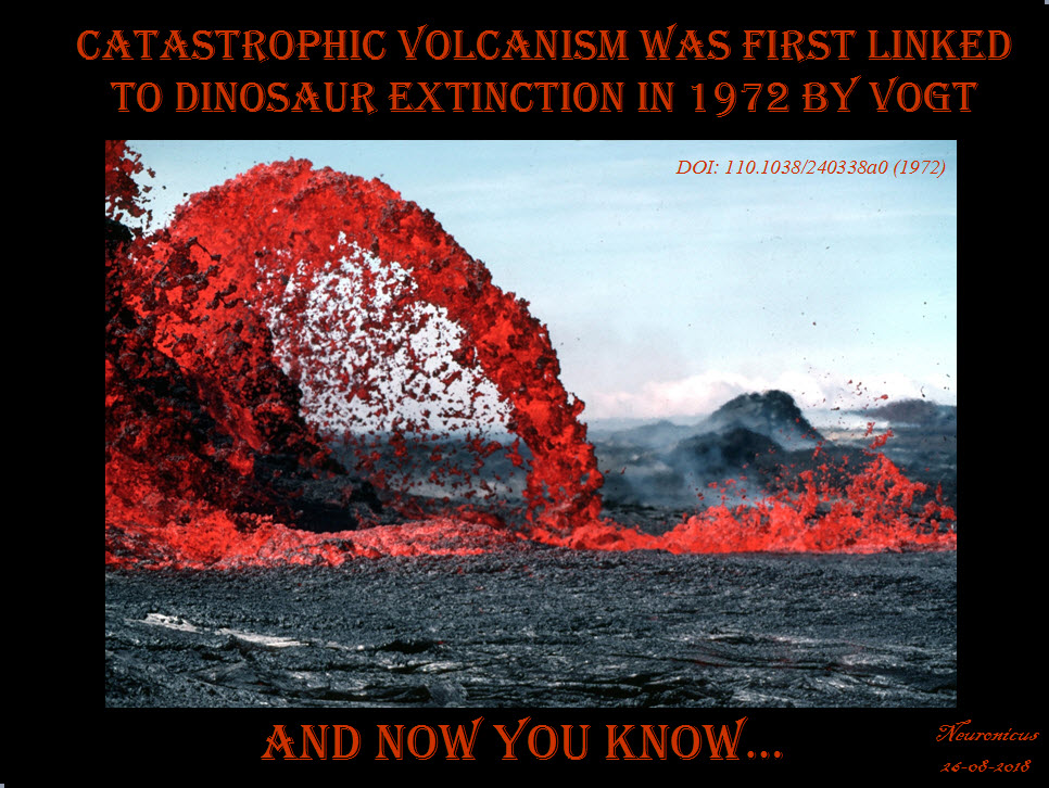
Well, The Atlantic article showcases the struggles of a scientist – paleontologist and geologist Gerta Keller – who doesn’t believe the mainstream asteroid hypothesis; rather she thinks there is enough evidence to point out that extreme volcano eruptions, like really extreme, thousands of times more powerful than anything we know in the recorded history, put out so much poison (soot, dust, hydrofluoric acid, sulfur, carbon dioxide, mercury, lead, and so on) in the atmosphere that, combined with the consequent dramatic climate change, killed the dinosaurs. The volcanoes were located in India and they erupted for hundreds of thousands of years, but most violent eruptions, Keller thinks, were in the last 40,000 years before the extinction. This hypothesis is called the Deccan volcanism from the region in India where these nasty volcanoes are located, first proposed by Vogt (1972) and Courtillot et al. (1986).

138- Vogt - Copy.jpg

So which is true? Or, rather, because this is science we’re talking about, which hypothesis is more supported by the facts: the volcanism or the impact?

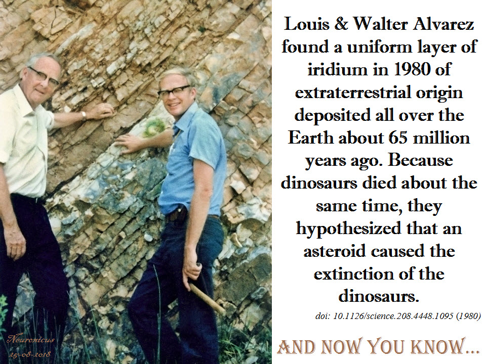
The impact hypothesis was put forward in 1980 when Walter Alvarez, a geologist, noticed a thin layer of clay in rocks that were about 65 million years old, which coincided with the time when the dinosaurs disappeared. This layer is on the KT boundary (sometimes called K-T, K-Pg, or KPB, looks like the biologists are not the only ones with acronym problems) and marks the boundary between the Cretaceous and Paleogenic geological periods (T is for Triassic, yeah, I know). Walter asked his father, the famous Nobel Prize physicist Louis Alvarez, to take a look at it and see what it is. Alvarez Sr. analyzed it and decided that the clay contains a lot of iridium, dozens of times more than expected. After gathering more samples from Europe and New Zealand, they published a paper (Alvarez et al., 1980) in which the scientists reasoned that because Earth’s iridium is deeply buried in its bowels and not in its crust, this iridium at the K-Pg boundary is of extraterrestrial origin, which could be brought here only by an asteroid/comet. This is also the paper in which it was put forth for the first time the conjecture that the asteroid impact killed the dinosaurs, based on the uncanny coincidence of timing.

138-alvarez - Copy

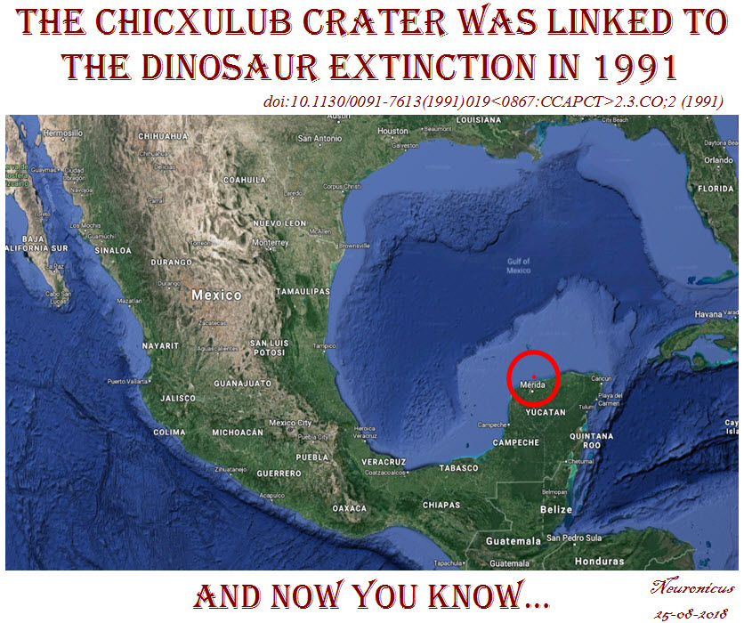
The discovery of the Chicxulub crater in Mexico followed a more sinuous path because the geophysicists who first discovered it in the ’70s were working for an oil company, looking for places to drill. Once the dinosaur-died-due-to-asteroid-impact hypothesis gained popularity outside academia, the geologists and the physicists put two-and-two together, acquired more data, and published a paper (Hildebrand et al., 1991) where the Chicxulub crater was for the first time linked with the dinosaur extinction. Although the crater was not radiologically dated yet, they had enough geophysical, stratigraphic, and petrologic evidence to believe it was as old as the iridium layer and the dinosaur die-out.

138-chicxulub - Copy

But, devil is in the details, as they say. Keller published a paper in 2007 saying the Chicxulub event predates the extinction by some 300,000 years (Keller et al., 2007). She looked at geological samples from Texas and found the glass granule layer (indicator of the Chicxhulub impact) way below the K-Pg boundary. So what’s up with the iridium then? Keller (2014) believes that is not of extraterrestrial origin and it might well have been spewed up by a particularly nasty eruption or the sediments got shifted. Schulte et al. (2010), on the other hand, found high levels of iridium in 85 samples from all over the world in the KPG layer. Keller says that some other 260 samples don’t have iridium anomalies. As a response, Esmeray-Senlet et al. (2017) used some fancy Mass Spectrometry to show that the iridium profiles could have come only from Chicxulub, at least in North America. They argue that the variability in iridium profiles around the world is due to regional geochemical processes. And so on, and so on, the controversy continues.

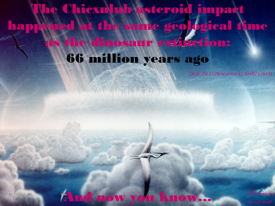
Actual radioisotope dating was done a bit later in 2013: date of K-Pg is 66.043 ± 0.043 MA (millions of years ago), date of the Chicxulub crater is 66.038 ±.025/0.049 MA. Which means that the researchers “established synchrony between the Cretaceous-Paleogene boundary and associated mass extinctions with the Chicxulub bolide impact to within 32,000 years” (Renne et al., 2013), which is a blink of an eye in geological times.

138-66 chixhulub - Copy

Now I want you to understand that often in science, though by far not always, matters are not so simple as she is wrong, he is right. In geology, what matters most is the sample. If the sample is corrupted, so will be your conclusions. Maybe Keller’s or Renne’s samples were affected by a myriad possible variables, some as simple as shifting the dirt from here to there by who knows what event. After all, it’s been 66 million years since. Also, methods used are just as important and dating something that happened so long ago is extremely difficult due to intrinsic physical methodological limitations. Keller (2014), for example, claims that Renne couldn’t have possibly gotten such an exact estimation because he used Argon isotopes when only U-Pb isotope dilution–thermal ionization mass spectrometry (ID-TIMS) zircon geochronology could be so accurate. But yet again, it looks like he did use both, so… I dunno. As the over-used always-trite but nevertheless extremely important saying goes: more data is needed.

Even if the dating puts Chicxulub at the KPB, the volcanologists say that the asteroid, by itself, couldn’t have produced a mass extinction because there are other impacts of its size and they did not have such dire effects, but were barely noticeable at the biota scale. Besides, most of the other mass extinctions on the planet have been already associated with extreme volcanism (Archibald et al., 2010). On the other hand, the circumstances of this particular asteroid could have made it deadly: it landed in the hydrocarbon-rich areas that occupied only 13% of the Earth’s surface at the time which resulted in a lot of “stratospheric soot and sulfate aerosols and causing extreme global cooling and drought” (Kaiho & Oshima, 2017). Food for thought: this means that the chances of us, humans, to be here today are 13%!…

I hope that you do notice that these are very recent papers, so the issue is hotly debated as we speak.

It is possible, nay probable, that the Deccan volcanism, which was going on long before and after the extinction, was exacerbated by the impact. This is exactly what Renne’s team postulated in 2015 after dating the lava plains in the Deccan Traps: the eruptions intensified about 50,000 years before the KT boundary, from “high-frequency, low-volume eruptions to low-frequency, high-volume eruptions”, which is about when the asteroid hit. Also, the Deccan eruptions continued for about half a million years after KPB, “which is comparable with the time lag between the KPB and the initial stage of ecological recovery in marine ecosystems” (Renne et al., 2016, p. 78).

Since we cannot get much more accurate dating than we already have, perhaps the fossils can tell us whether the dinosaurs died abruptly or slowly. Because if they got extinct in a few years instead of over 50,000 years, that would point to a cataclysmic event. Yes, but which one, big asteroid or violent volcano? Aaaand, we’re back to square one.

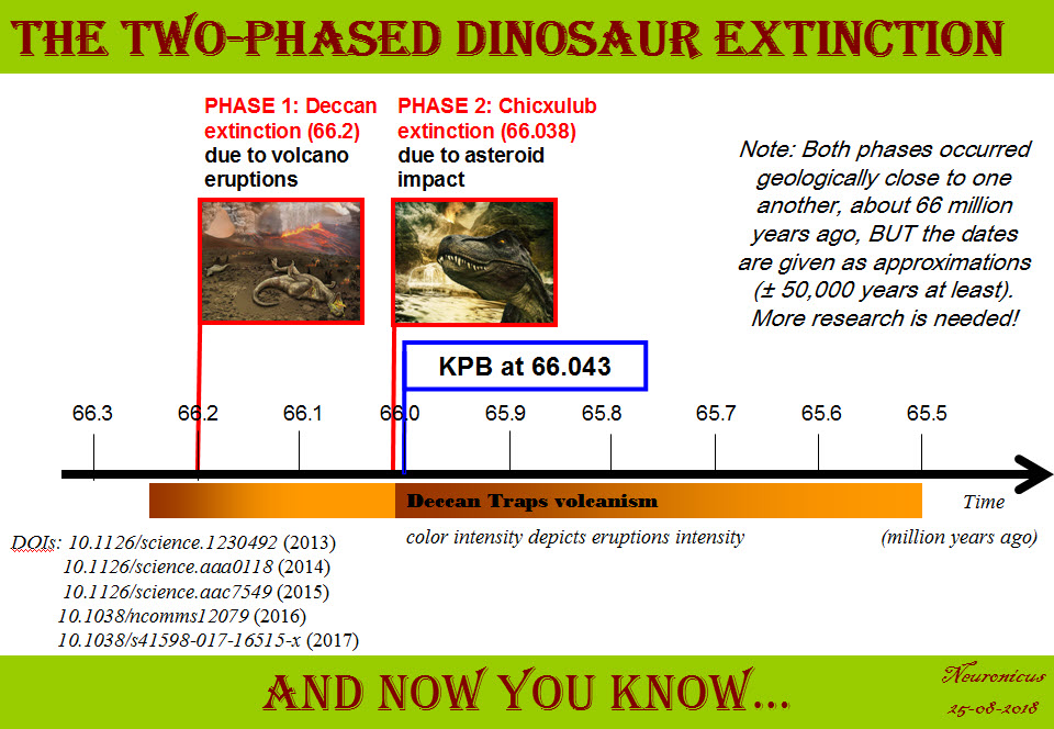
Actually, the last papers on the matter points to two extinctions: the Deccan extinction and the Chicxulub extinction. Petersen et al., (2016) went all the way to Antarctica to find pristine samples. They noticed a sharp increase in global temperatures by about 7.8 ºC at the onset of Deccan volcanism. This climate change would surely lead to some extinctions, and this is exactly what they found: out of 24 species of marine animals investigated, 10 died-out at the onset of Deccan volcanism and the remaining 14 died-out when Chicxulub hit.

In conclusion, because this post is already verrrry long and is becoming a proper college review, to me, a not-a-geologist/paleontologist/physicist-but-still-a-scientist, things happened thusly: first Deccan traps erupted and that lead to a dramatic global warming coupled with spewing poison in the atmosphere. Which resulted in a massive die-out (about 200,000 years before the bolide impact, says a corroborating paper, Tobin, 2017). The surviving species (maybe half or more of the biota?) continued the best they could for the next few hundred thousand years in the hostile environment. Then the Chicxulub meteorite hit and the resulting megatsunami, the cloud of super-heated dust and soot, colossal wildfires and earthquakes, acid rain and climate cooling, not to mention the intensification of the Deccan traps eruptions, finished off the surviving species. It took Earth 300,000 to 500,000 years to recover its ecosystem. “This sequence of events may have combined into a ‘one-two punch’ that produced one of the largest mass extinctions in Earth history” (Petersen et al., 2016, p. 6).

138-timeline dinosaur - Copy

By Neuronicus, 25 August 2018

P. S. You, high school and college students who will use this for some class assignment or other, give credit thusly: Neuronicus (Aug. 26, 2018). The FIRSTS: The cause(s) of dinosaur extinction. Retrieved from https://scientiaportal.wordpress.com/2018/08/26/the-firsts-the-causes-of-dinosaur-extinction/ on [date]. AND READ THE ORIGINAL PAPERS. Ask me for .pdfs if you don’t have access, although with sci-hub and all… not that I endorse any illegal and fraudulent use of the above mentioned server for the purpose of self-education and enlightenment in the quest for knowledge that all academics and scientists praise everywhere around the Globe!

EDIT March 29, 2019. Astounding one-of-a-kind discovery is being brought to print soon. It’s about a site in North Dakota that, reportedly, has preserved the day of the Chicxhulub impact in amazing detail, with tons of fossils of all kinds (flora, mammals, dinosaurs, fish) which seems to put the entire extinction of dinosaurs in one day, thus favoring the asteroid impact hypothesis. The data is not out yet. Can’t wait til it is! Actually, I’ll have to wait some more after it’s out for the experts to examine it and then I’ll find out. Until then, check the story of the discovery here and here.

REFERENCES:

1. Alvarez LW, Alvarez W, Asaro F, & Michel HV (6 Jun 1980). Extraterrestrial cause for the cretaceous-tertiary extinction. PMID: 17783054. DOI: 10.1126/science.208.4448.1095 Science, 208(4448):1095-1108. ABSTRACT | FULLTEXT PDF

2. Archibald JD, Clemens WA, Padian K, Rowe T, Macleod N, Barrett PM, Gale A, Holroyd P, Sues HD, Arens NC, Horner JR, Wilson GP, Goodwin MB, Brochu CA, Lofgren DL, Hurlbert SH, Hartman JH, Eberth DA, Wignall PB, Currie PJ, Weil A, Prasad GV, Dingus L, Courtillot V, Milner A, Milner A, Bajpai S, Ward DJ, Sahni A. (21 May 2010) Cretaceous extinctions: multiple causes. Science,328(5981):973; author reply 975-6. PMID: 20489004, DOI: 10.1126/science.328.5981.973-aScience. FULL REPLY

3. Courtillot V, Besse J, Vandamme D, Montigny R, Jaeger J-J, & Cappetta H (1986). Deccan flood basalts at the Cretaceous/Tertiary boundary? Earth and Planetary Science Letters, 80(3-4), 361–374. doi: 10.1016/0012-821x(86)90118-4. ABSTRACT

4. Esmeray-Senlet, S., Miller, K. G., Sherrell, R. M., Senlet, T., Vellekoop, J., & Brinkhuis, H. (2017). Iridium profiles and delivery across the Cretaceous/Paleogene boundary. Earth and Planetary Science Letters, 457, 117–126. doi:10.1016/j.epsl.2016.10.010. ABSTRACT

5. Hildebrand AR, Penfield GT, Kring DA, Pilkington M, Camargo AZ, Jacobsen SB, & Boynton WV (1 Sept. 1991). Chicxulub Crater: A possible Cretaceous/Tertiary boundary impact crater on the Yucatán Peninsula, Mexico. Geology, 19 (9): 867-871. DOI: https://doi.org/10.1130/0091-7613(1991)019<0867:CCAPCT>2.3.CO;2. ABSTRACT

6. Kaiho K & Oshima N (9 Nov 2017). Site of asteroid impact changed the history of life on Earth: the low probability of mass extinction. Scientific Reports,7(1):14855. PMID: 29123110, PMCID: PMC5680197, DOI:10.1038/s41598-017-14199-x. . ARTICLE | FREE FULLTEXT PDF

7. Keller G, Adatte T, Berner Z, Harting M, Baum G, Prauss M, Tantawy A, Stueben D (30 Mar 2007). Chicxulub impact predates K–T boundary: New evidence from Brazos, Texas, Earth and Planetary Science Letters, 255(3–4): 339-356. DOI: 10.1016/j.epsl.2006.12.026. ABSTRACT

8. Keller, G. (2014). Deccan volcanism, the Chicxulub impact, and the end-Cretaceous mass extinction: Coincidence? Cause and effect? Geological Society of America Special Papers, 505:57–89. doi:10.1130/2014.2505(03) ABSTRACT

9. Petersen SV, Dutton A, & Lohmann KC. (5 Jul 2016). End-Cretaceous extinction in Antarctica linked to both Deccan volcanism and meteorite impact via climate change. Nature Communications, 7:12079. doi: 10.1038/ncomms12079. PMID: 27377632, PMCID: PMC4935969, DOI: 10.1038/ncomms12079. ARTICLE | FREE FULLTEXT PDF 

10. Renne PR, Deino AL, Hilgen FJ, Kuiper KF, Mark DF, Mitchell WS 3rd, Morgan LE, Mundil R, & Smit J (8 Feb 2013). Time scales of critical events around the Cretaceous-Paleogene boundary. Science, 8;339(6120):684-687. doi: 10.1126/science.1230492. PMID: 23393261, DOI: 10.1126/science.1230492 ABSTRACT 

11. Renne PR, Sprain CJ, Richards MA, Self S, Vanderkluysen L, Pande K. (2 Oct 2015). State shift in Deccan volcanism at the Cretaceous-Paleogene boundary, possibly induced by impact. Science, 350(6256):76-8. PMID: 26430116. DOI: 10.1126/science.aac7549 ABSTRACT

12. Schoene B, Samperton KM, Eddy MP, Keller G, Adatte T, Bowring SA, Khadri SFR, & Gertsch B (2014). U-Pb geochronology of the Deccan Traps and relation to the end-Cretaceous mass extinction. Science, 347(6218), 182–184. doi:10.1126/science.aaa0118. ARTICLE

13. Schulte P, Alegret L, Arenillas I, Arz JA, Barton PJ, Bown PR, Bralower TJ, Christeson GL, Claeys P, Cockell CS, Collins GS, Deutsch A, Goldin TJ, Goto K, Grajales-Nishimura JM, Grieve RA, Gulick SP, Johnson KR, Kiessling W, Koeberl C, Kring DA, MacLeod KG, Matsui T, Melosh J, Montanari A, Morgan JV, Neal CR, Nichols DJ, Norris RD, Pierazzo E,Ravizza G, Rebolledo-Vieyra M, Reimold WU, Robin E, Salge T, Speijer RP, Sweet AR, Urrutia-Fucugauchi J, Vajda V, Whalen MT, Willumsen PS.(5 Mar 2010). The Chicxulub asteroid impact and mass extinction at the Cretaceous-Paleogene boundary. Science, 327(5970):1214-8. PMID: 20203042, DOI: 10.1126/science.1177265. ABSTRACT

14. Tobin TS (24 Nov 2017). Recognition of a likely two phased extinction at the K-Pg boundary in Antarctica. Scientific Reports, 7(1):16317. PMID: 29176556, PMCID: PMC5701184, DOI: 10.1038/s41598-017-16515-x. ARTICLE | FREE FULLTEXT PDF 

15. Vogt, PR (8 Dec 1972). Evidence for Global Synchronism in Mantle Plume Convection and Possible Significance for Geology. Nature, 240(5380), 338–342. doi:10.1038/240338a0 ABSTRACT

Interview with Jason D. Shepherd, PhD

During the first week of the publication, a Cell paper that I covered a couple of weeks ago has received a lot of attention from media outlets, like The Atlantic, Scicasts and Neuroscience News/University of Utah Press Release. It is not my intention to duplicate here their wonderfully done summaries and interviews; rather to provide answers to some geeky questions arisen from the minds of nerdy scientists like me.

Dr. Shepherd, you are the corresponding author of a paper published on Jan. 11 in Cell about a protein heavily involved in memory formation, called Arc. Your team and another team from University of Massachusetts, who published in the same issue of Cell, simultaneously discovered that Arc looks like and behaves like a virus. The protein “infects” nearby cells, in this case neurons, with instructions of how to make more of itself, i.e. it shuttles its own mRNA from one cell to another.

Neuronicus: Why is this discovery so important?

​Jason D. Shepherd: I think there’s a couple of big implications of this work:

  1. ​The so called “junk” DNA in our genomes that come from viruses and transposable elements actually provide source material for new genes. Arc isn’t the first example, but it’s the first prominent brain gene to have these kinds of origins.

  2. This is the first demonstration that cellular proteins are capable of assembling into capsid-like structures. This is a completely new way of thinking about communication between cells.

  3. We think there may be other genes that can also form capsids, suggesting this method of signaling is fairly common in organisms.

N: 2) When you and your colleagues compared Arc’s genetic sequence across species you concluded Arc comes from a virus that infected four-legged animals some time ago. A little time later the virus infected the flies too. When did these events occur?

​JDS: So we think the origins are from a retrotransposon not a virus. These are DNA sequences or elements that “jump” into the host genome. Think of them as primitive viruses. Indeed, these elements are thought to be the ancestors of retroviruses like HIV. The mammalian Arc gene seems to have originated ~400 million years ago, the fly about 150 million years ago. ​

N: 3) So, if Arc has been so successfully repurposed by the tetrapod and fly ancestors to add memory formation, what does that mean for the animals and insects before the infection? I understand that we move now in the realm of speculation, but who better to speculate on these things than the people who work on Arc? The question is: did these pre-infection creatures have bad and short memories? The alternate view would be that they had similar memory abilities due to a different mechanism that was replaced by Arc. Which one do you think is more likely?

​JDS: Good question. It’s certainly the case that memory capacity improved in tetrapods, but unclear if Arc is the sole reason. I suspect that Arc confers some unique aspects to brains, otherwise it would not have been so conserved after the initial insertion event, but I also think there are probably other Arc-like genes in other organisms that do not have Arc. I will also note that we are not even sure, yet, that the fly Arc is important for fly memory/learning.

N: 4) Remaining in the realm of speculation, if this intercellular mRNA transport proves to be ubiquitous for a variety of mRNAs, what does that say of the transcriptome of a cell at any given time? From a practical point of view, a cell is what is made off, meaning the ensemble of all its enzymes and proteins and so on, collectively termed transcriptome. So if a cell can just alter its neighbor’s transcriptome array, does that mean that it’s possible to alter also its function? Even more outrageously speculative, perhaps even its type? Can we make cancer cells commit suicide by shooting Arc capsules of mRNA at them?

​JDS: Yes! Cool ideas. I think this is quite likely, that these signaling extracellular vesicles can dramatically alter the state of a cell. We are obviously looking into this. ​

N: 5) Finally, in the paper, the Arc capsules containing mRNA are referred to as ACBAR (Arc Capsid Bearing Any RNA). At first I thought it was a reference to “Allahu akbar” which is Arabic for ‘God is greatest’, the allusion being “ACBAR! Our exosome is the greatest!” or “Arc Acbar! Our Arc is the greatest!”. Is this where the naming is coming from?

​JDS: No no. As I said on twitter, my lab came up with this acronym because we are all Star Wars nerds and the classic “It’s a trap!” line from general Ackbar seemed apt for something that was trapping RNA. ​

Below is the Twitter exchange Dr. Shepherd refers to:

129shepherd - Copy

Dr. Shepherd, thank you for your time! And congratulations on a well done paper and a well told story. Your Methods section is absolutely great; anybody can follow the instructions and replicate your data. Somebody in your lab must have kept great records. Congratulations again!

129_starwars - Copy
The ACBAR graphic is from the Cell’s abstract (©2017 Elsevier Inc.) but since it’s for comedic purposes, I’d say is fair use. Same for the Lego Ackbar.

By Neuronicus, 28 January 2018

P. S. Since I have obviously managed to annoy the #StarWars universe and twitterverse because I depicted General Ackbar using a Jedi sword when he’s not a Jedi, I thought only fair to annoy the other half of the world, the #trekkies. So here you go:

129_startrek - Copy.jpg


Arc: mRNA & protein from one neuron to another

EDIT 1 [Jan 17, 2018]: I promised four days ago that I will post this, while it was still hot, but my Internet was down, thanks to the only behemoth provider in USA. And rated the worst company in the Nation, too. You definitely know by now about whom I’m talking about. Grrrr…  Anyway, here is the paper:

As promised, today’s paper talks about mRNA transfer between neurons.

Pastuzyn et al. (2018) looked at the gene Arc in neurons because they thought its Gag sequence looks suspiciously similar to some retroviruses. Could it be possible that it also behaves like a virus?

Arc is heavily involved in the immune system, is essential for the formation of long-term memories, and is involved in all sorts of diseases, like schizophrenia and Alzheimer’s, among other things.

Pastuzyn et al. (2018) is a relatively long and dense paper, albeit well written. So, I thought that this time, instead of giving you a summary of their research it would be better to give you the authors’ story directly in their own words written as subtitles in the Results section (bold letters – the authors words, normal font – mine). Warning: this is a much more jargon-dense blog post than my previous one on the same topic and, because it is so much material, I will not explain every term.

  • Fly and Tetrapod (us) Arc Genes Independently Originated from Distinct Lineages of Ty3/gypsy Retrotransposons, the phylogenomic analyses tell us, meaning the authors have done a lot of computer-assisted comparisons of similar forms of the gene in hundreds of species.
  • Arc Proteins Self-Assemble into Virus-like Capsids. Arc likes to oligomerize spontaneously (dimers and trimers). The oligomers resemble virus-like capsids, similar to HIV.
  • Arc Binds and Encapsulates RNA. Although it loves its own RNA about 10 times more than other RNAs, it’s a promiscuous protein (doesn’t care which RNA as long as it follows the rules of stoichiometry). Arc capsids encapsulate both the Arc protein (maybe other proteins too?), its mRNA, and whatever mRNA happened to be in the vicinity at the time of encapsulation. Arc capsids are able to protect the mRNA from RNAases.
  • Arc Capsid Assembly Requires RNA. If there is no RNA around, the capsids are few and poorly formed.
  • Arc Protein and Arc mRNA Are Released by Neurons in Extracellular Vesicles. Arc capsid packages Arc protein & Arc mRNA into extracellular vesicles (EV). The size of these EVs is < 100nm, putting them in the exosome category. This exosome, which the authors gave the unfortunate name of ACBAR (Arc Capsid Bearing Any RNA), is being expelled from cortical neurons in an activity-dependent manner. In other words, when neurons are stimulated, they release ACBARs.
  • Arc Mediates Intercellular Transfer of mRNA in Extracellular Vesicles. ACBARs dock to the host cell and then undergo clathrin-dependent endocytosis, meaning they expel their cargo in the host cell. The levels of Arc protein and Arc mRNA peaks in a host hippocampal cell in four hours from incubation. The ACBARs tend to congregate around donor cell’s dendrites.
  • Transferred Arc mRNA Can Undergo Activity-Dependent Translation. Activating the group 1 metabotropic glutamate receptor (mGluR1/5) by application of the agonist DHPG induces a significant increase of the amount of Arc protein in the host neurons.

This is a veritable tour de force paper. The Results section has 7 sub-sections, each with multiple experiments to dot every i and cross every t. I’m eyeballing about 40 experiments. It is true that there are 13 authors on the paper from different institutions – yeay for collaboration! – but c’mon! Is this what you need to get in Cell these days? Apparently so. Don’t get me wrong, this is an outstanding paper. But in the end it is still only one paper, which means only one first author. The rest are there for the ride because for a tenure track application nobody cares about your papers in CNS (Cell, Nature, Science = The Central Nervous System of the scientific community, har, har) if you’re not the first author. It looks like the increasing amount of work you need to be published in top tier journals these days is becoming a pet peeve of mine as I keep mentioning it (for example, here).

My pet peeves aside, Pastuzyn et al. (2018) is an excellent paper that opens interesting practical (drug delivery) and theoretical (biological repurpose of ancient invaders) gates. Kudos!

128-1 - Copy

REFERENCE: Pastuzyn ED, Day CE, Kearns RB, Kyrke-Smith M, Taibi AV, McCormick J, Yoder N, Belnap DM, Erlendsson S, Morado DR, Briggs JAG, Feschotte C, & Shepherd JD. (11 Jan 2018). The Neuronal Gene Arc Encodes a Repurposed Retrotransposon Gag Protein that Mediates Intercellular RNA Transfer. Cell, 172(1-2):275-288.e18. PMID: 29328916. doi: 10.1016/j.cell.2017.12.024. ARTICLE | FULLTEXT PDF via ResearchGate

P.S. I said that ACBAR is an unfortunate acronym because I don’t know about you but I for one wouldn’t want my discovery to be linked either with a religion or with terrorist cries, even if that link is done only by a small fraction of the population. Although I can totally see the naming-by-committee going: “ACBAR! Our exosome is the greatest! Yeay!” or “Arc Acbar! Our Arc is the greatest. Double yeay!”. On a second thought, it’s kindda nerdy geeky neat. I still wouldn’t have done it though…

By Neuronicus, 14 January 2018

EDIT 2 [Jan 22, 2018]: There is another paper that discovered that Arc forms capsids that encapsulate RNA and then shuttles it across the neuromuscular junction in Drosophila (fly). To their credit, Cell published both these papers back-to-back so no researcher gets scooped of their discovery. From what I can see, the discovery really happened simultaneously, so I modified my infopic to reflect that (both papers were submitted in January 2017, received in revised version on August 15, 2017 and published in the same issue on January 11, 2018). Here is the reference to the other article:

Ashley J, Cordy B, Lucia D, Fradkin LG, Budnik V, & Thomson T (11 Jan 2018). Retrovirus-like Gag Protein Arc1 Binds RNA and Traffics across Synaptic Boutons, Cell. 172(1-2): 262-274.e11. PMID: 29328915. doi: 10.1016/j.cell.2017.12.022. ARTICLE

EDIT 3 [Jan 29, 2018]: Dr. Shepherd, the last author of the paper I featured, was kind enough to answer a few of my questions about the implications of his and his team’s findings, answers which you will find here.

By Neuronicus, 22 January 2018

The FIRSTS: mRNA from one cell can travel to another cell and be translated there (2006)

I’m interrupting the series on cognitive biases (unskilled-and-unaware, superiority illusion, and depressive realism) to tell you that I admit it, I’m old. -Ish. Well, ok, I’m not that old. But this following paper made me feel that old. Because it invalidates some stuff I thought I knew about molecular cell biology. Mind totally blown.

It all started with a paper freshly published two days ago and that I’ll cover tomorrow. It’s about what the title says: mRNA can travel between cells packaged nicely in vesicles and once in a target cell can be made into protein there. I’ll explain – briefly! – why this is such a mind-blowing thing.

Central_dogma - Copy
Fig. 1. Illustration of the central dogma of biology: information transfer between DNA, RNA, and protein. Courtesy of Wikipedia, PD

We’ll start with the central dogma of molecular biology (specialists, please bear with me): the DNA is transcribed into RNA and the RNA is translated into protein (see Fig. 1). It is an oversimplification of the complexity of information flow in a biological system, but it’ll do for our purposes.

DNA needs to be transcribed into RNA because RNA is a much more flexible molecule and thus can do many things. So RNA is the traveling mule between DNA and the place where its information becomes protein, i.e. ribosome. Hence the name mRNA. Just kidding; m stands for messenger RNA (not that I will ever be able to call that ever again: muleRNA is stuck in my brain now).

There are many kinds of RNA: some don’t even get out of the nucleus, some are chopped and re-glued (alternative splicing), some decide which bits of DNA (genes) are to be expressed, some are busy housekeepers and so on. Once an RNA has finished its business it is degraded in many inventive ways. It cannot leave the cell because it cannot cross the cell membrane. And that was that. Or so I’ve been taught.

Exceptions from the above were viruses whose ways of going from cell to cell are very clever. A virus is a stretch of nucleic acids (DNA and/or RNA) and some proteins encapsulated in a blob (capsid). Not a cell!

In the ’90s several groups were looking at some blobs (yes, most stuff in biology can be defined by the all-encompassing and enlightening term of ‘blob’) that cells spew out every now and then. These were termed extracellular vesicles (EV) for obvious reasons. Turned out that many kinds of cells were doing it and on a much more regular basis than previously thought. The contents of these EVs varied quite a bit, based on the type of cells studied. Proteins, mostly, and maybe some cytoplasmic debris. In the ’80s it was thought that this was one way for a cell to get rid of trash. But in 1982, Stegmayr & Ronquist showed that prostate cells release some EVs that result in sperm cell motility increase (Raposo & Stoorvogel, 2013) so, clearly, the EVs were more than trash. Soon it became evident that EVs were another way of cell-to-cell communication. (Note to self: the first time intercellular communication by EVs was demonstrated was in 1982, Stegmayr & Ronquist. Maybe I’ll dig out the paper to cover it sometime).

So. In 2005, Baj-Krzyworzeka et al. (2006) looked at some human cancer cells to see what they spew out and for what purpose. They saw that the cancer cells were transferring some of the tumor proteins packaged in EVs to monocytes. For devious purposes, probably. And then they made to what it looks to me like a serious leap in reasoning: since the EVs contain tumor proteins, why wouldn’t they also contain the mRNA for those proteins? My first answer to that would have been: “because it would be rapidly degraded”. And I would have been wrong. To my credit, if the experiment wouldn’t take up too many resources I still would have done it, especially if I would have some random primers lying around the lab. Luckily for the world, I was not in charge with this particular experiment and Baj-Krzyworzeka et al. (2005) proceeded with a real-time PCR (polymerase chain reaction) which showed them that the EVs released by the tumor cells also contained mRNA.

Now the 1 million dollar, stare-in-your-face question was: is this mRNA functional? Meaning, once delivered to the host cell, would it be translated into protein?

Six months later the group answered it. Ratajcza et al. (2006) used embryonic stem cells as the donor cells and hematopoietic progenitor cells as host cells. First, they found out that if you let the donors spit EVs at the hosts, the hosts are faring much better (better survival, upregulated good genes, phosphorylated MAPK to induce proliferation etc.). Next, they looked at the contents of EVs and found out that they contained proteins and mRNA that promote those good things (Wnt-3 protein, mRNA for transcription factors etc.). Next, to make sure that the host cells don’t show this enrichment all of a sudden out of the goodness of their little pluripotent hearts but is instead due to the mRNA from the donor cells, the authors looked at the expression of one of the transcription factors (Oct-4) in the hosts. They used as host a cell line (SKL) that does not express the pluripotent marker Oct-4. So if the hosts express this protein, it must have come only from outside. Lo and behold, they did. This means that the mRNA carried by the EVs is functional (Fig. 2).

128-1 - Copy
Fig. 2. Cell-to-cell mRNA transfer via extracellular vesicles (EVs). DNA is translated into RNA. A portion of RNA is transcribed into protein and another portion remains untranscribed. Both resultant protein and mRNA can get packaged into a vesicle: either a repackage into a microvesicle (a budding off of the cell membrane that shuttles cargo to and forth, about the size of 100-300nm) or packaged in a newly formed exosome (<100 nm) inside a multivesicular endosome (the yellow circle). The cell releases these vesicles in the intercellular space. The vesicles dock onto the host cell’s membrane and empty their cargo.

What bugs me is that these papers came out in a period where I was doing some heavy reading. How did I miss this?! Probably because they were published in cancer journals, not my field. But this is big enough you’d think others would mention it. (If you’re a recurrent reader of my blog, by now you should be familiarized with my stream-of-consciousness writing and my admittedly sometimes annoying in-parenthesis-meta-cognitions :D). So how did I miss this? How many more great discoveries have I missed? Am I the only one to discover such fundamental gaps in my knowledge? And thus the imposter syndrome takes root.

Just kidding, I don’t have the imposter syndrome. If anything, I got a superiority illusion complex. And I am absolutely sure that many, many scientists read things they consider fundamental to their way of thinking about the world all the time and wonder what other truly great discoveries are out there already that they missed.

Frankly, I should probably be grateful to this blog – and my friend GT who made me do it – because without nosing outside my field in search of material for it I would have probably remained ignorant of this awesome discovery. So, even if this is a decade old discovery for you, for me is one day old and I am a bit giddy about it.

This is a big deal because of the theoretical implications: a cell’s transcriptome (all the mRNA expressed in a cell) varies not only due to its needs, activity, and experiences, but also due to its neighbors’! A cell is, more or less, its transcriptome. Soooo… if we can change that at will, does that means we can change the type or function of the cell too? There are so many questions that such a discovery raises! And possibilities.

This is also a big deal because it opens up not a new therapy, or a new therapy direction, or a new drug class, but a new DELIVERY METHOD, the Holy Grail of Pharmacopeia. You just put your drug in one of these vesicles and let nature take its course. Of course, there are all sorts of roadblocks to overcome, like specificity, toxicity, etc. Looks like some are already conquered as there are several clinical trials out there that take advantage of this mechanism and I bet there will be more.

Stop by tomorrow for a freshly published paper on this mechanism in neurons.

127 - Copy

REFERENCES:

1) Baj-Krzyworzeka M, Szatanek R, Weglarczyk K, Baran J, Urbanowicz B, Brański P, Ratajczak MZ, & Zembala M. (Jul. 2006, Epub 9 Nov 2005). Tumour-derived microvesicles carry several surface determinants and mRNA of tumour cells and transfer some of these determinants to monocytes. Cancer Immunology, Immunotherapy, 55(7):808-818. PMID: 16283305, DOI: 10.1007/s00262-005-0075-9. ARTICLE

2) Ratajczak J, Miekus K, Kucia M, Zhang J, Reca R, Dvorak P, & Ratajczak MZ (May 2006). Embryonic stem cell-derived microvesicles reprogram hematopoietic progenitors: evidence for horizontal transfer of mRNA and protein delivery. Leukemia, 20(5):847-856. PMID: 16453000, DOI: 10.1038/sj.leu.2404132. ARTICLE | FREE FULLTEXT PDF 

Bibliography:

Raposo G & Stoorvogel W. (18 Feb. 2013). Extracellular vesicles: exosomes, microvesicles, and friends. The Journal of Cell Biology, 200(4):373-383. PMID: 23420871, PMCID: PMC3575529, DOI: 10.1083/jcb.201211138. ARTICLE | FREE FULLTEXT PDF

By Neuronicus, 13 January 2018

The FIRSTS: Dinosaurs and reputation (1842)

‘Dinosaur’ is a common noun in most languages of the Globe and, in its weak sense, it means “extinct huge reptile-like animal that lived a long-time ago”. The word has been in usage for so long that it can be used also for describing something “impractically large, out-of-date, or obsolete” (Merriam-Webster dictionary). “Dinosaur” is a composite of two ancient Greek words (“deinos”, “sauros”) and it means “terrible lizard”.

But, it turns out that the word hasn’t been in usage for so long, just for a mere 175 years. Sir Richard Owen, a paleontologist that dabbled in many disciplines, coined the term in 1842. Owen introduced the taxon Dinosauria as if it was always called thus, no fuss: “The present and concluding part of the Report on British Fossil Reptiles contains an account of the remains of the Crocodilian, Dinosaurian, Lacertian, Pterodactylian, Chelonian, Ophidian and Batrachian reptiles.” (p. 60). Only later in the Report does he tell us his paleontological reasons for the baptism, namely some anatomical features that distinguish dinosaurs from crocodiles and other reptiles.

“…The combination of such characters, some, as the sacral ones, altogether peculiar among Reptiles, others borrowed, as it were, from groups now distinct from each other, and all manifested by creatures far surpassing in size the largest of existing reptiles, will, it is presumed, be deemed sufficient ground for establishing a distinct tribe or sub-order of Saurian Reptiles, for which I would propose the name of Dinosauria.” (p.103)

At the time he was presenting this report to the British Association for the Advancement of Science, other giants of biology were running around the same halls, like Charles Darwin and Thomas Henry Huxley. Indisputably, Owen had a keen observational eye and a strong background in comparative anatomy that resulted in hundreds of published works, some of them excellent. That, in addition to establishing the British Museum of Natural History.

Therefore, Owen had reasons to be proud of his accomplishments and secure in his influence and legacy, and yet his contemporaries tell us that he was an absolutely vicious man, spiteful to the point of obsession, vengeful and extremely jealous of other people’s work. Apparently, he would steal the work of the younger people around him, never give credit, lie and cheat at every opportunity, and even write lengthy anonymous letters to various printed media to denigrate his contemporaries. He seemed to love his natal city of Lancaster and his family though (Wessels & Taylor, 2015).

121Richard-owen _PD
Sir Richard Owen (20 July 1804 – 18 December 1892). PD, courtesy of Wikipedia.

Owen had a particular hate for Darwin. They had been close friends for 20 years and then Darwin published the “Origin of Species”. The book quickly became widely read and talked about and then poof: vitriol and hate. Darwin himself said the only reason he could think of for Owen’s hatred was the popularity of the book.

Various biographies and monographers seem to agree on his unpleasant personality (see his entry in The Telegraph, Encyclopedia.com, Encylopaedia Britannica, BBC). On a side note, should you be concerned about your legacy and have the means to persuade The Times to write you an obituary, by all means, do so. In all the 8 pages of obituary written in 1896 you will not find a single blemish on the portrait of Sir Richard Owen.

This makes me ponder on the judgement of history based not on your work, but on your personality. As I said, the man contributed to science in more ways than just naming the dinosaur and having spats with Darwin. And yet it seems that his accomplishments are somewhat diminished by the way he treated others.

This reminded me of Nicolae Constantin Paulescu, a Romanian scientist who discovered insulin in 1916 (published in 1921). Yes, yes, I know all about the controversy with the Canadians that extracted and purified the insulin in 1922 and got the Nobel for it in 1923. Paulescu did the same, even if Paulescu’s “pancreatic extract” from a few years earlier was insufficiently purified; it still successfully lowered the glicemic index in dogs. He even obtained a patent for the “fabrication of pancrein” (his name for insulin, because he obtained it from the pancreas) in April 1922 from the Romanian Government (patent no. 6255). The Canadian team was aware of his work, but because it was published in French, they had a poor translation and they misunderstood his findings, so, technically, they didn’t steal anything. Or so they say. Feel free to feed the conspiracy mill. I personally don’t know, I haven’t looked at the original work to form an opinion because it is in French and my French is non-existent.

Annnywaaaay, whether or not Paulescu was the first in discovering the insulin is debatable, but few doubt that he should have shared the Nobel at least.

Rumor has it that Paulescu did not share the Nobel because he was a devout Nazi. His antisemitic writings are remarkably horrifying, even by the standards of the extreme right. That’s also why you won’t hear about him in medical textbooks or at various diabetes associations and gatherings. Yet millions of people worldwide may be alive today because of his work, at least partly.

How should we remember? Just the discoveries and accomplishments with no reference to the people behind them? Is remembering the same as honoring? “Clara cells” were lung cells discovered by the infamous Nazi anatomist Max Clara by dissecting prisoners without consent. They were renamed by the lung community “club cells” in 2013. We cannot get rid of the discovery, but we can rename the cells, so it doesn’t look like we honor him. I completely understand that. And yet I also don’t want to lose important pieces of history because of the atrocities (in the case of Nazis) or unsavory behavior (in the case of Owen) committed by our predecessors. I understand why the International Federation of Diabetes does not wish to give awards in the name of Paulescu or have a Special Paulescu lecture. Perhaps the Romanians should take down his busts and statues, too. But I don’t understand why (medical) history books should exclude him.

In other words, don’t honor the unsavories of history, but don’t forget them either. You never know what we – or the future generations – may learn by looking back at them and their actions.

123 - Copy.jpg

By Neuronicus, 19 October 2017

References:

1) Owen, R (1842). “Report on British Fossil Reptiles”. Part II. Report of the Eleventh Meeting of the British Association for the Advancement of Science; Held at Plymouth in July 1841. London: John Murray. p. 60–204. Google Books Fulltext 

2) “Eminent persons: Biographies reprinted from the Times, Vol V, 1891–1892 – Sir Richard Owen (Obituary)” (1896). Macmillan & Co., p. 291–299. Google Books Fulltext

3) Wessels Q & Taylor AM (28 Oct 2015). Anecdotes to the life and times of Sir Richard Owen (1804-1892) in Lancaster. Journal of Medical Biography. pii: 0967772015608053. PMID: 26512064, DOI: 10.1177/0967772015608053. ARTICLE

Midichlorians, midichloria, and mitochondria

Nathan Lo is an evolutionary biologist interested in creepy crawlies, i.e. arthropods. Well, he’s Australian, so I guess that comes with the territory (see what I did there?). While postdoc’ing, he and his colleagues published a paper (Sassera et al., 2006) that would seem boring for anybody without an interest in taxonomy, a truly under-appreciated field.

The paper describes a bacterium that is a parasite for the mitochondria of a tick species called Ixodes ricinus, the nasty bugger responsible for Lyme disease. The authors obtained a female tick from Berlin, Germany and let it feed on a hamster until it laid eggs. By using genetic sequencing (you can use kits these days to extract the DNA, do PCR, gels and cloning, pretty much everything), electron microscopy (real powerful microscopes) and phylogenetic analysis (using computer softwares to see how closely related some species are) the authors came to the conclusion that this parasite they were working on is a new species. So they named it. And below is the full account of the naming, from the horse’s mouth, as it were:

“In accordance with the guidelines of the International Committee of Systematic Bacteriology, unculturable bacteria should be classified as Candidatus (Murray & Stackebrandt, 1995). Thus we propose the name ‘Candidatus Midichloria mitochondrii’ for the novel bacterium. The genus name Midichloria (mi.di.chlo′ria. N.L. fem. n.) is derived from the midichlorians, organisms within the fictional Star Wars universe. Midichlorians are microscopic symbionts that reside within the cells of living things and ‘‘communicate with the Force’’. Star Wars creator George Lucas stated that the idea of the midichlorians is based on endosymbiotic theory. The word ‘midichlorian’ appears to be a blend of the words mitochondrion and chloroplast. The specific epithet, mitochondrii (mi.to′chon.drii. N.L. n. mitochondrium -i a mitochondrion; N.L. gen. n. mitochondrii of a mitochondrion), refers to the unique intramitochondrial lifestyle of this bacterium. ‘Candidatus M. mitochondrii’ belongs to the phylum Proteobacteria, to the class Alphaproteobacteria and to the order Rickettsiales. ‘Candidatus M. mitochondrii’ is assigned on the basis of the 16S rRNA (AJ566640) and gyrB gene sequences (AM159536)” (p. 2539).

George Lucas gave his blessing to the Christening (of course he did).

119-midi - Copy1 - Copy.jpg

Acknowledgements: Thanks go to Ms. BBD who prevented me from making a fool of myself (this time!) on the social media by pointing out to me that midichloria are real and that they are a mitochondrial parasite.

REFERENCE: Sassera D, Beninati T, Bandi C, Bouman EA, Sacchi L, Fabbi M, Lo N. (Nov. 2006). ‘Candidatus Midichloria mitochondrii’, an endosymbiont of the tick Ixodes ricinus with a unique intramitochondrial lifestyle. International Journal of Systematic and Evolutionary Microbiology, 56(Pt 11): 2535-2540. PMID: 17082386, DOI: 10.1099/ijs.0.64386-0. ABSTRACT | FREE FULLTEXT PDF 

By Neuronicus, 29 July 2017

The third eye

The pineal gland has held fascination since Descartes’ nefarious claim that it is the seat of the soul. There is no evidence of that; he said it might be where the soul resides because he thought the pineal gland was the only solitaire structure in the brain so it must be special. By ‘solitaire’ I mean that all other brain structures come in doublets: 2 amygdalae, 2 hippocampi, 2 thalami, 2 hemispheres etc. He was wrong about that as well, in that there are some other singletons in the brain besides the pineal, like the anterior or posterior commissure, the cerebellar vermis, some deep brainstem and medullary structures etc.

Descartes’ dualism was the only escape route the mystics at the time had from the demands for evidence by the budding natural philosophers later known as scientists. So when some scientists noted that some lizards have a third eye on top of their head connected to the pineal gland, the mystics and, later, the conspiracy theorists went nuts. Here, see, if the seat of the soul is linked with the third eye, then the awakening of this eye in people would surely result in heightened awareness, closeness to the Divinity, oneness with Universe and other similar rubbish that can otherwise easily and reliably be achieved by a good dollop of magic mushrooms. Cheaper, too.

Back to the lizards. Yes, you read right: some lizards and frogs have a third eye. This eye is not exactly like the other two, but it has cells sensitive to light, even if they are not perceiving light in the same way the retinal cells from the lateral eyes are. It is located on the top of the skull, so sometimes it is called the parietal organ (because it is in-between the parietal skull bones, see pic).

Anolis_carolinensis_parietal_eye.CC BY-SA 3.0jpg
Dorsal view of the head of the adult Carolina anole (Anolis carolinensis) clearly showing the parietal eye (small gray/clear oval) at the top of its head. Photo by TheAlphaWolf. License: CC BY-SA 3.0, courtesy of Wikipedia.

It is believed to be a vestigial organ, meaning that primitive vertebrates might have had it as a matter of course but it disappeared in the more recently evolved animals. Importantly, birds and mammals don’t have it. Not at all, not a bit, not atrophied, not able to be “awakened” no matter what your favorite “lemme see your chakras” guru says. Go on, touch your top of the skull and see if you have some peeking soft tissue there. And no, the soft tissue that babies are born with right there on the top of the skull is not a third eye; it’s a fontanelle that allows for the rapid expansion of the brain during the first year of life.

The parietal organ’s anatomical connection to the pineal gland is not surprising at all for scientists because the pineal’s role in every single animal that has it is the regulation of some circadian rhythms by the production of melatonin. In humans, the eyes tell the pineal that is day or night and the pineal adjusts the melatonin production accordingly, i.e. less melatonin produced during the day and more during the night. Likewise, the lizards’ third eye’s main role is to provide information to the pineal about the ambient light for thermoregulatory purposes.

After this long introduction, here is the point: almost twenty years ago Xiong et al. (1998) looked at how this third eye perceives light. In the human eye, light hitting the rods and cones in the retina (reception) launches a biochemical cascade (transduction) that results in seeing (coding of the stimulus in the brain). Briefly, transduction goes thusly: the photon(s) cause(s) a special protein sensitive to light (e.g. rhodopsin) in the photoreceptor cells in the retina to split into its components (photobleaching), one of these components (11-cis-retinal) changes its conformation (photoisomerization), then activates a G-protein (transducin), which then activates the enzyme phosphodiesterase (PDE), which then destroys a nucleotide called cyclic guanosine monophosphate (cGMP), which results in the closing of the cell’s sodium ion channels, which leads to less neurotransmitter released (glutamate), which causes the nearby cells (bipolar cells) to release the same neurotransmitter, which now has the opposite effect, meaning it increases the firing rate of another set of cells (ganglion cells) and from there to the brain we go. Phew, visual transduction IS difficult. And this is the brief version.

It turns out that the third eye’s retina doesn’t have all the types of cells that the normal eyes have. Specifically, it misses the bipolar, horizontal and amacrine cells, having only ganglion and photoreception cells. So how goes the phototransduction in the third eye’s retina, if at all?

Xiong et al. (1998) isolated photoreceptor cells from the third eyes of the lizard Uta stansburiana. And then they did a bunch of electrophysiological recording on those cells under different illumination and chemical conditions.

They found that the phototransduction in the third eye is different from the lateral eyes in that when they expected to see hyperpolarization of the cell, they observed depolarization instead. Also, when they expected the PDE to break down cGMP they found that PDE is inhibited thereby increasing the amount of cGMP.  The fact that G-protein can inhibit PDE was totally unexpected and showed a novel way of cellular signaling. Moreover, they speculate that their results can make sense only if not one, but two G-proteins with opposite actions work in tandem.

A probably dumb technical question though: the human rhodopsin takes about 30 minutes to restore itself from photobleaching. Xiong et al. (1998) let the cells adapt to dark for 10 minutes before recordings. So I wonder if the results would have been slightly different if they allowed the cell more time to adapt? But I’m not an expert in retina science, you’ve seen how difficult it is, right? Maybe the lizard proteins are different or rhodopsin adaptation time has little or nothing to do with their experiments? After all, later research has shown that the third eye has its own unique opsins, like the green-sensitive parietopsin discovered by Su et al. (2006).

115 third eye - Copy

REFERENCE:  Xiong WH, Solessio EC, & Yau KW (Sep 1998). An unusual cGMP pathway underlying depolarizing light response of the vertebrate parietal-eye photoreceptor. Nature Neuroscience, 1(5): 359-365. PMID: 10196524, DOI: 10.1038/1570. ARTICLE

Additional bibliography: Su CY, Luo DG, Terakita A, Shichida Y, Liao HW, Kazmi MA, Sakmar TP, Yau KW (17 Mar 2006). Parietal-eye phototransduction components and their potential evolutionary implications. Science, 311(5767): 1617-1621. PMID: 16543463, DOI: 10.1126/science.1123802. ARTICLE

By Neuronicus, 30 March 2017

Save

Save

Save

The FIRSTS: Melatonin (1958)

By the late 18th and beginning of 19th century, some scientists were busily investigating how animals get their colors and how do they change colour in response to the environment. They identified several types of chromatophores, i.e. cells that contain pigments. Biological pigments are called melanins (don’t confuse them with melatonin). One of these cells is the melanophore which contains black pigments, called this way in the very typical scientist unimaginative style because “melas” in Greek means black or dark and “phoros” means carrier.

A couple of these scientists, McCord & Allen (1917), thought that the pineal gland from the brain might contain some substance that might interact with the melanophores. How did they get this idea is unclear from their paper; seems like a logical outcome of their contemporaries’ discussions and experiments, though they do not explain it in detail. They hint of other experiments where various glands have been fed to amphibians and then noticed their color change. So McCord & Allen obtained cow brains, extracted the pineal glands and fed them to tadpoles. Within 30 to 60 minutes, depending on the concentration, the tadpoles fed with pineal extract changed color from dark to light (see picture).

mccord1917 - Copy
Excerpt from McCord & Allen (1917, doi: 10.1002/jez.1400230108) showing the change in tadpole skin appearance after application of bovine pineal gland extract.

Fast forward now to 1958 when an MD PhD called Aaron B. Lerner with an interest in dermatology thought that whatever was responsible for the skin color changes in the McCord & Allen (1917) paper might be useful in treating skin diseases. But first he had to extract the substance from the pineal glands and he and his colleagues had better tools for this task than the mere alcohol and acetone of his brethren of 40 years ago.

Lerner et al. (1958) made full use of the then-recently discovered paper chromatography and some standard biochemistry techniques for the time like Soxhlet extraction and fluorescence spectroscopy and discovered a substance that can lighten frog skin color and can inhibit the melanocyte stimulating hormone (MSH). “It is suggested that this substance be called melatonin” (p. 2587). Lerner and his colleagues also isolated the MSH and cryoglobulin.

Changing skin color is one of melatonin’s minor roles; its main function is to regulate circadian rhythms like sleep and awake cycles in animals (it has an oxidative stress protection in plants). Melatonin, in animals, is produced by the pineal gland only, more during the night, less during the day. Pineal gets information about the day/night cycles from the eyes. In some countries melatonin is sold as an over the counter soporific, i.e. sleeping pill.

Melatonin - Copy

REFERENCES:

1) McCord CP & Allen FP (Jan 1917). Evidences associating pineal gland function with alterations in pigmentation. Journal of Experimental Zoology, Part A, 23 (1):  207–224, DOI: 10.1002/jez.1400230108 ARTICLE 

2) Lerner AB, Case JD, Takahashi Y, Lee TH, & Mori W (May 1958). Isolation of Melatonin, the Pineal Gland Factor that Lightens Melanocytes. Journal of the American Chemical Society, 80 (10), p. 2587–2587, DOI: 10.1021/ja01543a060 ARTICLE (although JACS gives access only to the first page of a paper, the fact that this article is only half a page makes their endeavour useless in this case)

By Neuronicus, 18 March 2017

Save

Save

Vanity and passion fruit

Ultraviolet irradiation exposure from our sun accelerates the skin aging, process called photoaging. It can even cause skin cancers. There has been some considerable research on how our beloved sun does that.

For example, one way the UV radiation leads to skin damage is by promoting the production of free radicals as reactive oxygen species (ROS), which do many bad things, like direct DNA damage. Another bad thing done by ROS is the upregulation of the mitogen-activated protein kinase (MAPK) signaling pathway which activates all sorts of transcription factors which, in turn, produce proteins that lead to collagen degradation and voilà, aged skin. I know I lost some of you at the MAPK point; you can think of MAPK as a massive proteinaceous hub, a multi-button console with many inputs and outputs. A very sensitive and incredibly complex hub that controls nearly all important aspects of cell function, with many feedback loops, so if you mess with it, cell Armageddon may be happening. Or nothing at all. It’s that complex.

But I digress. What MAPK is doing is less relevant for the paper I am introducing to you today than the fact that we have physiological markers for skin aging due to UV. Bravo et al. (2017) cultured human skin cells in a Petri dish, treated them with various concentrations of an extract of passion fruit (Passiflora tarminiana) and then bombarded them with UV (the B type, 280–315 nm). The authors made the extract themselves, is not something you just buy (yet).

The UV produced the expected damage, translated as increased matrix mettoproteinase-1 (MMP-1), collagenase, and ROS production and decreased procollagen. Pretreatment with passion fruit extract significantly mitigated these UV effects in a dose-dependant manner. The concentration of their concoction that worked best was 10 μg/mL. Then the authors did some more chemistry to figure out what in their concoction is responsible, or at least probably responsible, for the observed wonderful effects. The authors believe the procyianidins and flavonoids are the culprits because 1) they have been proven to be strong antioxidants before and 2) this plant has them in very high amounts.

Good news then for the antiaging cosmetics industry. Perhaps even for dermatologists and their patients.

113passion-copy

Reference: Bravo K, Duque L, Ferreres F, Moreno DA, & Osorio E. (EPUB ahead of print: 3 Feb 2017). Passiflora tarminiana fruits reduce UVB-induced photoaging in human skin fibroblasts. Journal of Photochemistry and Photobiology, 168: 78-88. PMID: 28189068, DOI: 10.1016/j.jphotobiol.2017.01.023. ARTICLE

By Neuronicus, 13 February 2017

Save

The FIRSTS: The Name of Myelin (1854)

One reason why I don’t post more often is that I have such a hard time deciding what to cover (Hint: send me stuff YOU find awesome). Most of the cool and new stuff is already covered by big platforms with full-time employees and I try to stay away of the media-grabbers. Mostly. Some papers I find so cool that it doesn’t matter that professional science journalists have already covered them and I too jump on the wagon with my meager contribution. Anyway, here is a glimpse on how my train of thought goes on inspiration-less days.

Inner monologue: Check the usual journals’ current issues. Nothing catches my eye. Maybe I’ll feature a historical. Open Wikipedia front page and see what happened today throughout history. Aha, apparently Babinski died in 1932. He’s the one who described the Babinski’s sign. Normally, when the sole of the foot is stroked, the big toe flexes inwards, towards the sole. If it extends upwards, then that’s a sure sign of neurological damage, the Babinski’s sign. But healthy infants can have that sign too not because they have neurological damage, but because their corticospinal neurons are not fully myelinated. Myelin, who discovered that? Probably Schwann. Quick search on PubMed. Too many. Restrict to ‘history”. I hate the search function on PubMed, it brings either to many or no hits, no matter the parameters. Ah, look, Virchow. Interesting. Aha. Find the original reference. Aha. Springer charges 40 bucks for a paper published in 1854?! The hell with that! I’m not even going to check if I have institutional access. Get the pdf from other sources. It’s in German. Bummer. Go to Highwire. Find recent history of myelin. Mielinization? Myelination? Myelinification? All have hits… Get “Fundamental Neuroscience” off of the shelf and check… aha, myelination. Ok. Look at the pretty diagram with the saltatory conduction! Enough! Go back to Virchow. Does it have pictures, maybe I can navigate the legend? Nope. Check if any German speaking friends are online. Nope, they’re probably asleep, which is what I should be doing. Drat. Refine Highwire search. Evrika! “Hystory of Myelin” by Boullerne, 2016. Got the author manuscript. Hurray. Read. Write.

Myelinated fibers, a.k.a. white matter has been observed and described by various anatomists, as early as the 16th century, Boullerne (2016) informs us. But the name of myelin was given only in 1854 by Rudolph Virchow, a physician with a rich academic and public life. Although Virchow introduced the term to distinguish between bone marrow and the medullary substance, paradoxically, he managed to muddy waters even more because he did not restrict the usage of the term mylein to … well, myelin. He used it also to refer to substances in blood cells and egg’s yolk and spleen and, frankly, from the quotes provided in the paper, I cannot make heads or tails of what Virchow thought myelin was. The word myelin comes form the Greek myelos or muelos, which means marrow.

Boullerne (2016) obviously did a lot of research, as the 53-page account is full of quotes from original references. Being such a scholar on the history of myelin I have no choice but to believe her when she says: “In 1868, the neurologist Jean-Martin Charcot (1825-1893) used myelin (myéline) in what can be considered its first correct attribution.”

So even if Virchow coined the term, he was using it incorrectly! Nevertheless, in 1858 he correctly identified the main role of myelin: electrical insulation of the axon. Genial insight for the time.

103-copy

I love historical reviews of sciency stuff. This one is a ‘must-have’ for any biologist or neuroscientist. Chemists and physicists, too, don’t shy away; the paper has something for you too, like myelin’s biochemistry or its birefringence properties.

Reference: Boullerne, AI (Sep 2016, Epub 8 Jun 2016). The history of myelin. Experimental Neurology, 283(Pt B): 431-45. doi: 10.1016/j.expneurol.2016.06.005. ARTICLE

Original Reference: Virchow R. (Dec 1854). Ueber das ausgebreitete Vorkommen einer dem Nervenmark analogen Substanz in den thierischen Geweben. Archiv für pathologische Anatomie und Physiologie und für klinische Medicin, 6(4): 562–572. doi:10.1007/BF02116709. ARTICLE

P.S. I don’t think is right that Springer can retain the copyright for the Virchow paper and charge $39.95 for it. I don’t think they have the copyright for it anyway, despite their claims, because the paper is 162 years old. I am aware of no German or American copyright law that extends for so long. So, if you need it for academic purposes, write to me and thou shalt have it.

By Neuronicus, 29 October 2016

Save

The FIRSTS: The rise and fall of Pokemon (2001-2005?)

90pok - Copy

Few people know that Pokemon refers not only to a game, but also to a gene. An oncogene, to be precise, with a rather strange story.

An oncogene is a gene that promotes cancer (from oncology). Conventionally, a gene name is written in lowercase italicized letters (pokemon), whereas the protein the gene makes is not italicized (POKEMON, Pokemon, or pokemon, depending on the species). Maeda et al. (2005) first established in a Petri dish that the Pokemon is required for the growth of malignant tumors. Then, through a series of classic molecular biology experiments, the scientists found out how exactly Pokemon acts to accomplish this (by suppressing the expression of anti-cancer genes). Next, they engineered mice with pokemon overexpressed and saw that the mice with a lot of Pokemon “developed aggressive tumours” (p. 282). Then the authors checked how is this gene behaving in human cancers and found out that “Pokemon is expressed at very high levels in a subset of human lymphomas” (p. 284).

And here is how the gene got its name, according to Pier Paolo Pandolfi, the leader of the research group. Bear with me because it’s complicated. [*Takes deep breath*]: PO in POK stands for POZ domain (poxvirus and zinc finger) and K in POK stands for Krüppel (zinc finger transcription factor) whereas EMON stands for erythroid myeloid ontogenic factor. POK-EMON. Simple, eh? Phew…

Truth be told, Pandolfi first named the gene pokemon at a conference in 2001 (Simonite, 2005). Then the name has been used by researchers at various scientific meetings and poster presentations.

But when the Maeda et al. paper was published in Nature in 2005 which discovered the mechanism through which the gene promotes cancer, a lot of people, scientists and journalists alike, in an attempt at humour, flooded the internet with eye-catching titles along the lines of “Pokemon causes cancer”, “Pokemon kills you” and the like. I mean, even the researchers themselves in the abstract of the paper state: “Pokemon is aberrantly overexpressed in human cancers”. In response, The Pokémon Company threatened to sue for trademark copyright infringement because they didn’t want the game to be associated with cancer, like the gene is, even if the researches said the name is an acronym (maybe they meant backronym?). In the end, the researchers changed the name of the pokemon gene to the far less enticing zbtb7.

As the question mark in the title of the post suggests, the pokeman gene may not be entirely dead yet because there are stubborn scientists that still use the name pokemon and not zbtb7. I hope they have the cash to take on Nintendo if they decide to sue after all.

Too bad the zbtb7 (a.k.a. pokemon) gene was not a beneficial gene… Because another group of researchers named their new-found gene in 2008 pikachurin and so far, Nintendo din not make any waves… That is, probably, because Pikachurin is a protein in the eye retina that is required for proper vision by speeding the electric signals. Zip zip zip Pikachurin goes…

References:

  1. Maeda T, Hobbs RM, Merghoub T, Guernah I, Zelent A, Cordon-Cardo C, Teruya-Feldstein J, & Pandolfi PP (20 Jan 2005). Role of the proto-oncogene Pokemon in cellular transformation and ARF repression. Nature, 433(7023):278-85. PMID: 15662416, DOI: 10.1038/nature03203. ARTICLE | FULLTEXT PDF at Univ. Barcelona
  2. Simonite T (15 Dec 2005). Pokémon blocks gene name. Nature, 438(7070):897. PMID: 16355177, DOI: 10.1038/438897a. ARTICLE 

By Neuronicus, 18 July 2016

Eating high-fat dairy may lower your risk of being overweight

Many people buy low-fat dairy, like 2% milk, in the hopes that ingesting less fat means that they will be less fattier.

Contrary to this popular belief, a new study found that consumption of high-fat dairy lowers the risk of weight gain by 8% in middle-aged and elderly women.

Rautiainen et al. (2016) studied 18 438 women over 45 years old who did not have cancer, diabetes or cardiovascular diseases. They collected data on the women’s weight, eating habits, smoking, alcohol use, physical activity, medical history, hormone use, and vitamin intake for  8 to 17 years. “Total dairy product intake was calculated by summing intake of low-fat dairy products (skim and low-fat milk, sherbet, yogurt, and cottage and ricotta cheeses) and high-fat dairy products (whole milk, cream, sour cream, ice cream, cream cheese, other cheese, and butter)” (p. 980).

At the beginning of the study, all women included in the analyses were normal weight.

Over the course of the study, all women gained some weight, probably as a result of normal aging.

Women who ate more dairy gained less weight than women who didn’t. This finding is due to the high-fat dairy intake; in other words, women who ate high-fat dairy gained less weight compared to the women who consumed low-fat dairy. Skimmed milk seemed to be the worst for weight gain compared to low-fat yogurt.

I did not notice any speculation as to why this may be the case, so I’ll offer one: maybe the people who eat high-fat dairy get more calories from the same amount of food so maybe they eat less overall.

84 - Copy

Reference: Rautiainen S, Wang L, Lee IM, Manson JE, Buring JE, & Sesso HD (Apr 2016, Epub 24 Feb 2016). Dairy consumption in association with weight change and risk of becoming overweight or obese in middle-aged and older women: a prospective cohort study. The American Journal of Clinical Nutrition, 103(4): 979-988. doi: 10.3945/ajcn.115.118406. Article | FREE FULLTEXT PDF | SuppData

By Neuronicus, 7 April 2016

Pic of the day: The most prevalent infection

00cat toxo - Copy (2)

Reference: Flegr J, Prandota J, Sovickova M, Israili ZH (2014). Toxoplasmosis – A Global Threat. Correlation of Latent Toxoplasmosis with Specific Disease Burden in a Set of 88 Countries. PLoS ONE, 9(3): e90203. doi:10.1371/journal.pone.0090203. Article | FREE fulltext PDF

By Neuronicus, 22 March 2016

Now, isn’t that sweet?

80sugar - Copy

When I opened one of my social media pages today, I saw a message from a friend of mine which was urging people to not believe everything they read, particularly when it comes to issues like safety and health. Instead, one should go directly at the original research articles on a particular issue. In case the reader is not familiar with the scientific jargon, the message was accompanied by one of the many very useful links to blogs that teach a non-scientist how to cleverly read a scientific paper without any specific science training.

Needless to say, I had to spread the message, as I believe in it wholeheartedly. All good and well, but what happens when you encounter two research papers with drastically opposite views on the same topic? What do you do then? Who do you believe?

So I thought pertinent to tell you my short experience with one of these issues and see if we can find a way out of this conundrum. A few days ago, the British Chancellor of the Exchequer (the rough equivalent of a Secretary of the Treasury or Minister of Finance in other countries) announced the introduction of a new tax on sugary drinks: the more sugar a company puts in its drinks, the more taxes it would pay. In his speech announcing the law, Mr. George Osborne was saying that the reason for this law is that there is a positive association between sugar consumption and obesity, meaning the more sugar you eat, the fatter you get. Naturally, he did not cite any studies (he would be a really odd politician if he did so).

Therefore, I started looking for these studies. As a scientist, but not a specialist in nutrition, the first thing I did was searching for reviews on the association between sugar consumption and obesity on peer-reviewed databases (like the Nature journals, the US NIH Library of Medicine, and the Stanford Search Engine). My next step would have been skimming a handful of reviews and then look at their references and select some dozens or so of research papers and read those. But I didn’t get that far and here is why.

At first glance (that is, skimming about a hundred abstracts or so), it seems there are overwhelmingly more papers out there that say there is a positive correlation between sugar intake and obesity in both children and adults. But, when looking at reviews, there are plenty of reviews on both sides of the issue! Usually, the reviews tend to reflect the compounded data, that’s what they are for and that’s why is a good idea to start with a review on a subject, if one knows nothing about it. So this dissociation between research data and reviews seemed suspicious. Among the reviews in question, the ones that seemed more systematic than others are this one and this one, with obvious opposite conclusions.

And then, instead of going for the original research and leave the reviews alone, I did something I am trying like hell not to do: I looked the authors and their affiliations up. Those who follow my blog might have noticed that very rarely do I mention where the research has taken place and, except in the Reference section, I almost never mention the name of the journal where the research was published in the main body of the text. And I do this quite intentionally as I am trying – and urge the readers to do the same thing – to not judge the book by the cover. That is, not forming a priori expectations based on the fame/prestige (or lack thereof) of the institution or journal in which the research was conducted and published, respectively. Judge the work by its value, not by its authors; and this paid off many times during my career, as I have seen crappy-crappity-crap papers published in Nature or Science, bloopers of cosmic proportions coming from NASA (see arsenic-DNA incorporation), or really big names screwing up big time. On the other hand, I have seen some quite interesting work, admittedly rare, done in Thailand, Morocco or other countries not known for their expensive research facilities.

But even in research the old dictum “follow the money” is, unfortunately, valid. Because a quick search showed that most of the nay-sayers (i.e. sugar does not cause weight gain) were 1) from USA and 2) had been funded by the food and beverage industry. Luckily for everybody, enter the scene: Canada. Leave it for the Canadians to set things straight. In other words, a true rara avis poked its head amidst this controversy: a meta-review. Lo and behold – a review of reviews! Massougbodji et al. (2014) found all sorts of things, from the lack of consensus on the strength of the evidence on causality to the quality of these reviews. But the one finding that was interesting to me was:

“reviews funded by the industry were less likely to conclude that there was a strong association between sugar-sweetened beverages consumption and obesity/weight gain” (p. 1103).

In conclusion, I would add a morsel of advice to my friend’s message: in addition to looking up the original research on a topic, also look where the money funding that research is coming from. Money with no strings attached usually comes only from governments. Usually is the word, there may be exceptions, I am sure I am not well-versed in the behind-the-scenes money politics. But if you see Marlboro paying for “research” that says smoking is not causing lung cancer or the American Beverage Association funding studies to establish daily intake limits for high-fructose corn syrup, for sure you should cock an eyebrow before reading further.

Reference: Massougbodji J, Le Bodo Y, Fratu R, & De Wals P (2014). Reviews examining sugar-sweetened beverages and body weight: correlates of their quality and conclusions. The American Journal of Clinical Nutrition, 99:1096–1104. doi: 10.3945/ajcn.113.063776. Article | FREE PDF

By Neuronicus, 20 March 2016