The Kallmann syndrome should be Maestre syndrome (or MSJJ)

Few of the Generation X people are unfamiliar with the super-hit/movie cult Twin Peaks. And from those, even fewer find the dwarf dancing in the Red Room not scary as hell. And yet even fewer know who the man singing “Sycamore Trees” in that room is. For memory refreshment, here is the clip.

The man with the unusual voice, to match all the rest of the… um… “unusualness”, is none other than Jimmy Scott, a phenomenal jazz singer. If you listen to him, particularly in the hit “I’m Afraid The Masquerade Is Over“, you’ll notice that, if you close your eyes, you might be confused on whether the voice belongs to an adult man or a very gifted prepubertal boy.

And that is because Jimmy Scott suffered his entire life from a rare and obscure disease, called the “Kallmann syndrome”. This is a genetic disorder that prevents a person to start or to fully complete puberty. And that is because they have low circulating sex hormones: testosterone in males and estrogen and progesterone in females. And that is because their hypothalamus does not produce enough or at the proper times gonadotropin-releasing hormone (GnRH). And that is because sometime during the first trimester of pregnancy, there was an abnormality in the development of the olfactory fibers. You might ask what does smell development has with sex hormones? Well, I’m glad you asked. The cells that will end up releasing GnRH during puberty need to migrate from where they are born (nasal epithelium) to hypothalamus. If something impedes said migration, like, say, a mass, then the cells cannot reach their destination and you get a person who cannot start or finish puberty, plus a few other symptoms (Teixeira et al., 2010).

As some of you might have already surmised, something else besides lack of puberty must happen to these people regarding their sense of smell. After all, their olfactory fibers got tangled during embryonic development, forming, probably, a benign tumor, a neuroma. Not surprisingly, the person ends up with severely diminished or absent sense of smell, information that can be clinically used for diagnosis (Yu et al., 2022).

The first one to notice that people with failure to start or fully complete puberty are also anosmic was Aureliano Maestre de San Juan, a Spanish scientist. Unfortunately, the syndrome he documented in 1856 was named not named after him, but after the German scientist Franz Joseph Kallmann who described it almost a century later, in 1944 (Martin et al., 2011). Kallmann not only he did not discover the syndrome himself, but he was a staunch supporter of “racial hygiene”, advocating for finding and sterilizing relatives of people with schizophrenia so to eradicate the disease from future generations. Ironically, he fled Germany in 1939 because he was of Jewish heritage into a country which enthusiastically embraced eugenics and performed their own sterilizations programs, the USA (Benbassat, 2016).

So, I hereby propose, in an obscure unvisited corner of the Internet, to rename the disease the Maestre syndrome or MSJJ, after the guy who actually noticed it the first time and published about it. Besides, it’s his birthday today, having been born on October 17, 1828.

REFERENCES (in order of appearance):

Yu B, Chen K, Mao J, Hou B, You H, Wang X, Nie M,Huang Q, Zhang R, Zhu Y, Sun B, Feng F, Zhou W, & Wu X (2022, Sep 22).The diagnostic value of the olfactory evaluation for congenital hypogonadotropic hypogonadism. Frontiers in Endocrinology (Lausanne). 2022; 13: 909623. doi: 10.3389/fendo.2022.909623, PMCID: PMC9523726, PMID: 36187095. ARTICLE | FREE FULLTEXT PDF

Teixeira L, Guimiot F, Dodé C, Fallet-Bianco C, Millar RP, Delezoide A-L, & Hardelin J-P (2010, Oct 1, Published online 2010 Sep 13). Defective migration of neuroendocrine GnRH cells in human arrhinencephalic conditions. Journal of Clinical Investigation, 120(10): 3668–3672. doi: 10.1172/JCI43699, PMCID: PMC2947242, PMID: 20940512. ARTICLE | FREE FULLTEXT PDF

Benbassat, CA (2016, Published online 2016 Apr 19). Kallmann Syndrome: Eugenics and the Man behind the Eponym. Rambam Maimonides Medical Journal, 7(2): e0015. doi: 10.5041/RMMJ.10242, PMCID: PMC4839542, PMID: 27101217. ARTICLE | FREE FULLTEXT ARTICLE

Martin C, Balasubramanian R, Dwyer AA, Au MG, Sidis Y, Kaiser UB, Seminara SB, Pitteloud N,  Zhou Q-Y, & Crowley, Jr WF (2011, Published online 2010 Oct 29). The Role of the Prokineticin 2 Pathway in Human Reproduction: Evidence from the Study of Human and Murine Gene Mutations. Endocrine Reviews, 32(2): 225–246. doi: 10.1210/er.2010-0007, PMCID: PMC3365793, PMID: 21037178. ARTICLE | FREE FULLTEXT PDF

Original references which I couldn’t find, as they appear in Martin et al. (2011):

Maestre de San Juan A. (1856). Teratologia: Falta total de los nervios olfactorios con anosmia en un individuo en quien existia una atrofia congénita de los testículos y miembro viril. Siglo Medico, 3:211–221 [Google Scholar]

Kallmann F, Schoenfeld W & Barrera S (1944). The genetic aspects of primary eunuchoidism. Am J Ment Defic, 48:203–236 [Google Scholar]

By Neuronicus, 17 October 2022

Drink before sleep

Among the many humorous sayings, puns, and jokes that one inevitably encounters on any social medium account, one that was popular this year was about the similarity between putting a 2 year old to bed and putting your drunk friend to bed, which went like this: they both sing to themselves, request water, mumble and blabber incoherently, do some weird yoga posses, cry, hiccup, and then they pass out. The joke manages to steal a smile only if someone has been through both situations, otherwise it looses its appeal.

Being exposed to both situations, I thought that while the water request from the drunk friend is a response to the dehydrating effects of alcohol, the water request from the toddler is probably nothing more than a delaying tactic to postpone bedtime. Whether there may or may not be some truth to my assumption in the case of the toddler, here is a paper to show that there is definitely more to the water request than meets the eye.

Generally, thirst is generated by the hypothalamus when its neurons and neurons from organum vasculosum lamina terminalis (OVLT) in the brainstem sense that the blood is either too viscous (hypovolaemia) or too salty (hyperosmolality), both phenomena indicating a need for water. Ingesting water would bring these indices to homeostatic values.

More than a decade ago, researchers observed that rodents get a good gulp of water just before going to sleep. This surge was not motivated by thirst because the mice were not feverish, were not hungry and they did not have a too viscous or a too salty blood. So why do it then? If the rodents are restricted from drinking the water they get dehydrated, so obviously the behavior has function. But is not motivated by thirst, at least not the way we know it. Huh… The authors call this “anticipatory thirst”, because it keeps the animal from becoming dehydrated later on.

Since the behavior occurs with regularity, maybe the neurons that control circadian rhythms have something to do with it. So Gizowski et al. (2016) took a closer look at  the activity of clock neurons from the suprachiasmatic nucleus (SCN), a well known hypothalamic nucleus heavily involved in circadian rhythms. The authors did a lot of work on SCN and OVLT neurons: fluorescent labeling, c-fos expression, anatomical tracing, optogenetics, genetic knockouts, pharmacological manipulations, electrophysiological recordings, and behavioral experiments. All these to come to this conclusion:

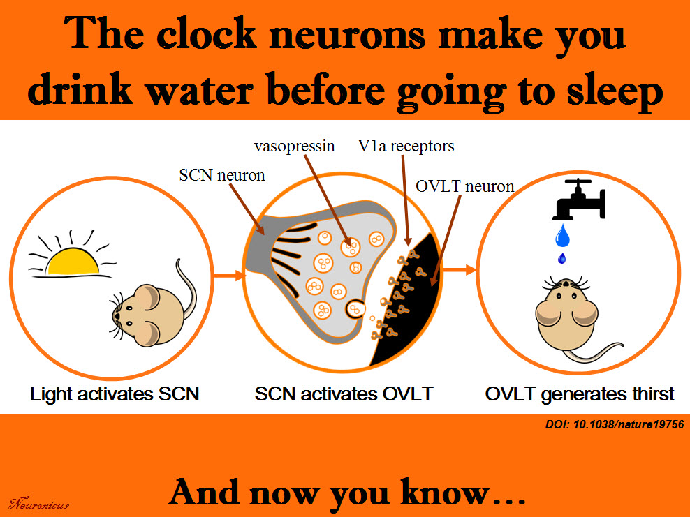
SCN neurons release vasopressin and that excites the OVLT neurons via V1a receptors. This is necessary and sufficient to make the animal drink the water, even if it’s not thirsty.

That’s a lot of techniques used in a lot of experiments for only three authors. Ten years ago, you needed only one, maybe two techniques to prove the same point. Either there have been a lot of students and technicians who did not get credit (there isn’t even an Acknowledgements section. EDIT: yes, there is, see the comments below or, if they’re missing, the P.S.) or these three authors are experts in all these techniques. In this day and age, I wouldn’t be surprised by either option. No wonder small universities have difficulty publishing in Big Name journals; they don’t have the resources to compete. And without publishing, no tenure… And without tenure, less research… And thus shall the gap widen.

Musings about workload aside, this is a great paper, shedding light on yet another mysterious behavior and elucidating the mechanism behind it. There’s still work to be done though, like answering how accurate is the SCN in predicting bedtime to activate the drinking behavior. Does it take its cues from light only? Does ambient temperature play a role and so on. This line of work can help people that work in shifts to prevent certain health problems. Their SCN is out of rhythm and that can influence deleteriously the activity of a whole slew of organs.

scn-h2o-copy
Summary of the doi: 10.1038/nature19756 findings. 1) The light is a cue for suprachiasmatic nulceus (SCN) that bedtime is near. 2) The SCN vasopressin neurons that project to organum vasculosum lamina terminalis (OVLT) are activated. 3) The OVLT generates the anticipatory thirst. 4) The animal drinks fluids.

Reference: Gizowski C, Zaelzer C, & Bourque CW (28 Sep 2016). Clock-driven vasopressin neurotransmission mediates anticipatory thirst prior to sleep. Nature, 537(7622): 685-688. PMID: 27680940. DOI: 10.1038/nature19756. ARTICLE

By Neuronicus, 5 October 2016

EDIT (12 Oct 2016): P.S. The blog comments are automatically deleted after a period of time. In case of this post that would be a pity because I have been fortunate to receive comments from at least one of the authors of the paper, the PI, Dr. Charles Bourque and, presumably under pseudonym, but I don’t know that for sure, also the first author, Claire Gizowski. So I will include here, in a post scriptum, the main idea of their comments. Here is an excerpt from Dr. Bourque’s comment:

“Let me state for the record that Claire accomplished pretty much ALL of the work in this paper (there is a description of who did what at the end of the paper). More importantly, there were no “unthanked” undergraduates, volunteers or other parties that contributed to this work.”

My hat, Ms. Gizowski. It is tipped. To you. Congratulations! With such an impressive work I am sure I will hear about you again and that pretty soon I will blog about Dr. Gizowski.

Fructose bad effects reversed by DHA, an omega-3 fatty acid

Despite alarm signals raised by various groups and organizations regarding the dangers of the presence of sugars – particularly fructose derived from corn syrup – in almost every food in the markets, only in the past decade there has been some serious evidence against high consumption of fructose.

A bitter-sweet (sic!) paper comes from Meng et al. (2016) who, in addition to showing some bad things that fructose does to brain and body, it also shows some rescue from its deleterious effects by DHA (docosahexaenoic acid), an omega-3 fatty acid.

The authors had 3 groups of rodents: one group got fructose in their water for 6 weeks, another group got fructose and DHA, and another group got their normal chow. The amount of fructose was calculated to be ecologically valid, meaning that they fed the animals the equivalent of 1 litre soda bottle per day (130 g of sugar for a 60 Kg human).

The rats that got fructose had worse learning and memory performance at a maze test compared to the other two groups.

The rats that got fructose had altered gene expression in two brain areas: hypothalamus (involved in metabolism) and hippocampus (involved in learning and memory) compared to the other two groups.

The rats that got fructose had bad metabolic changes that are precursors for Type 2 diabetes, obesity and other metabolic disorders (high blood glucose, triglycerides, insulin, and insulin resistance index) compared to the other two groups.

86 - Copy.jpg

The genetic analyses that the researchers did (sequencing the RNA and analyzing the DNA methylation) revealed a whole slew of the genes that had been affected by the fructose treatment. So, they did some computer work that involved Bayesian modeling  and gene library searching and they selected two genes (Bgn and Fmod) out of almost a thousand possible candidates who seemed to be the drivers of these changes. Then, they engineered mice that lacked these genes. The resultant mice had the same metabolic changes as the rats that got fructose, but… their learning and memory was even better than that of the normals? I must have missed something here. EDIT: Well… yes and no. Please read the comment below from the Principal Investigator of the study.

It is an ok paper, done by the collaboration of 7 laboratories from 3 countries. But there are a few things that bother me, as a neuroscientist, about it. First is the behavior of the genetic knock-outs. Do they really learn faster? The behavioral results are not addressed in the discussion. Granted, a genetic knockout deletes that gene everywhere in the brain and in the body, whereas the genetic alterations induced by fructose are almost certainly location-specific.

Which brings me to the second bother: nowhere in the paper (including the supplemental materials, yeas, I went through those) are any brain pictures or diagrams or anything that can tell us which nuclei of the hypothalamus the samples came from. Hypothalamus is a relatively small structure with drastically different functional nuclei very close to one another. For example, the medial preoptic nucleus that deals with sexual hormones is just above the suprachiasmatic nucleus that deals with circadian rhythms and near the preoptic is the anterior nucleus that deals mainly with thermoregulation. The nuclei that deal with hunger and satiety (the lateral and the ventromedial nucleus, respectively) are located in different parts of the hypothalamus. In short, it would matter very much where they got their samples from because the transcriptome and methylome would differ substantially from nucleus to nucleus. Hippocampus is not so complicated as that, but it also has areas with specialized functions. So maybe they messed up the identification of the two genes Bgn and Fmod as drivers of the changes; after all, they found almost 1 000 genes altered by fructose. And that mess-up might have been derived by their blind hypothalamic and hippocampal sampling. EDIT: They didn’t mess up,  per se. Turns out there were technical difficulties of extracting enough nucleic acids from specific parts of hypothalamus for analyses. I told you them nuclei are small…

Anyway, the good news comes from the first experiment, where DHA reverses the bad effects of fructose. Yeay! As a side note, the fructose from corn syrup is metabolized differently than the fructose from fruits. So you are far better off consuming the equivalent amount on fructose from a litre of soda in fruits. And DHA comes either manufactured from algae or extracted from cold-water oceanic fish oils (but not farmed fish, apparently).

If anybody that read the paper has some info that can help clarify my “bothers”, please do so in the Comment section below. The other media outlets covering this paper do not mention anything about the knockouts. Thanks! EDIT: The last author of the paper, Dr. Yang, was very kind and clarified a bit of my “bothers” in the Comments section. Thanks again!

Reference: Meng Q, Ying Z, Noble E, Zhao Y, Agrawal R, Mikhail A, Zhuang Y, Tyagi E, Zhang Q, Lee J-H, Morselli M, Orozco L, Guo W, Kilts TM, Zhu J, Zhang B, Pellegrini M, Xiao X, Young MF, Gomez-Pinilla F, Yang X (2016). Systems Nutrigenomics Reveals Brain Gene Networks Linking Metabolic and Brain Disorders. EBioMedicine, doi: 10.1016/j.ebiom.2016.04.008. Article | FREE fulltext PDF | Supplementals | Science Daily cover | NeuroscienceNews cover

By Neuronicus, 24 April 2016

Prostaglandins in the sickness syndrome

63woman-698962_960_720When you’re sick you also feel awful: no appetite, weak, sleepy, feverish, achy, and so on. This is called, appropriately so, the sickness syndrome.

Saper, Romanovsky & Scammell (2012) wrote a beautiful review of the neural circuits underlying this collection of symptoms. In a nutshell, the immune system releases cytokines to fight the inflammation, which in turn stimulate the release of prostaglandins. Prostaglandins bind to various areas in the brain to produce the sickness syndrome symptoms. Below are outlined four simplified brain circuits which the non-specialists can skip entirely.

  1. Prostaglandins in the median preoptic nucleus lead to a cascade involving dorsomedial hypothalamus, rostral medullary raphe and finally the spinal cord to produce fever by activating the brown adipose tissue.
  2. Prostaglandins in the preoptic area lead to the inhibition of the brain’s analgesic system involving the descending projections of the periaqueductal grey to spinal cord, thus promoting achiness.
  3. Prostaglandins in the meninges result in adenosine release in nucleus accumbens and ventrolateral preoptic nucleus which, downstream, end in inhibiting the arousal system to produce sleepiness.
  4. Prostaglandins in the arcuate nucleus lead to inhibition of several hypothalamic nuclei involved in promoting feeding, thereby producing anorexia.

The sickness syndrome and the role prostaglandins play in it has tremendous adaptive role, as it promotes rest and recuperation. So don’t blame them too much. And if you’re really done feeling sick, take some non-steroid anti-inflammatory drugs, like aspirin, which inhibit the prostaglandins’ synthesis very effectively. That’s how and why NSAIDs work.

Reference: Saper CB, Romanovsky AA & Scammell TE (26 Jul 2012). Neural Circuitry Engaged by Prostaglandins during the Sickness Syndrome. Nature Neuroscience, 15(8):1088-95. doi: 10.1038/nn.3159. Article | FREE Fulltext PDF

By Neuronicus, 21 December 2015