Tomato transcriptome

As most children, growing up I showed little appreciation for what I had, coveting instead what I did not. Now I realize how fortunate I have been to have grown up half the time in a metropolis and the other half at the countryside. At the farm. A subsistence farm, although I truly loathe the term because we were not just subsisting but thriving off the land, as we planted and harvested a bit of everything and we had a specimen or four of almost all the farm animals, from bipeds to quadrupeds.

I got on this memory lane after reading the paper of Shinozaki et al. (2018) on tomatoes. It was a difficult read for me as it was punctured by many term definition lookups since botany evolved quite steeply since the last time I checked, about 25 years or so.

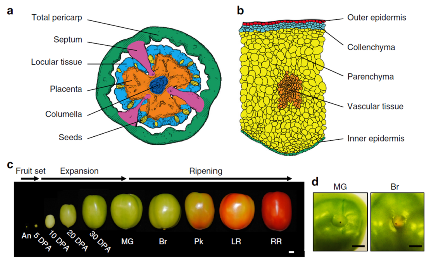
Briefly, the scientists grew tomato plants in a greenhouse at Cornell, NY. They harvested the fruit from 60 plants about 5 to 50 days after the flower was at its peak (DPA, days post anthesis) following this chart:

  • Expanding [fruit] stage (harvested at 5, 10, 20, or 30 DPA)
  • Mature Green stage (full-size green fruit, ≈ 39 DPA),
  • Breaker stage (definite break in color from green to tannish-yellow with less than 10% of the surface, ≈ 42 DPA),
  • Pink stage (50% pink or red color, ≈ 44 DPA),
  • Light red stage (100% light red, ≈ 46 DPA),
  • Red ripe stage (full red for 8 days, ≈ 50 DPA).

(simplified from the Methods section, p. 10, see pic)

130-shinozaki - Copy
Fig. 1 (partial from Shinozaki et al., 2018). A tissue/cell-based transcript profiling of developing tomato fruit. a Traced image of six targeted fruit tissues. Shaded areas of the total pericarp and the placenta were not harvested. b Traced image of five pericarp cells. c Representative pictures of harvested fruit spanning ten developmental stages. d Representative pictures of the stylar end of MG and Br stage fruit. DPA, days post anthesis; MG, mature green; Br, breaker; Pk, pink; LR, light red; RR, red ripe. Credit: DOI: 10.1038/s41467-017-02782-9. License: CC BY 4.0 IL.

Immediately after harvesting, the tomato was scanned with a micro-computed tomograph (micro-CT) to generate a 3D image of the fruit, including its internal structures. Then, the fruit was dissected by hand or laser, depending of its size, divided into various tissue types and then preserved either via snap-freezing in liquid nitrogen or standard tissue fixation for light or transmission electron microscopy. Finally, the researchers used kits to extract and analyze the RNA from their samples. And, last but not least, a lot of math & stats.

This is what I got out of it:

  1. A total of 24,660 genes were uniquely expressed in various tomato cell types and at various stages of development.
  2. The tomato ripens from within, meaning from the interior to the exterior and not the other way around.
  3. The ripening seems to be a continuous process, starting before the ‘Breaker’ stage.
  4. The ripening signals originate in the locular tissues (the goo around the seeds; it’s possible that the seeds themselves send the signals to the locular tissue to start the ripening process).
  5. The flesh of the fruit is only one part of the tomato and the most investigated, but the other types of tissue are also important. For example, some genes responsible for aroma and flavor (CTOMT1, TOMLOXC) are predominantly or even exclusively expressed in the flesh, but some genes that improve the nutritional value (SlGAD3) are expressed mostly in the placenta.
  6. The fruit can do photosynthesis, probably for the benefit of its seeds.
  7. Each developmental stage is characterized by a distinct transcriptome profile (by inference, also a distinct proteomic profile, although not necessarily in exact correspondence)
  8. Botany, like any serious science, is complicated.

Ah, I have been vindicated. By science, nonetheless! You see, in my pursuit to recapture the tomato taste of my childhood I sample various homegrown exemplars of Solanum lycopersicum derived both from more or less failed personal attempts with pots on the balcony and from various farmer’s market vendors. While I can understand – though not approve of – the industrial scale agro-growers’ practice to pick the tomatoes green, unripe and then artificially injecting them with ethylene to prolong shelf life, I completely fail to understand the picking them up when green by the sellers in the farmer’s markets. I had many surreal conversations with such vendors (I cannot call them farmers for the life of me) who more than once attempted to reassure me that 1) Everybody’s picking tomatoes green off the vine because that’s how it’s done and 2) Ripening happens on the window sill. In vain have I tried to explain the difference between ripen and rotten; in vain have I pointed out that color is only one indicator of ripening; in vain did I explain that during ripening on the vine the plant delivers certain substances to the fruit that lead to changes in the flesh composition to make it more nutritious for the future seedling, process that the aforesaid window sill does nor partake in. Alas, ultimately, my arguments (and my family’s last 400 years of farming experience) hit the wall of “I am growing tomatoes for three years now and I know what I’m doing. Are you buying or not?” As you might imagine, I end up going home frustrated and yet staring at some exorbitantly expensive and looking as sad as I feel greenish tomatoes.

For me, this is what Shinozaki et al. (2018) validated: Ripening is a complex process that involves a lot of physiological changes in the fruit, not merely some extra production of ethylene that can be conveniently supplied externally by a syringe or rotting on the window sill. Of course, there is nowhere in the paper that Shinozaki et al. (2018) say that. What they do say is this: “The ripening program is revealed as comprising gradients of gene expression, initiating in internal tissues then radiating outward, and basipetally along a latitudinal axis. We also identify spatial variations in the patterns of epigenetic control superimposed on ripening gradients” (Abstract). Tomayto, tomahto…

Now we know that… simply put, I’m right. Sometimes is good to be right. I am old enough to prefer happiness and tranquility over rightness & righteousness, but still young enough that sometimes, just sometimes, it feels good to be right. Yes, the Shinozaki et al. (2018) paper exists only for my vindication in my farmer’s market squabbles and not for providing a huge comprehensive atlas on the tomato transcriptome, along with an awesome spatiotemporal map showing the place and time of the expression of genes responsible for fruit ripening, quality traits and so on.

Good job, Shinozaki et al. (2018)!

130-tomato - Copy

REFERENCE: Shinozaki Y, Nicolas P, Fernandez-Pozo N, Ma Q, Evanich DJ, Shi Y, Xu Y, Zheng Y, Snyder SI, Martin LBB, Ruiz-May E, Thannhauser TW, Chen K, Domozych DS, Catalá C, Fei Z, Mueller LA, Giovannoni JJ, & Rose JKC (25 Jan 2018). High-resolution spatiotemporal transcriptome mapping of tomato fruit development and ripening. Nature Communications, 9(1):364. PMID: 29371663, PMCID: PMC5785480, DOI: 10.1038/s41467-017-02782-9. ARTICLE | FREE FULLTEXT PDF | The Tomato Expression Atlas database

By Neuronicus, 7 February 2018

Interview with Jason D. Shepherd, PhD

During the first week of the publication, a Cell paper that I covered a couple of weeks ago has received a lot of attention from media outlets, like The Atlantic, Scicasts and Neuroscience News/University of Utah Press Release. It is not my intention to duplicate here their wonderfully done summaries and interviews; rather to provide answers to some geeky questions arisen from the minds of nerdy scientists like me.

Dr. Shepherd, you are the corresponding author of a paper published on Jan. 11 in Cell about a protein heavily involved in memory formation, called Arc. Your team and another team from University of Massachusetts, who published in the same issue of Cell, simultaneously discovered that Arc looks like and behaves like a virus. The protein “infects” nearby cells, in this case neurons, with instructions of how to make more of itself, i.e. it shuttles its own mRNA from one cell to another.

Neuronicus: Why is this discovery so important?

​Jason D. Shepherd: I think there’s a couple of big implications of this work:

  1. ​The so called “junk” DNA in our genomes that come from viruses and transposable elements actually provide source material for new genes. Arc isn’t the first example, but it’s the first prominent brain gene to have these kinds of origins.

  2. This is the first demonstration that cellular proteins are capable of assembling into capsid-like structures. This is a completely new way of thinking about communication between cells.

  3. We think there may be other genes that can also form capsids, suggesting this method of signaling is fairly common in organisms.

N: 2) When you and your colleagues compared Arc’s genetic sequence across species you concluded Arc comes from a virus that infected four-legged animals some time ago. A little time later the virus infected the flies too. When did these events occur?

​JDS: So we think the origins are from a retrotransposon not a virus. These are DNA sequences or elements that “jump” into the host genome. Think of them as primitive viruses. Indeed, these elements are thought to be the ancestors of retroviruses like HIV. The mammalian Arc gene seems to have originated ~400 million years ago, the fly about 150 million years ago. ​

N: 3) So, if Arc has been so successfully repurposed by the tetrapod and fly ancestors to add memory formation, what does that mean for the animals and insects before the infection? I understand that we move now in the realm of speculation, but who better to speculate on these things than the people who work on Arc? The question is: did these pre-infection creatures have bad and short memories? The alternate view would be that they had similar memory abilities due to a different mechanism that was replaced by Arc. Which one do you think is more likely?

​JDS: Good question. It’s certainly the case that memory capacity improved in tetrapods, but unclear if Arc is the sole reason. I suspect that Arc confers some unique aspects to brains, otherwise it would not have been so conserved after the initial insertion event, but I also think there are probably other Arc-like genes in other organisms that do not have Arc. I will also note that we are not even sure, yet, that the fly Arc is important for fly memory/learning.

N: 4) Remaining in the realm of speculation, if this intercellular mRNA transport proves to be ubiquitous for a variety of mRNAs, what does that say of the transcriptome of a cell at any given time? From a practical point of view, a cell is what is made off, meaning the ensemble of all its enzymes and proteins and so on, collectively termed transcriptome. So if a cell can just alter its neighbor’s transcriptome array, does that mean that it’s possible to alter also its function? Even more outrageously speculative, perhaps even its type? Can we make cancer cells commit suicide by shooting Arc capsules of mRNA at them?

​JDS: Yes! Cool ideas. I think this is quite likely, that these signaling extracellular vesicles can dramatically alter the state of a cell. We are obviously looking into this. ​

N: 5) Finally, in the paper, the Arc capsules containing mRNA are referred to as ACBAR (Arc Capsid Bearing Any RNA). At first I thought it was a reference to “Allahu akbar” which is Arabic for ‘God is greatest’, the allusion being “ACBAR! Our exosome is the greatest!” or “Arc Acbar! Our Arc is the greatest!”. Is this where the naming is coming from?

​JDS: No no. As I said on twitter, my lab came up with this acronym because we are all Star Wars nerds and the classic “It’s a trap!” line from general Ackbar seemed apt for something that was trapping RNA. ​

Below is the Twitter exchange Dr. Shepherd refers to:

129shepherd - Copy

Dr. Shepherd, thank you for your time! And congratulations on a well done paper and a well told story. Your Methods section is absolutely great; anybody can follow the instructions and replicate your data. Somebody in your lab must have kept great records. Congratulations again!

129_starwars - Copy
The ACBAR graphic is from the Cell’s abstract (©2017 Elsevier Inc.) but since it’s for comedic purposes, I’d say is fair use. Same for the Lego Ackbar.

By Neuronicus, 28 January 2018

P. S. Since I have obviously managed to annoy the #StarWars universe and twitterverse because I depicted General Ackbar using a Jedi sword when he’s not a Jedi, I thought only fair to annoy the other half of the world, the #trekkies. So here you go:

129_startrek - Copy.jpg


Arc: mRNA & protein from one neuron to another

EDIT 1 [Jan 17, 2018]: I promised four days ago that I will post this, while it was still hot, but my Internet was down, thanks to the only behemoth provider in USA. And rated the worst company in the Nation, too. You definitely know by now about whom I’m talking about. Grrrr…  Anyway, here is the paper:

As promised, today’s paper talks about mRNA transfer between neurons.

Pastuzyn et al. (2018) looked at the gene Arc in neurons because they thought its Gag sequence looks suspiciously similar to some retroviruses. Could it be possible that it also behaves like a virus?

Arc is heavily involved in the immune system, is essential for the formation of long-term memories, and is involved in all sorts of diseases, like schizophrenia and Alzheimer’s, among other things.

Pastuzyn et al. (2018) is a relatively long and dense paper, albeit well written. So, I thought that this time, instead of giving you a summary of their research it would be better to give you the authors’ story directly in their own words written as subtitles in the Results section (bold letters – the authors words, normal font – mine). Warning: this is a much more jargon-dense blog post than my previous one on the same topic and, because it is so much material, I will not explain every term.

  • Fly and Tetrapod (us) Arc Genes Independently Originated from Distinct Lineages of Ty3/gypsy Retrotransposons, the phylogenomic analyses tell us, meaning the authors have done a lot of computer-assisted comparisons of similar forms of the gene in hundreds of species.
  • Arc Proteins Self-Assemble into Virus-like Capsids. Arc likes to oligomerize spontaneously (dimers and trimers). The oligomers resemble virus-like capsids, similar to HIV.
  • Arc Binds and Encapsulates RNA. Although it loves its own RNA about 10 times more than other RNAs, it’s a promiscuous protein (doesn’t care which RNA as long as it follows the rules of stoichiometry). Arc capsids encapsulate both the Arc protein (maybe other proteins too?), its mRNA, and whatever mRNA happened to be in the vicinity at the time of encapsulation. Arc capsids are able to protect the mRNA from RNAases.
  • Arc Capsid Assembly Requires RNA. If there is no RNA around, the capsids are few and poorly formed.
  • Arc Protein and Arc mRNA Are Released by Neurons in Extracellular Vesicles. Arc capsid packages Arc protein & Arc mRNA into extracellular vesicles (EV). The size of these EVs is < 100nm, putting them in the exosome category. This exosome, which the authors gave the unfortunate name of ACBAR (Arc Capsid Bearing Any RNA), is being expelled from cortical neurons in an activity-dependent manner. In other words, when neurons are stimulated, they release ACBARs.
  • Arc Mediates Intercellular Transfer of mRNA in Extracellular Vesicles. ACBARs dock to the host cell and then undergo clathrin-dependent endocytosis, meaning they expel their cargo in the host cell. The levels of Arc protein and Arc mRNA peaks in a host hippocampal cell in four hours from incubation. The ACBARs tend to congregate around donor cell’s dendrites.
  • Transferred Arc mRNA Can Undergo Activity-Dependent Translation. Activating the group 1 metabotropic glutamate receptor (mGluR1/5) by application of the agonist DHPG induces a significant increase of the amount of Arc protein in the host neurons.

This is a veritable tour de force paper. The Results section has 7 sub-sections, each with multiple experiments to dot every i and cross every t. I’m eyeballing about 40 experiments. It is true that there are 13 authors on the paper from different institutions – yeay for collaboration! – but c’mon! Is this what you need to get in Cell these days? Apparently so. Don’t get me wrong, this is an outstanding paper. But in the end it is still only one paper, which means only one first author. The rest are there for the ride because for a tenure track application nobody cares about your papers in CNS (Cell, Nature, Science = The Central Nervous System of the scientific community, har, har) if you’re not the first author. It looks like the increasing amount of work you need to be published in top tier journals these days is becoming a pet peeve of mine as I keep mentioning it (for example, here).

My pet peeves aside, Pastuzyn et al. (2018) is an excellent paper that opens interesting practical (drug delivery) and theoretical (biological repurpose of ancient invaders) gates. Kudos!

128-1 - Copy

REFERENCE: Pastuzyn ED, Day CE, Kearns RB, Kyrke-Smith M, Taibi AV, McCormick J, Yoder N, Belnap DM, Erlendsson S, Morado DR, Briggs JAG, Feschotte C, & Shepherd JD. (11 Jan 2018). The Neuronal Gene Arc Encodes a Repurposed Retrotransposon Gag Protein that Mediates Intercellular RNA Transfer. Cell, 172(1-2):275-288.e18. PMID: 29328916. doi: 10.1016/j.cell.2017.12.024. ARTICLE | FULLTEXT PDF via ResearchGate

P.S. I said that ACBAR is an unfortunate acronym because I don’t know about you but I for one wouldn’t want my discovery to be linked either with a religion or with terrorist cries, even if that link is done only by a small fraction of the population. Although I can totally see the naming-by-committee going: “ACBAR! Our exosome is the greatest! Yeay!” or “Arc Acbar! Our Arc is the greatest. Double yeay!”. On a second thought, it’s kindda nerdy geeky neat. I still wouldn’t have done it though…

By Neuronicus, 14 January 2018

EDIT 2 [Jan 22, 2018]: There is another paper that discovered that Arc forms capsids that encapsulate RNA and then shuttles it across the neuromuscular junction in Drosophila (fly). To their credit, Cell published both these papers back-to-back so no researcher gets scooped of their discovery. From what I can see, the discovery really happened simultaneously, so I modified my infopic to reflect that (both papers were submitted in January 2017, received in revised version on August 15, 2017 and published in the same issue on January 11, 2018). Here is the reference to the other article:

Ashley J, Cordy B, Lucia D, Fradkin LG, Budnik V, & Thomson T (11 Jan 2018). Retrovirus-like Gag Protein Arc1 Binds RNA and Traffics across Synaptic Boutons, Cell. 172(1-2): 262-274.e11. PMID: 29328915. doi: 10.1016/j.cell.2017.12.022. ARTICLE

EDIT 3 [Jan 29, 2018]: Dr. Shepherd, the last author of the paper I featured, was kind enough to answer a few of my questions about the implications of his and his team’s findings, answers which you will find here.

By Neuronicus, 22 January 2018